. 2013:3:1932.
doi: 10.1038/srep01932.

Intravital analysis of vascular permeability in mice using two-photon microscopy

Affiliations

Intravital analysis of vascular permeability in mice using two-photon microscopy

Gyohei Egawa et al. Sci Rep. 2013.

Abstract

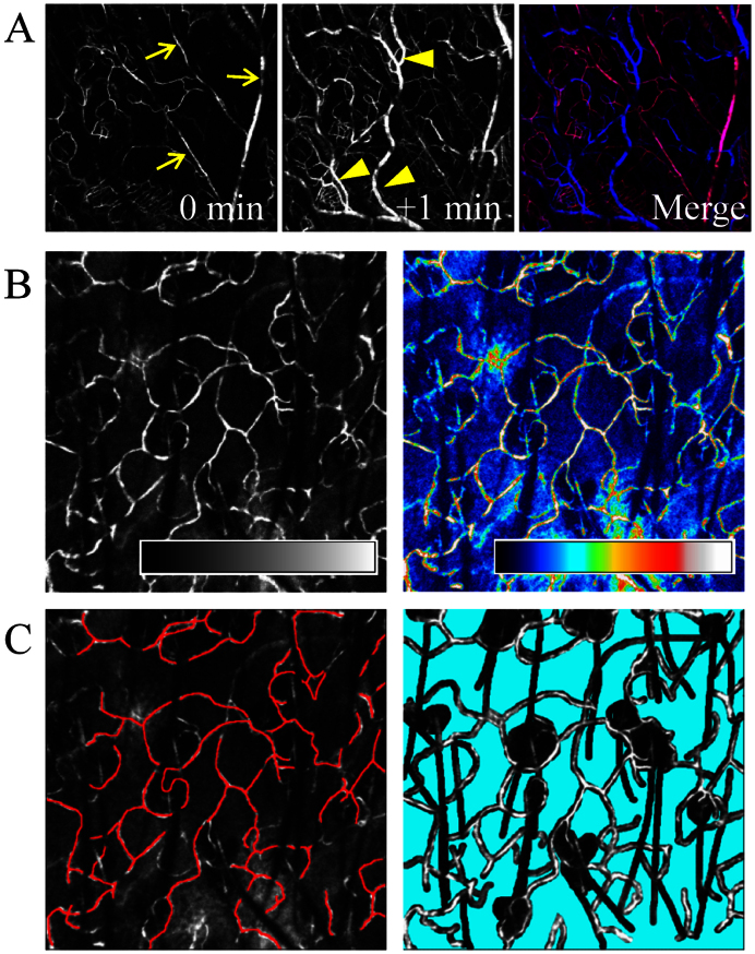
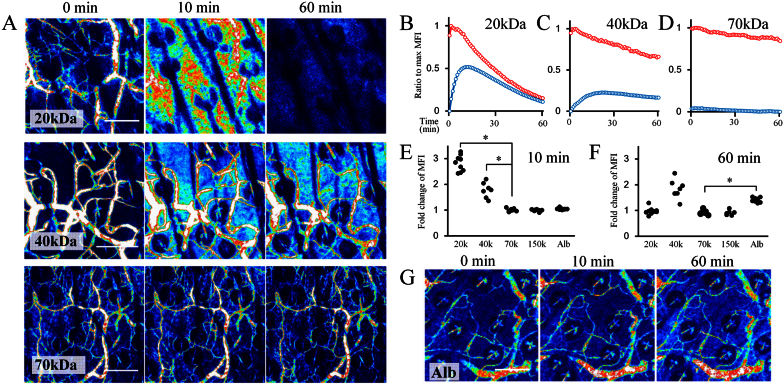
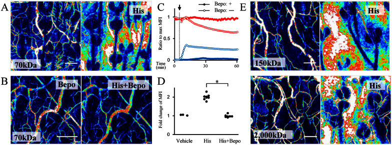
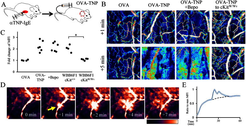
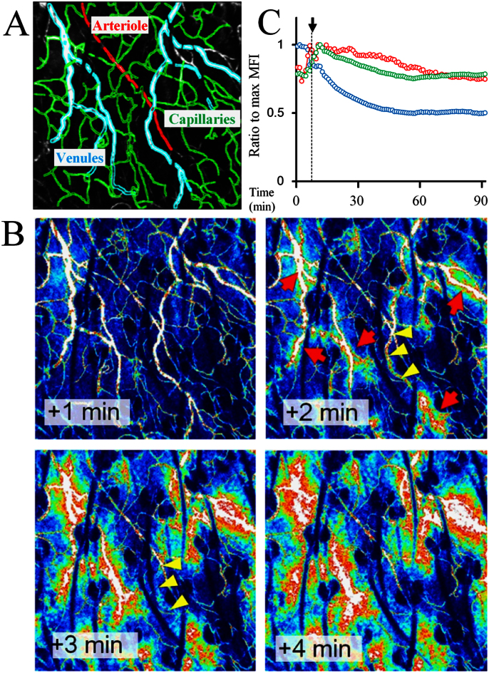
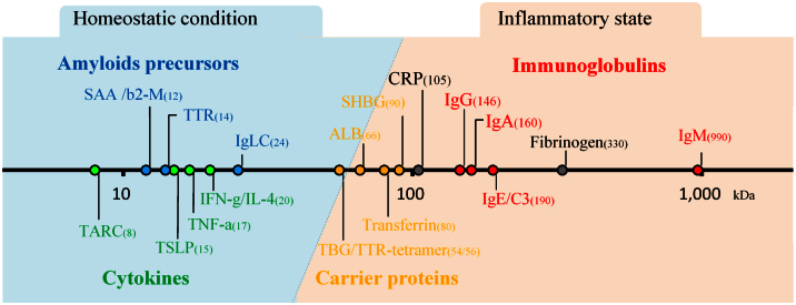
Blood vessel endothelium forms a semi-permeable barrier and its permeability controls the traffics of plasma contents. Here we report an intravital evaluation system for vascular permeability in mice using two-photon microscopy. We used various sizes of fluorescein-conjugated dextran as a tracer and its efflux was quantified by measuring the changes of fluorescent intensity both on the blood vessel area and the interstitial space. Using this system, we demonstrated that skin blood vessels limited the passage of dextran larger than 70 kDa under homeostatic conditions. We evaluated the kinetics of vascular permeability in histamine- or IgE-induced type I allergic models and a hapten-induced type IV allergic model. In such inflammatory conditions, the hyperpermeability was selectively induced in the postcapillary venules and dextran as large as 2000-kDa leaked from the bloods. Taken together, our study provides a convenient method to characterize the skin blood vessels as a traffic barrier in physiological conditions.


Figures

Figure 1
Figure 1. Bloodstream visualization with fluorescent-dextran.
(A) FITC-dextran injection first delineated arterioles (arrows) and then capillaries and postcapillary venules (arrow heads). The merged image depicts vessels at 0 min (red) and 1 min (blue) after the injection, respectively. (B) The black-and-white images (left) were converted to rainbow color-scale images (right) according to fluorescent intensity. (C) The blood vessel area (left: red) and the interstitial space (right: cyan) were separately circumscribed to monitor the fluorescent intensity.
Figure 2
Figure 2. Vascular permeability under basal conditions.
(A) Sequential images taken 0, 10, and 60 min after FITC-dextran injection. The molecular sizes of the injected dextran are shown in the bottom left of each image. (B–D) Kinetics of the MFI in the blood vessel area (red) and interstitial space area (blue) following injection of 20 kDa , 40 kDa, and 70 kDa dextrans, respectively. All experiments were performed at least three times with similar findings. (E, F) Fold change of the MFI in the interstitial space at 10 min or 60 min after the injection of FITC-dextran (20–150 kDa) or albumin. MFI in the interstitial space at 1 min was used as basis. *P < 0.05. (G) Sequential images of FITC-albumin injection. Scale bar = 100 μm.
Figure 3
Figure 3. Vascular hyper-permeability induced by injection of histamine.
(A, B) Sequential images before (left) and 5 min after (right) histamine injection. Mice were pretreated with an antihistamine agent, bepotastine besilate or vehicle 1 hour before injection. (C) Kinetics of the MFI in the blood vessel area (red) and in the interstitial space (blue). The arrow denotes the timepoint of histamine injection. Mice were pretreated with bepotastine (closed circles) or vehicle (open circles). (D) Fold changes of the MFI in the interstitial space 5 min after the injection of vehicle or histamine with or without antihistamine pretreatment. MIF at 1 min before the injection was used as basis. *P < 0.05. (E) Sequential images before (left) and 5 min after (right) histamine injection. The molecular sizes of the injected dextran are shown in the bottom left of each image. His; histamine, Bepo; bepotastine besilate. Scale bar = 100 μm.
Figure 4
Figure 4. Vascular hyper-permeability in models of anaphylaxis and CHS.
(A) Passive anaphylaxis model. Anti-TNP IgE antibody was injected intraperitoneally into mice. The following day, vascular permeability was analyzed in the ear after an intravenous injection of OVA-TNP. (B) Sequential images 1 and 5 minutes after the injection of OVA and OVA-TNP with or without bepotastine in the passive anaphylaxis model. In the rightmost panel, OVA-TNP was injected into WBB6F1-kitW/Wv mice. (C) Fold change of the MFI in the interstitial area 5 min after the OVA or OVA-TNP injection. MIF at 1 min before the injection was used as basis. *P < 0.05. (D) Serial images of the bloodstream in CHS-induced inflamed skin. The arrow indicates transient hyper-permeability in a localized area. Elapsed time is shown in the bottom right of each image. (E) Kinetics of the MFI in the interstitial space in (D). The dotted line represents the predicted baseline in this area.
Figure 5
Figure 5. Hyper-permeability occurs in postcapillary venules.
(A) Microvasculature in the ear skin. Arteriole (red), capillaries (green), and post capillary venules (blue) are shown. (B) Sequential images after histamine injection. Elapsed time is shown in the bottom left of each image. Red arrows indicate initiation sites of leakage from postcapillary venules. Some areas of postcapillary venules became undetectable thereafter (yellow arrowheads). (C) Kinetics of the MFI in the area of arterioles (red), capillaries (green), and post capillary venules (blue) after histamine injection. Arrow denotes the timepoint of histamine injection.
Figure 6
Figure 6. Molecular size and the permeability of plasma proteins.
Molecular size of plasma proteins. TARC: thymus and activation-regulated chemokine; SSA: serum amyloid A; b2-M: b2-microglobulin; TTR: transthyretin; TSLP: thymic stromal lymphopoietin; Ig-LC: immunoglobulin light chain; TBG: thyroxin-binding globulin; ALB: albumin; SHBG: sex hormone binding globulin; CRP: c-reactive protein.

References

    1. Mehta D. & Malik A. B. Signaling mechanisms regulating endothelial permeability. Physiol Rev 86, 279–367 (2006). - PubMed
    1. Vandenbroucke E., Mehta D., Minshall R. & Malik A. B. Regulation of endothelial junctional permeability. Ann N Y Acad Sci 1123, 134–145 (2008). - PubMed
    1. Nagy J. A., Benjamin L., Zeng H., Dvorak A. M. & Dvorak H. F. Vascular permeability, vascular hyperpermeability and angiogenesis. Angiogenesis 11, 109–119 (2008). - PMC - PubMed
    1. Albelda S. M., Sampson P. M., Haselton F. R., McNiff J. M., Mueller S. N. et al. Permeability characteristics of cultured endothelial cell monolayers. J Appl Physiol 64, 308–322 (1988). - PubMed
    1. Del Vecchio P. J., Siflinger-Birnboim A., Shepard J. M., Bizios R., Cooper J. A. et al. Endothelial monolayer permeability to macromolecules. Fed Proc 46, 2511–2515 (1987). - PubMed

Publication types

MeSH terms