. 2013 Jun;18(6):067003.
doi: 10.1117/1.JBO.18.6.067003.

Dual-wavelength photoacoustic technique for monitoring tissue status during thermal treatments

Affiliations

Dual-wavelength photoacoustic technique for monitoring tissue status during thermal treatments

Yi-Sing Hsiao et al. J Biomed Opt. 2013 Jun.

Abstract

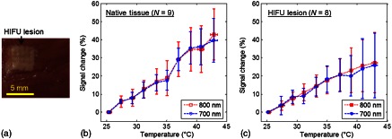
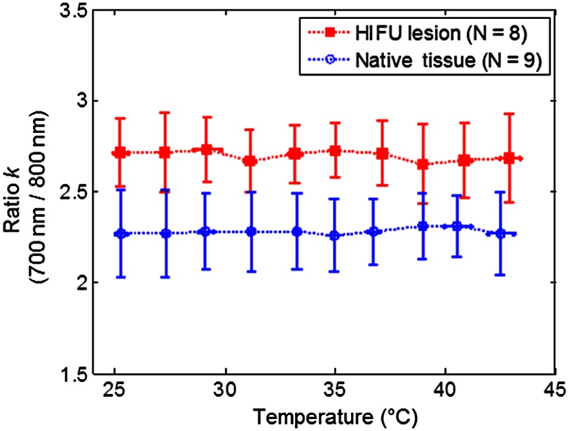
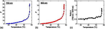
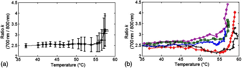
Photoacoustic (PA) techniques have been exploited for monitoring thermal treatments. However, PA signals depend not only on tissue temperature but also on tissue optical properties which indicate tissue status (e.g., native or coagulated). The changes in temperature and tissue status often occur simultaneously during thermal treatments, so both effects cause changes to PA signals. A new dual-wavelength PA technique to monitor tissue status independent of temperature is performed. By dividing the PA signal intensities obtained at two wavelengths at the same temperature, a ratio, which only depends on tissue optical properties, is obtained. Experiments were performed with two experimental groups, one with untreated tissue samples and the other with high-intensity focused ultrasound treated tissue samples including thermal coagulated lesion, using ex vivo porcine myocardium specimens to test the technique. The ratio of PA signal intensities obtained at 700 and 800 nm was constant for both groups from 25 to 43°C, but with distinct values for the two groups. Tissue alteration during thermal treatment was then studied using water bath heating of tissue samples from 35 to 60°C. We found that the ratio stayed constant before it exhibited a marked increase at around 55°C, indicating tissue changes at this temperature.


Figures

Fig. 1
Fig. 1
Schematic illustration of experimental setup for (a) HIFU ablation and (b) PA sensing in a temperature-controlled water bath. (c) and (d) Typical PA signal and photodiode signal. Vp-p denotes the peak-to-peak amplitude.
Fig. 2
Fig. 2
(a) Gross photo of a tissue specimen with an HIFU lesion generated before PA experiment. (b) and (c) The relative signal change (%) of the averaged normalized PA amplitude P as a function of temperature in native tissues and in HIFU lesions. T0=25.3±0.3°C.
Fig. 3
Fig. 3
Comparison of the dual-wavelength ratio k (700  nm/800  nm) in HIFU lesions and native tissues as a function of temperature.
Fig. 4
Fig. 4
An example of coagulation during water bath heating. (a) and (b) The averaged normalized PA amplitude P as a function of temperature at 700 and 800 nm wavelengths. (c) Corresponding dual-wavelength ratio k (700  nm/800  nm).
Fig. 5
Fig. 5
Dual-wavelength ratio k (700  nm/800  nm) during the coagulation process: (a) Mean±S.D. (N=5) and (b) individual plots as a function of temperature (°C).

Similar articles

Cited by

References

    1. Fennessy F. M., Tempany C. M., “A review of magnetic resonance imaging-guided focused ultrasound surgery of uterine fibroids,” Top. Magn. Reson. Imag. 17(3), 173–179 (2006).TMRIEY10.1097/RMR.0b013e3180337e1f - DOI - PubMed
    1. Anand A., Savery D., Hall C., “Three-dimensional spatial and temporal temperature imaging in gel phantoms using backscattered ultrasound,” IEEE Trans. Ultrason. Ferroelectr. Freq. Control 54(1), 23–31 (2007).ITUCER10.1109/TUFFC.2007.208 - DOI - PubMed
    1. Arthur R. M., et al. , “3-D in vitro estimation of temperature using the change in backscattered ultrasonic energy,” IEEE Trans. Ultrason. Ferroelectr. Freq. Control 57(8), 1724–1733 (2010).ITUCER10.1109/TUFFC.2010.1611 - DOI - PubMed
    1. Hynynen K., “MRI-guided focused ultrasound treatments,” Ultrasonics 50(2), 221–229 (2010).ULTRA310.1016/j.ultras.2009.08.015 - DOI - PubMed
    1. Miller N. R., Bamber J. C., Meaney P. M., “Fundamental limitations of noninvasive temperature imaging by means of ultrasound echo strain estimation,” Ultrasound Med. Biol. 28(10), 1319–1333 (2002).USMBA310.1016/S0301-5629(02)00608-7 - DOI - PubMed

Publication types