. 2013 Aug 1;122(5):749-58.
doi: 10.1182/blood-2013-01-480129. Epub 2013 Jun 3.

Acute myeloid leukemia creates an arginase-dependent immunosuppressive microenvironment

Affiliations

Acute myeloid leukemia creates an arginase-dependent immunosuppressive microenvironment

Francis Mussai et al. Blood. .

Abstract

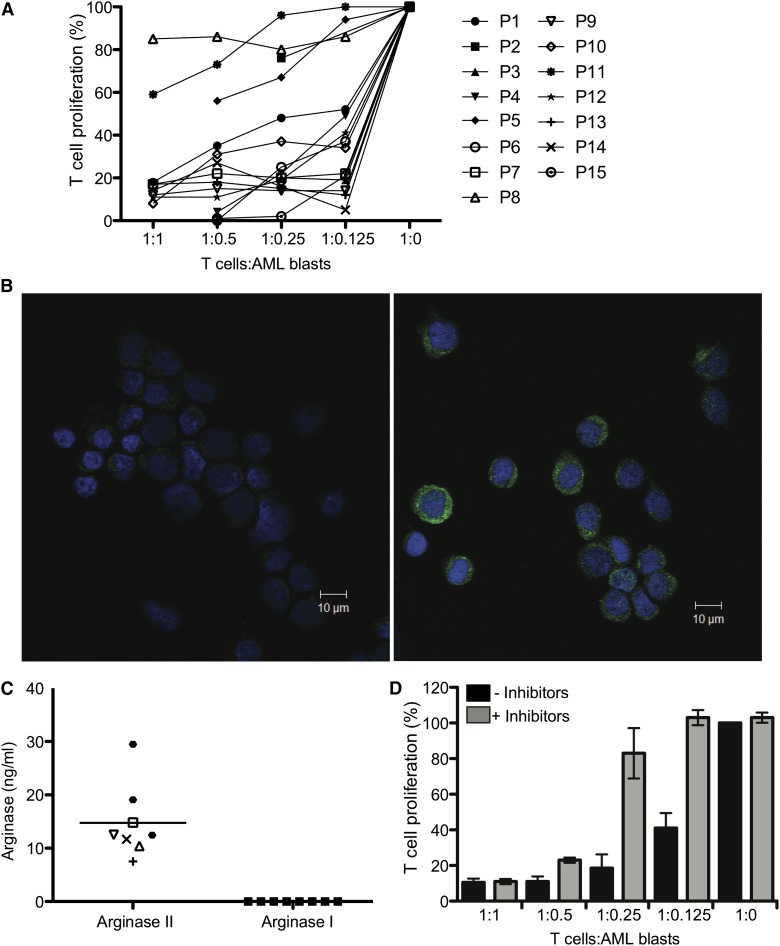
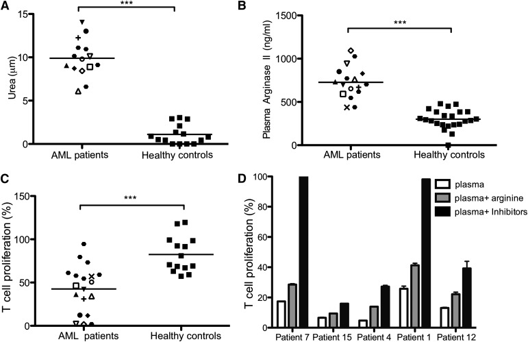
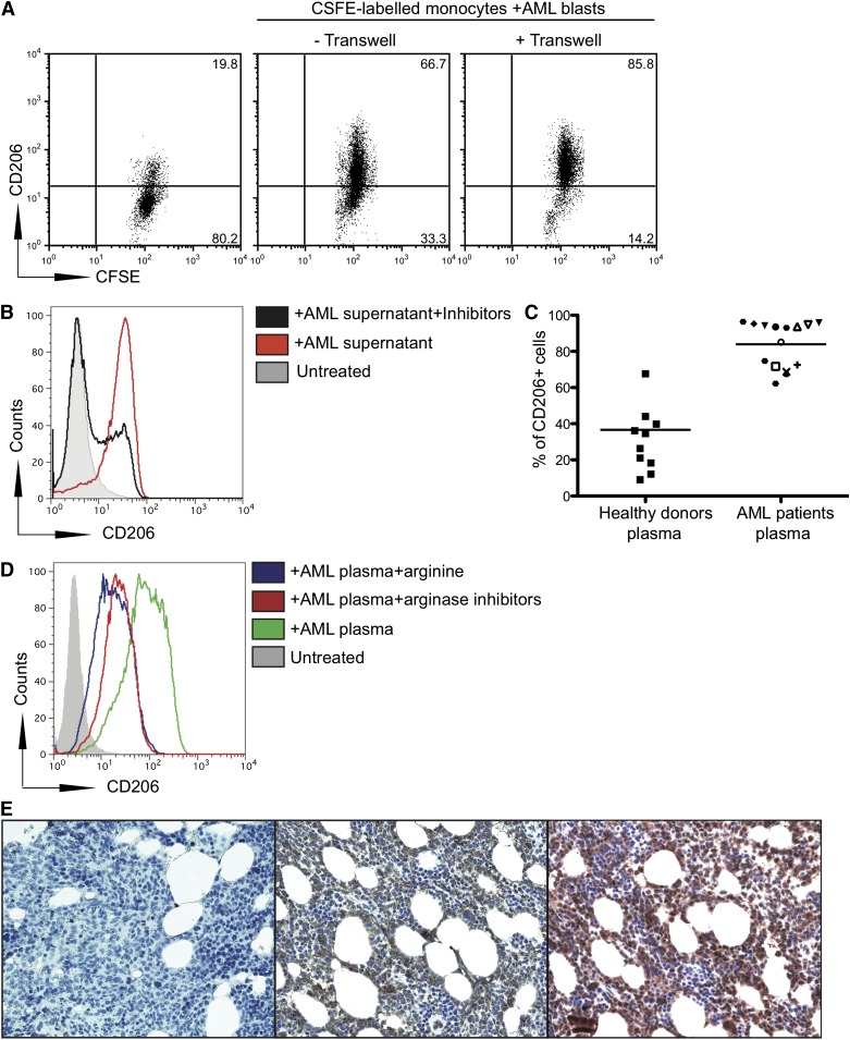
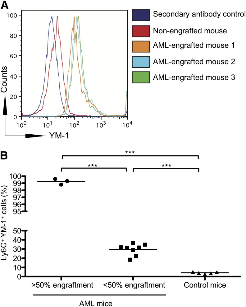
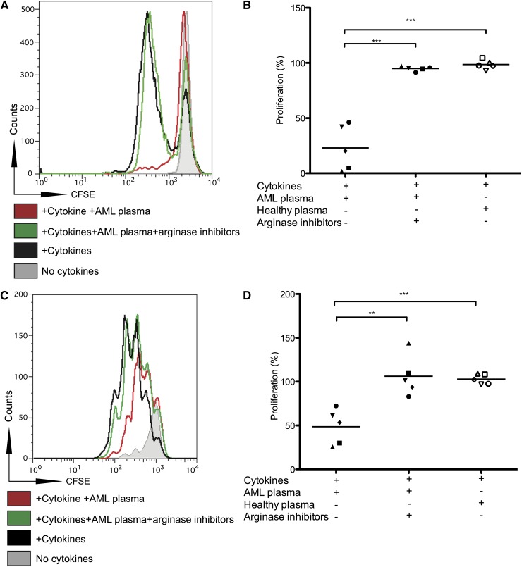
Acute myeloid leukemia (AML) is the most common acute leukemia in adults and the second most common frequent leukemia of childhood. Patients may present with lymphopenia or pancytopenia at diagnosis. We investigated the mechanisms by which AML causes pancytopenia and suppresses patients' immune response. This study identified for the first time that AML blasts alter the immune microenvironment through enhanced arginine metabolism. Arginase II is expressed and released from AML blasts and is present at high concentrations in the plasma of patients with AML, resulting in suppression of T-cell proliferation. We extended these results by demonstrating an arginase-dependent ability of AML blasts to polarize surrounding monocytes into a suppressive M2-like phenotype in vitro and in engrafted nonobese diabetic-severe combined immunodeficiency mice. In addition, AML blasts can suppress the proliferation and differentiation of murine granulocyte-monocyte progenitors and human CD34(+) progenitors. Finally, the study showed that the immunosuppressive activity of AML blasts can be modulated through small-molecule inhibitors of arginase and inducible nitric oxide synthase, suggesting a novel therapeutic target in AML. The results strongly support the hypothesis that AML creates an immunosuppressive microenvironment that contributes to the pancytopenia observed at diagnosis.


Figures

Figure 1
Figure 1
Arginine metabolism regulates the suppressive activity of AML blasts. (A) AML blasts from 15 patients were cultured with allogeneic T cells in a MLR. The ratio of AML cells:T cells ranged from 1:1 to 1:0. T-cell proliferation was measured by 3H-thymidine incorporation after 4 days. AML blasts from 12/15 patients strongly suppressed T-cell proliferation. Patients are identified by unique symbols, which are used consistently throughout the manuscript. (B) Expression of arginase II in blasts from patients with AML was confirmed by confocal microscopy. (Left) Staining with DAPI (nuclear stain) and secondary antibody alone; (right) staining with DAPI, anti-human arginase II antibody, and secondary antibody. Scale bar = 10 μm. (C) AML blasts release arginase II into the microenvironment. Supernatants from cultures (24 hours) of patients’ AML blasts were analyzed by ELISA for arginase II and arginase I (***P = .0001). (D) AML blasts from patients were cultured with allogeneic T cells in a MLR in the presence of the enzyme-specific inhibitors for arginase (NOHA) and iNOS (L-NMMA). The ratio of AML cells:T cells ranged from 1:1 to 1:8. T-cell proliferation was measured by 3H-thymidine incorporation after 4 days. Culture with the enzyme inhibitors restores T-cell proliferation. Data are representative of 5 independent experiments (error bars, SD).
Figure 2
Figure 2
AML blasts extend their suppressive microenvironment through high concentrations of active arginase II in patient plasma. (A) Arginase activity from plasma of 15 patients with AML and 15 healthy donors was analyzed (***P = .0001). Fifty microliters of patient plasma was tested for the ability to convert arginine into urea, using a colorimetric assay. (B) Plasma (50 μL) from 17 patients with AML and 21 healthy donors was analyzed for arginase II concentration by ELISA (***P = .0001). (C) T-cell proliferation of alloreactive T cells stimulated by allogeneic DC in a total volume of 200 μL, with 50 μL of plasma from patients with AML collected at time of diagnosis or from healthy donors (***P = .0001). (D) T cells from healthy donors were cultured in a MLR in the presence of plasma from patients with AML and the enzyme-specific inhibitors for arginase (NOHA) and iNOS (L-NMMA) or arginine. T-cell proliferation was measured by 3H-thymidine incorporation after 4 days. Culture with the enzyme inhibitors or arginine replacement restores T-cell proliferation. Data are representative of 5 independent experiments (error bars, SD).
Figure 3
Figure 3
AML blasts increase CD206 expression on monocytes. (A) Transwell culture of CFSE-labeled monocytes from healthy donors with AML blasts from patients upregulates CD206 expression on monocytes. Monocytes from healthy donors were placed in the lower well and AML blasts from patients were placed in the upper well of a transwell assay system. Representative flow cytometry plots from 1 patient of 11 tested are shown. (B) Coculture of CFSE-labeled monocytes from healthy patients with supernatants (50% of final volume) from AML blasts upregulates CD206 on monocytes, as analyzed by flow cytometry. (C) Upregulation of CD206 on monocytes cultured with plasma of 15 patients with AML or 15 healthy donors (P < .0001). Percentage of CD206+ cells is shown. (D) Upregulation of CD206 on monocytes cultured with plasma of patients with AML with or without arginine (100 ng/mL) and inhibitors (0.5 mM). Data are representative of 3 independent experiments (E) Staining of bone marrow from patients with AML at diagnosis with anti-human CD206 (center), with DAPI alone (left), and with anti-arginase II (right). Representative marrow from a single patient with AML shown of 6 (samples obtained from University of Oxford Biobank).
Figure 4
Figure 4
Monocytes from NOD-SCID mice engrafted with human AML express increased YM-1. (A) NOD-SCID mice were injected with AML cells from patients. After engraftment, bone marrow was harvested from the femurs, and the expression of YM-1 on murine monocytes (Ly6C+) was assessed by flow cytometry. (B) Increased percentage of YM-1 positive monocytes in NOD-SCID mice engrafted with human AML. NOD-SCID mice were injected with AML cells from patients. After engraftment, bone marrow was harvested from the femurs, and the percentage of YM-1+ Ly6C+ murine monocytes was assessed by flow cytometry (***P = .0001).
Figure 5
Figure 5
The AML microenvironment suppresses GMP and CD34+ progenitors. (A) plasma from patients with AML suppresses murine GMP proliferation. CFSE-labeled murine GMPs were isolated and cultured in the presence of plasma from patients with AML and in the presence of L-NMMA (0.5 mM) and NOHA (0.5 mM). A representative histogram plot of 5 patient AML plasma experiments is shown. Independent experiments were performed on 2 separate occasions. (B) AML-induced GMP suppression is overcome by the addition of L-NMMA and NOHA. GMPs cultured in the presence of AML plasma (50% of final volume) have significantly reduced proliferation compared with those cultured in the presence of healthy donor plasma (P = .0001) or AML plasma with L-NMMA (0.5 mM) and NOHA (0.5 mM) (P = .0001). (C) Plasma from patients with AML suppresses human CD34+ HSC proliferation. CFSE-labeled human CD34+ HSCs were isolated and cultured in the presence of plasma from AML patients and in the presence of L-NMMA (0.5 mM) and NOHA (0.5 mM). Data are representative of 5 experiments with plasma from patients with AML. Independent experiments were performed on 2 separate occasions. (D) AML-induced human CD34+ HSC suppression is overcome by the addition of L-NMMA and NOHA. CD34+ HSCs cultured in the presence of AML plasma have significantly reduced proliferation compared with those cultured in the presence of healthy donor plasma (P = .0002) or AML plasma with L-NMMA and NOHA (P = .003). Independent experiments were performed on 2 separate occasions.

Comment in

References

    1. Marcucci G, Haferlach T, Döhner H. Molecular genetics of adult acute myeloid leukemia: prognostic and therapeutic implications. J Clin Oncol. 2011;29(5):475–486. - PubMed
    1. Woods WG, Neudorf S, Gold S, et al. Children’s Cancer Group. A comparison of allogeneic bone marrow transplantation, autologous bone marrow transplantation, and aggressive chemotherapy in children with acute myeloid leukemia in remission. Blood. 2001;97(1):56–62. - PubMed
    1. Horowitz MM, Gale RP, Sondel PM, et al. Graft-versus-leukemia reactions after bone marrow transplantation. Blood. 1990;75(3):555–562. - PubMed
    1. Gibson BE, Wheatley K, Hann IM, et al. Treatment strategy and long-term results in paediatric patients treated in consecutive UK AML trials. Leukemia. 2005;19(12):2130–2138. - PubMed
    1. Orleans-Lindsay JK, Barber LD, Prentice HG, Lowdell MW. Acute myeloid leukaemia cells secrete a soluble factor that inhibits T and NK cell proliferation but not cytolytic function—implications for the adoptive immunotherapy of leukaemia. Clin Exp Immunol. 2001;126(3):403–411. - PMC - PubMed

Publication types

MeSH terms