. 2014 May;102(5):1361-9.
doi: 10.1002/jbm.a.34814. Epub 2013 Jun 11.

One-dimensional patterning of cells in silicone wells via compression-induced fracture

Affiliations

One-dimensional patterning of cells in silicone wells via compression-induced fracture

Angela R Dixon et al. J Biomed Mater Res A. 2014 May.

Abstract

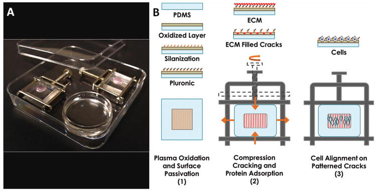
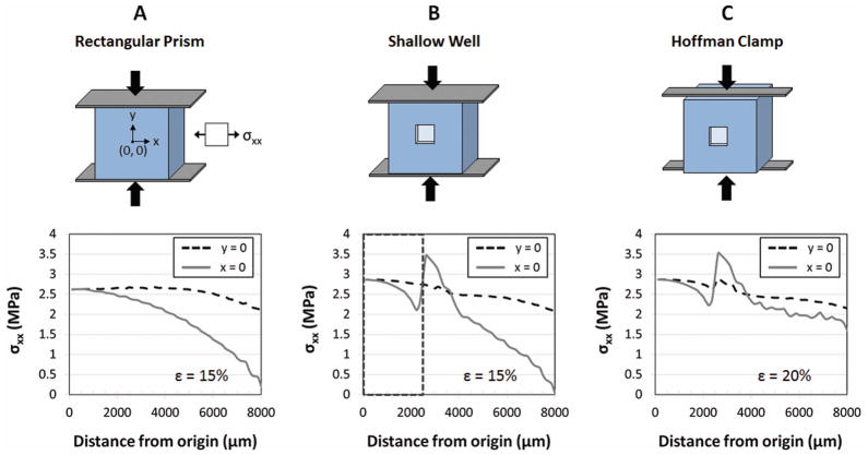
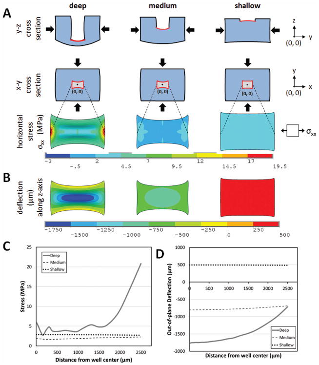
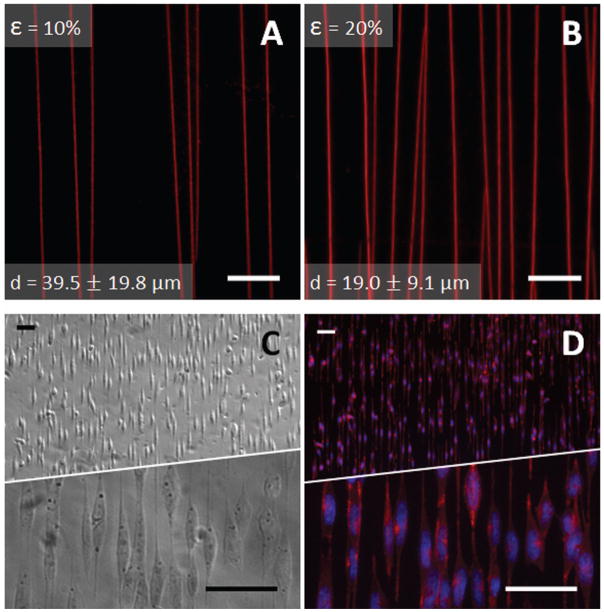
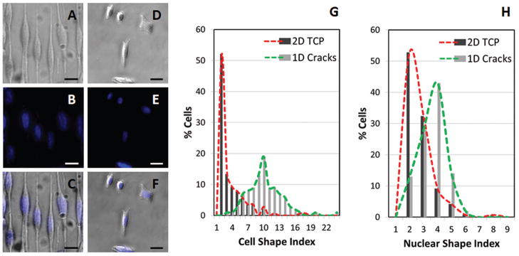
We have adapted our existing compression-induced fracture technology to cell culture studies by generating linear patterns on a complex cell culture well structure rather than on simple solid constructs. We present a simple method to create one-dimensional (1D), submicron, and linear patterns of extracellular matrix on a multilayer silicone material. We identified critical design parameters necessary to optimize compression-induced fracture patterning on the wells, and applied stresses using compression Hoffman clamps. Finite-element analyses show that the incorporation of the well improves stress homogeneity (stress variation = 25%), and, thus, crack uniformity over the patterned region. Notably, a shallow well with a thick base (vs. deeper wells with thinner bases) reduces out-of-plane deflections by greater than a sixth in the cell culture region, improving clarity for optical imaging. The comparison of cellular and nuclear shape indices of a neuroblast line cultured on patterned 1D lines and unpatterned 2D surfaces reveals significant differences in cellular morphology, which could impact many cellular functions. Because 1D cell cultures recapitulate many important phenotypical traits of 3D cell cultures, our culture system offers a simple means to further study the relationship between 1D and 3D cell culture environments, without demanding expensive engineering techniques and expertise.

Keywords: biomaterial design; compression; fracture; patterning; polydimethyl siloxane.


Figures

Figure 1
Figure 1
Device set-up and cell alignment scheme. (A) Wells mounted in Hoffman clamps are housed in square petri dishes, containing an open petri dish (round) of water for humidification. (B) Plasma oxidized wells are coated with silane that serves as a binding site for Pluronic blocking agents. The passivated well is filled with extracellular matrix (ECM) protein solution and mounted in a Hoffman clamp. Cracks form in the oxidized surface layer when pressure is applied by tightening the screw clamp, and protein fills the cracks, presenting an adhesive surface for cells.
Figure 2
Figure 2
Comparison of lateral surface stresses (σxx) generated perpendicular to applied compressive stresses in various culture systems. (A) Compression of a simple rectangular prism, under ideal no-slip clamping conditions, yields stresses ranging from 0 to 2.6 MPa across the strained surface for an applied compression of 15%. (B) Incorporating a shallow well, with ideal clamping conditions, limits cells to an area (dotted box) in which stresses range from 2.1 to 2.7 MPa for similar applied strains. (C) Utilizing a Hoffman clamp, which allows some freedom of the clamped ends, can yield stresses, within the cell culture region, that are similar in magnitude and uniformity to those in (B) when the applied strain is increased to 20%. Lateral stress (σxx) is shown along line y = 0 and line x = 0 at a distance from the origin (0, 0). Cell culture region spans 2500 μm from the origin in all directions along x and y.
Figure 3
Figure 3
Effect of geometry on mechanical behavior of culture wells under compression. (A) Lateral stresses generated perpendicular to applied compressive strains of 15% in deep (0.5 mm thick base), medium (5 mm thick base) and shallow (9.5 mm thick base) wells. (B) Out-of-plane deflection of the wells resulting from applied compression. (C) Lateral stresses and (D) out-of-plane deflection across the center of the wells (centered at origin x = 0, y = 0) demonstrate an increase in stress uniformity and decrease in magnitude of out-of-plane displacement with decreasing well depth. Compressive stress, tensile stress, and deflection are reported along the y-axis, x-axis, and z-axis respectively.
Figure 4
Figure 4
Crack spacing, protein adherence, and staining of aligned cells. Cracks were visualized with TRITC-BSA (red), and the average spacing resulting from (A) 10% and (B) 20% compressive loads was respectively 39.5 ± 19.8 μm and 19.0 ± 9.1 μm (mean ± S.D.). Data was collected from five different fields in the well region of each of three cuboids for each compressive load. Scale bar = 25 μm (C) Phase-contrast image of N27 cells aligned on cracks. (D) Aligned N27 cells were counterstained with FITC-labeled Phalloidin (red) and DAPI (blue, to label nuclei) revealing aligned actin filaments and elongated nuclei. Scale Bar = 50 μm.
Figure 5
Figure 5
Comparison of patterned 1D and unpatterned 2D N27 cells quantified by Cell Shape Index (CSI) and Nuclear Shape Index (NSI). Images of cells (phase-contrast) and nuclei (stained with DAPI, blue) and overlay of cells and nuclei for cells aligned on 1D crack substrates (A, B, C) and on 2D tissue culture plastic (TCP) substrates (D, E, F). Scale bar = 25 μm. Histogram comparisons of patterned and nonpatterned cells for CSI (G) and NSI (H). Total cells for 1D crack = 100. Total cells for 2D TCP = 142. Cell culture duration = 8 hrs. Significant differences exist between both the CSI and NSI mean values when comparing morphologies on the two culture surfaces (p < 0.001).

References

    1. Smalley K, Lioni M, Herlyn M. Life isn’t flat: taking cancer biology to the next dimension. In Vitro Cell Dev Biol Anim. 2006;42:242–247. - PubMed
    1. Pampaloni F, Reynaud E, Stelzer E. The third dimension bridges the gap between cell culture and live tissue. Nature Rev Mol Cell Biol. 2007;8:839–845. - PubMed
    1. Justice B, Badr N, Felder R. 3D cell culture opens new dimensions in cell-based assays. Drug Discov Today. 2009;14:102–107. - PubMed
    1. Yamada K, Cukierman E. Modeling tissue morphogenesis and cancer in 3D. Cell. 2007;130:601–610. - PubMed
    1. Kraning-Rush C, Carey S, Califano J, Smith B, Reinhart-King C. The role of the cytoskeleton in cellular force generation in 2D and 3D environments. Phys Biol. 2011;8(1):015009. - PMC - PubMed

Publication types