. 2014 Jan;472(1):98-104.
doi: 10.1007/s11999-013-2898-6.

Constitutional varus does not affect joint line orientation in the coronal plane

Affiliations

Constitutional varus does not affect joint line orientation in the coronal plane

Jan M K Victor et al. Clin Orthop Relat Res. 2014 Jan.

Abstract

Background: In a previous study, we described the distribution of coronal alignment in a normal asymptomatic population and recognized the occurrence of constitutional varus in one of four individuals. It is important to further investigate the influence of this condition on the joint line orientation and how the latter is affected by the onset and progression of arthritis.

Questions/purposes: The purposes of this study are (1) to describe the distribution of joint line orientation in the coronal plane in the normal population; (2) to compare joint line orientation between patients with constitutional varus and neutral mechanical alignment; and (3) to compare joint line orientation between a cohort of patients with prearthritic constitutional varus and a cohort of patients with established symptomatic varus arthritis.

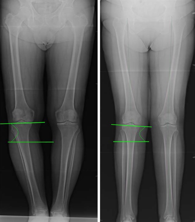
Methods: Full-leg standing hip-to-ankle digital radiographs were performed in 248 young healthy individuals and 532 patients with knee arthritis. Hip-knee-ankle (HKA) angle and tibial joint line angle (TJLA) were measured in the coronal plane. Patients were subdivided into varus (HKA ≤ -3°), neutral, and valgus (HKA ≥ 3°).

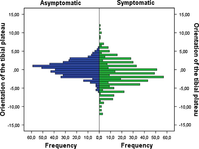
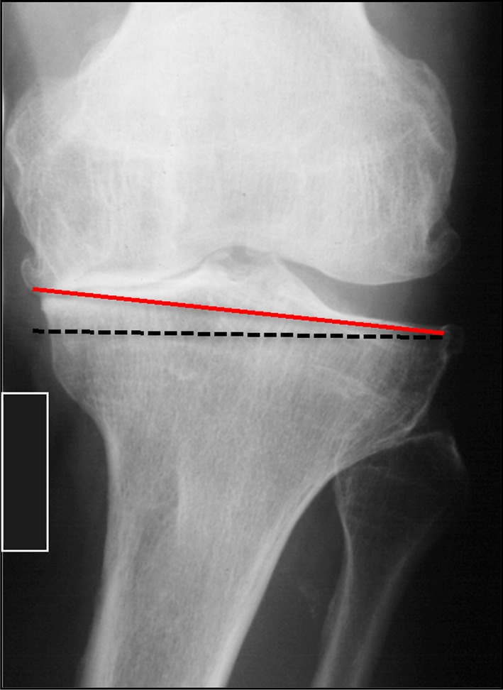
Results: The mean TJLA in healthy subjects was 0.3° (SD 2.0°). TJLA was parallel to the floor in healthy subgroups with neutral alignment (TJLA 0.3°, SD 1.9) and constitutional varus (TJLA 0.2°, SD 2.2°). In patients with symptomatic arthritis and varus alignment, the TJLA opened medially (mean -1.9°, SD 3.5°).

Conclusions: Constitutional varus does not affect joint line orientation. Advanced medial arthritis causes divergence of the joint line from parallel to the floor. These findings influence decision-making for osteotomy and alignment in total knee arthroplasty.


Figures

Fig. 1
Fig. 1
An example is shown of a medial open tibial joint line angle (left) and a lateral open tibial joint line angle (right).
Fig. 2
Fig. 2
Distribution of the joint line orientation angles in the symptomatic and asymptomatic cohorts is demonstrated.
Fig. 3
Fig. 3
Distribution of the joint line orientation angles in the symptomatic and asymptomatic cohorts for the subgroup with constitutional varus is demonstrated.
Fig. 4A–C
Fig. 4A–C
(A) Geometric illustration of normal alignment. When the angle a between the floor and the mechanical axis of the tibia equals the angle formed between the mechanical axis of the tibia and the tibial plateau, the joint is parallel to the floor. (B) Geometric illustration of constitutional varus. The proximal tibia displays an increased varus configuration, reducing the angle between the tibial plateau and the mechanical axis of the tibia to a′. As the tibia is pushed outward, the angle between the floor and the mechanical axis of the tibia is also reduced to a′ and the joint line remains parallel to the floor. (C) Geometric illustration of constitutional varus with advanced arthritis. When bone loss is occurring at the level of the distal femur, the intrinsic geometry of the tibia remains unchanged and the angle between the tibial plateau and the mechanical axis remains a′. However, as the tibia is further pushed outward, the angle between the tibial mechanical axis and the floor is reduced to a″ resulting in a medial opening of TJLA (see also Fig. 5). TMA = tibial mechanical axis; FMA = femoral mechanical axis.
Fig. 5
Fig. 5
Example of an arthritic knee with constitutional varus (taken from a full-leg standing radiograph). The loss of distal femoral bone pushes the knee further outward and changes the joint line orientation. The red line represents the tangential to the proximal tibia; the dotted black line is the parallel to the floor. Compare the radiographic image with Fig. 4C.

References

    1. Ahlbäck S. Osteoarthrosis of the knee: a radiographic investigation. Acta Radiol Diagn (Stockh). 1968;Suppl 277:7–72. - PubMed
    1. Bellemans J, Colyn W, Vandenneucker H, Victor J. The Chitranjan Ranawat award: is neutral mechanical alignment normal for all patients? The concept of constitutional varus. Clin Orthop Relat Res. 2012;470:45–53. doi: 10.1007/s11999-011-1936-5. - DOI - PMC - PubMed
    1. Belo JN, Berger MY, Reijman M, Koes BW, Bierma-Zeinstra SMA. Prognostic factors of progression of osteoarthritis of the knee: a systematic review of observational studies. Arthritis Rheum. 2007;57:13–26. doi: 10.1002/art.22475. - DOI - PubMed
    1. Birmingham TB, Kramer JF, Kirkley A, Inglis JT, Spaulding SJ, Vandervoort AA. Association among neuromuscular and anatomic measures for patients with knee osteoarthritis. Arch Phys Med Rehabil. 2001;82:1112–1118. - PubMed
    1. Brouwer GM, van Tol AW, Bergink AP, Belo JN, Bernsen RMD, Reijman M. Association between valgus and varus alignment and the development and progression of radiographic osteoarthritis of the knee. Arthritis Rheum. 2007;56:1204–1211. doi: 10.1002/art.22515. - DOI - PubMed