Review
. 2011 Jul;1(3):1175-232.
doi: 10.1002/cphy.c100049.

Pathophysiology of the diabetic kidney

Affiliations
Review

Pathophysiology of the diabetic kidney

Volker Vallon et al. Compr Physiol. 2011 Jul.

Abstract

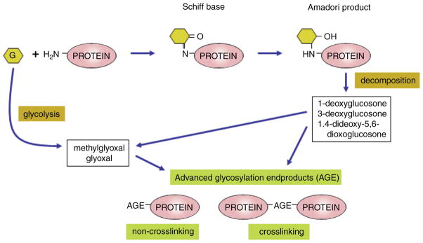
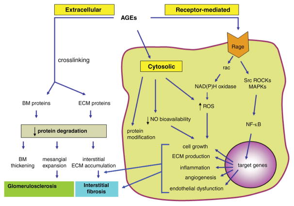
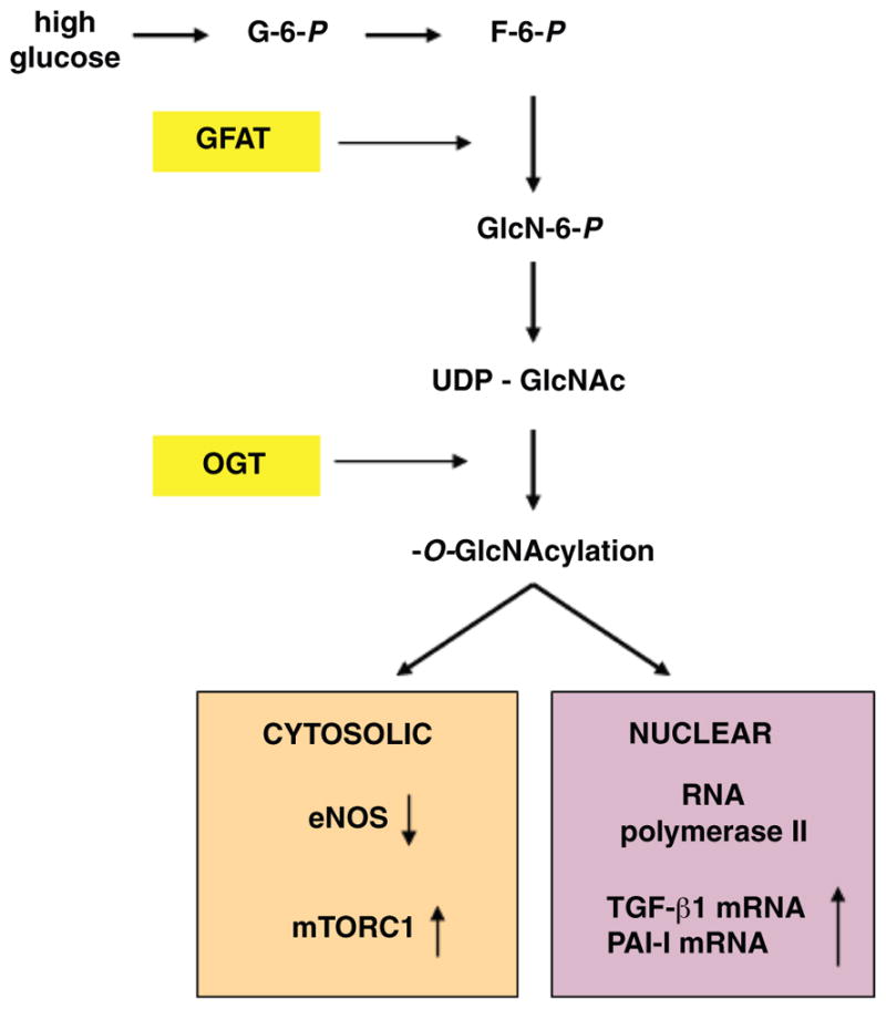
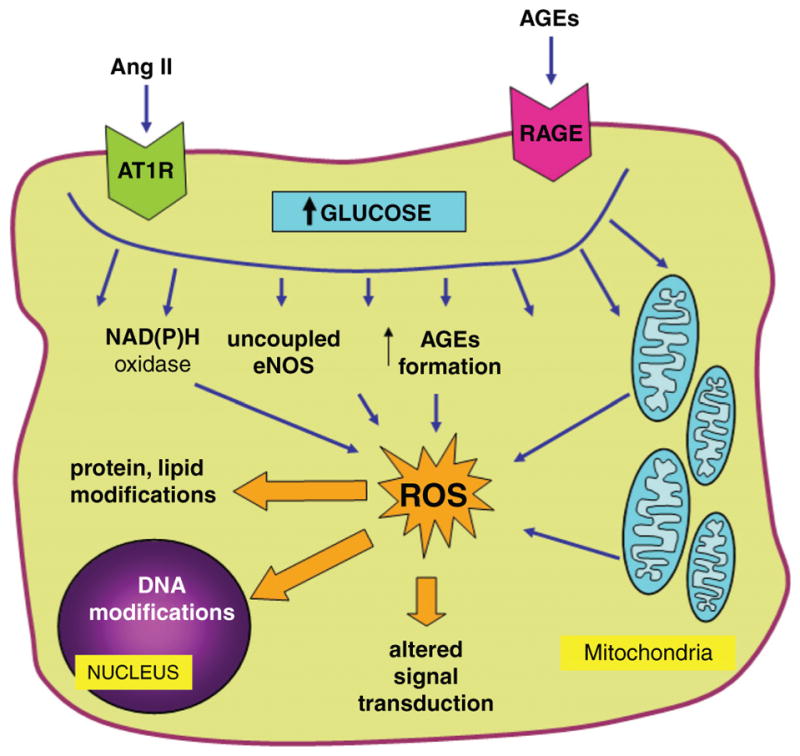
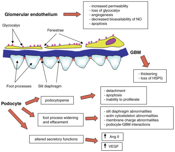
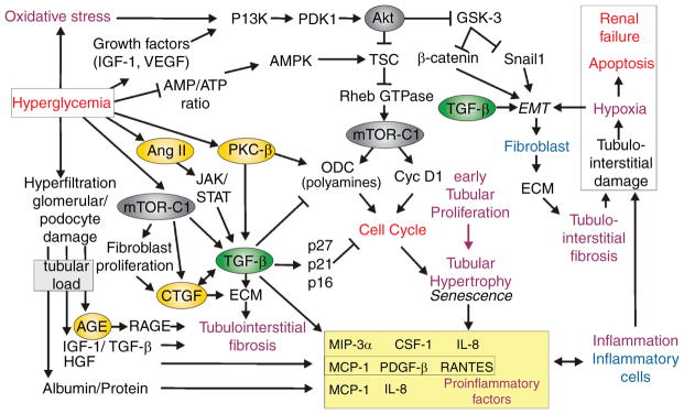
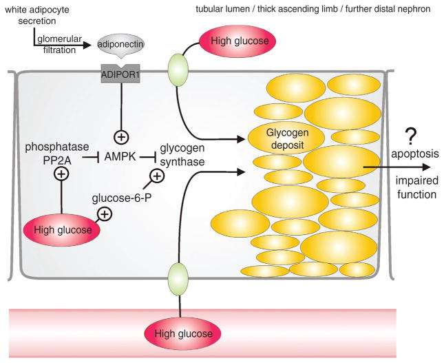
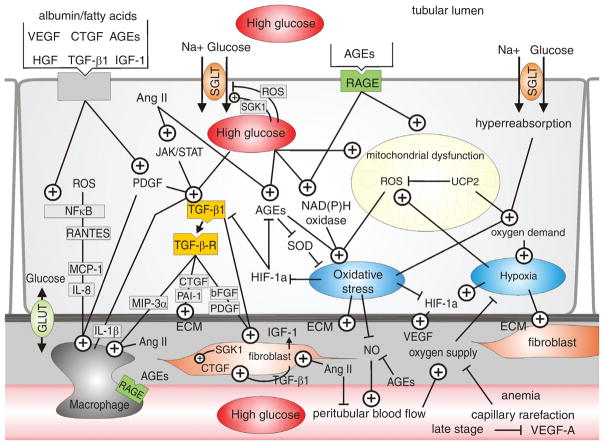
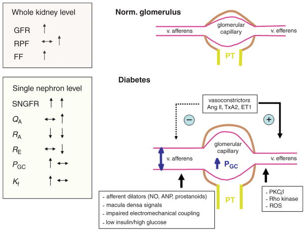
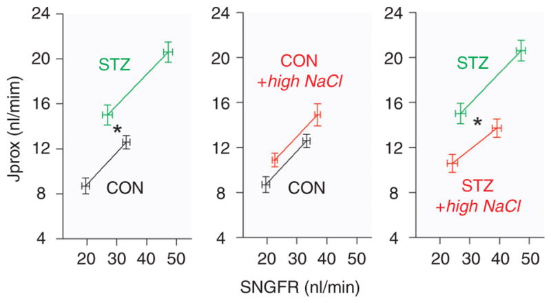
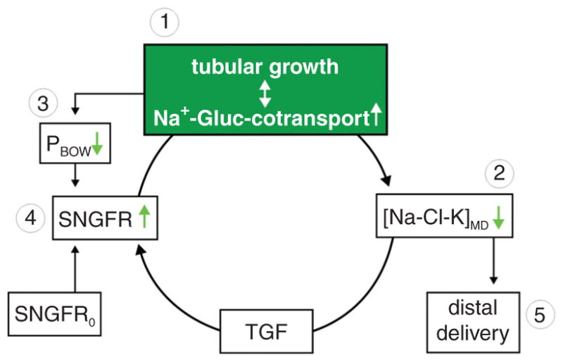
Diabetes mellitus contributes greatly to morbidity, mortality, and overall health care costs. In major part, these outcomes derive from the high incidence of progressive kidney dysfunction in patients with diabetes making diabetic nephropathy a leading cause of end-stage renal disease. A better understanding of the molecular mechanism involved and of the early dysfunctions observed in the diabetic kidney may permit the development of new strategies to prevent diabetic nephropathy. Here we review the pathophysiological changes that occur in the kidney in response to hyperglycemia, including the cellular responses to high glucose and the responses in vascular, glomerular, podocyte, and tubular function. The molecular basis, characteristics, and consequences of the unique growth phenotypes observed in the diabetic kidney, including glomerular structures and tubular segments, are outlined. We delineate mechanisms of early diabetic glomerular hyperfiltration including primary vascular events as well as the primary role of tubular growth, hyperreabsorption, and tubuloglomerular communication as part of a "tubulocentric" concept of early diabetic kidney function. The latter also explains the "salt paradox" of the early diabetic kidney, that is, a unique and inverse relationship between glomerular filtration rate and dietary salt intake. The mechanisms and consequences of the intrarenal activation of the renin-angiotensin system and of diabetes-induced tubular glycogen accumulation are discussed. Moreover, we aim to link the changes that occur early in the diabetic kidney including the growth phenotype, oxidative stress, hypoxia, and formation of advanced glycation end products to mechanisms involved in progressive kidney disease.


Figures

Figure 1
Figure 1
Simplified biochemistry of advanced glycation end-product formation. Prolonged hyperglycemia, but also dyslipidemia and oxidative stress in diabetes result in the production and accumulation of advanced glycation end products (AGEs) in the kidney, and at other sites of diabetic complications. AGEs are formed by nonenzymatic Maillard or “browning” reaction between carbonyl groups of reducing sugars, like glucose, and amino groups on proteins, lipids, or nucleic acids. The first stable adduct between glucose and protein is the Amadori product. In addition, the glycation of intracellular proteins is initiated by the elevation of intracellular glucose degradation products or decomposition of early glycation products leading to formation of intermediates such as glyoxal or methylglyoxal. Subsequent rearrangement and fragmentation reactions lead to the formation of AGEs. Under physiological conditions these reactions are slow. However, in diabetes, persistent hyperglycemia, dyslipidemia, and oxidative stress all act to hasten the formation of AGEs.
Figure 2
Figure 2
Consequences of accelerated formation of advanced glycation end products (AGEs) in the diabetic kidney. The schematic shows major mechanisms whereby AGEs exert their harmful effects in the diabetic kidney. AGEs typically crosslink and alter protein structure and function in extracellular compartment, modify cytosolic molecules and exert receptor-mediated effects that lead to activation of multiple signaling pathways and genes implicated in a variety of pathophysiological mechanisms in the diabetic kidney. RAGE, receptor for AGEs; ROS, reactive oxygen species; MAPKs, mitogen-activated protein kinases; ROCKs, Rho kinases; NO, nitric oxide; BM, basement membrane; ECM, extracellular matrix; NF-κB, nuclear factor kappa B.
Figure 3
Figure 3
Activation and downstream targets of the hexosamine pathway in diabetes. When the intracellular concentrations of glucose are high, fructose-6-phosphate, an intermediate in the glycolytic pathway, is partially diverted into the hexosamine pathway. After a series of enzymatic steps glucosamine (GlcNAc) is transferred in O-linkage to specific serine/threonine residues of numerous proteins, including transcription factors as well as cytosolic and nuclear enzymes, with patho-physiological consequences in affected cells. Key enzymes in the pathway are highlighted in yellow. More detailed description is provided in the text. G-6-P, fructose-6-phosphate; F-6-P, fructose-6-phosphate; GFAT, glutamine:fructose 6-phosphate amidotransferase; GlcN-6-P, glucosamine 6-phosphate; UDP-GlcNAc, UDP-N-acetylglucosamine; OGT, O-GlcNAc transferase; eNOS, endothelial NO synthase; mTOR, mammalian target of rapamycin; TGF-β, transforming growth factor-β; PAI-I, plasminogen activator inhibitor-I.
Figure 4
Figure 4
Mechanisms of enhanced formation of reactive oxygen species (ROS) in renal cells in diabetes. High intracellular glucose, stimulation of RAGE, as well as increased activity of vasoactive and pro-growth factors, such as the renin-angiotensin system, lead to formation of cytosolic and mitochondrial ROS via multiple mechanisms. ROS alter protein, lipid and DNA structure, and act as signaling molecules in pathways implicated in the pathophysiology of diabetic complications. See text for further details. Ang II, angiotensin II; AT1R, angiotensin II type 1 receptor; AGEs, advanced glycation end products; RAGE, receptor for AGEs.
Figure 5
Figure 5
Diabetes-induced alterations of glomerular filtration barrier. The schematic presentation describes major consequences of diabetes-induced changes in glomerular endothelial cells, glomerular basement membrane, and podocytes, as discussed in more detail in the text.
Figure 6
Figure 6
Podocyte foot process effacement and glomerular basement membrane (GBM) thickening in experimental diabetes. Transmission electron microscopy was used to investigate the glomerular filtration barrier (GFB) in normal (C) and STZ-diabetic DBA/2J mice (V, L, DL, L + DL, and L + DH). Compare the appearance of the GFB in normal (C) and diabetic mice (V), and the effects of nephroprotective treatments [L, DL, L + DL, and L + DH; for details on treatments see (741)]. Stars indicate the areas of podocyte foot process effacement; arrow pairs point to both sides of the glomerular basement membrane (GBM). Adapted with permission from (741).
Figure 7
Figure 7
Regulation of tubular growth in the diabetic kidney. Illustrated is a conceptional frame work that links hyperglycemia to tubular growth including an early phase of hyperplasia that is followed by G1 cell-cycle arrest and development of hypertrophy and a senescence-like phenotype. Potential links to enhanced formation of extracellular matrix, inflammation, and tubulointerstitial injury are shown. See text for further explanations. ECM, extracellular matrix; EMT, epithelial mesenchymal transition; TSC, tuberous sclerosis complex.
Figure 8
Figure 8
Hyperglycemia induces glycogen accumulation in thick ascending limb and further distal cortical nephron segments. Glycogen accumulation, also know as glycogen nephrosis or Armanni-Ebstein lesions, may relate to increased delivery of glucose via the tubular fluid or peritubular capillaries. Glycogen accumulation may also reflect activation of glycogen synthase, due to accumulation of the allosteric activator, glucose-6-phosphate. Adiponectin is secreted from white adipocytes and signals through its receptor, ADIPOR1, to activate AMP-activated protein kinase (AMPK) thereby inhibiting glycogen synthase. Thus, glycogen synthase can be activated as a consequence of reduced adiponectin levels or adiponectin resistance with impaired activation of AMPK due to activation of phosphatase PP2A. See text for further details.
Figure 9
Figure 9
Mechanisms of tubulointerstitial injury in the diabetic kidney. Illustrated is the interplay of hyperglycemia, luminal factors (derived from glomerular filtration and tubular release), reabsorption, and blood flow in the interaction of tubular cells with fibroblasts and inflammatory cells. TGF-β, chemokines, and the complex interactions between advanced glycation end products (AGE), hypoxia and oxidative stress play key roles in the development of diabetic tubulointerstitial injury. ECM, extracellular matrix. See text for further details.
Figure 10
Figure 10
Renal hemodynamic changes in diabetes at the whole kidney and single nephron level. Early stages of diabetes are associated with specific renal hemodynamic changes schematically presented in this figure. The schematic shows regulators of afferent and efferent arteriolar tone, which have been implicated in these changes. GFR, glomerular filtration rate; RPF, renal plasma flow; SNGFR, single nephron glomerular filtration rate; QA, glomerular plasma flow rate; RA, afferent arteriolar resistance; RE, efferent arteriolar resistance; PGC, glomerular capillary pressure; Kf, ultrafiltration coefficient.
Figure 11
Figure 11
Primary increase in proximal tubular reabsorption and inhibition by high NaCl diet in the early diabetic kidney. Absolute proximal fluid reabsorption (Jprox) shown as a function of SNGFR. SNGFR was manipulated by perfusing Henle’s loop to activate TGF to characterize proximal reabsorption as a function of SNGFR in control rats (CON) and diabetic rats (STZ) on normal versus high NaCl diet. *P < 0.05 for a GFR-independent effect of STZ (left panel) or high NaCl diet (right panel) on proximal tubular reabsorption. Adapted with permission from (651).
Figure 12
Figure 12
Tubular basis of glomerular hyperfiltration in the early diabetic kidney. Hyperglycemia causes a primary increase in proximal tubular reabsorption through enhanced tubular growth and Na+-glucose (Gluc) cotransport (1). The enhanced reabsorption reduces the signal of the tubuloglomerular feedback (TGF) at the macula densa ([Na-Cl-K]MD) (2) and via TGF increases SNGFR (4). Enhanced growth and tubular reabsorption also reduce the hydrostatic pressure in Bowman space (PBOW) (3), which by increasing effective filtration pressure can also increase SNGFR (4). The resulting increase in SNGFR serves to partly restore the fluid and electrolyte load to the distal nephron (5). SNGFR0 is the input to SNGFR independent of TGF. Adapted with permission from (648).
Figure 13
Figure 13
Lower efficiency of tubuloglomerular feedback (TGF) in the diabetic kidney. (A) In vivo free-flow perturbation analysis of tubular flow rate in control and STZ-diabetic rats. The fractional compensation profiles illustrate the ability of the TGF system to stabilize tubular flow in response to a perturbation in late proximal tubular flow (by adding or subtracting tubular fluid). The data show that the efficiency of the TGF system to stabilize tubular flow rate (as an indirect measure of stabilizing SNGFR or PGC) is reduced in diabetic rats with a compensation of small perturbations around the operating point of 40% versus 70% in control rats. (B) Data from perturbation analysis of late proximal tubular flow rate were combined with data for fractional proximal reabsorption to synthesize “traditional” TGF functions describing the dependence of SNGFR on late proximal tubular flow rate. The triangles indicate the operating points. The gain of the curve at the operating point is modestly reduced in diabetic rats (red lines). Adapted with permission from (647).
Figure 14
Figure 14
Tubular basis of the salt paradox in the early diabetic kidney. (A) In classical physiology, renal function and total body NaCl are linked by several parallel feedback loops. Paths from dietary NaCl to GFR and renal blood flow (RBF) are highlighted (black arrows). Each highlighted path contains only “+” signs or an even number of “−” signs, indicating a positive influence of dietary NaCl on GFR and RBF. Interfering with these processes can alter the strength of this influence, but cannot make it paradoxical. (B) Incorporating tubuloglomerular feedback (TGF) provides a pathway whereby dietary NaCl can inversely impact GFR and RBF via a primary change in proximal (i.e., upstream to macula densa) reabsorption (note the odd number of “−” signs along the dotted path). Thus, GFR and RBF are subject to competing influences in response to changes in dietary NaCl. The NaCl paradox arises when TGF prevails. Adapted with permission from (648).
Figure 15
Figure 15
Diabetes-induced upregulation of the renin-angiotensin-system in glomeruli. Illustrated is a proposed role of succinate acting on its receptor (SUCNR1) in glomerular endothelial cells and macula densa cells in the activation of renin. Renin and prorenin can induce angiotensin II-dependent and independent effects by activation of renin receptors on glomerular structures including mesangial cells and podocytes. See text for further details. ADO, adenosine; Ado A1, adenosine A1 receptor; Agten, angiotensinogen; RR, renin receptor.

References

    1. Aaltonen P, Luimula P, Astrom E, Palmen T, Gronholm T, Palojoki E, Jaakkola I, Ahola H, Tikkanen I, Holthofer H. Changes in the expression of nephrin gene and protein in experimental diabetic nephropathy. Lab Invest. 2001;81:1185–1190. - PubMed
    1. Abbate M, Remuzzi G. Proteinuria as a mediator of tubulointerstitial injury. Kidney Blood Press Res. 1999;22:37–46. - PubMed
    1. Abbate M, Zoja C, Corna D, Capitanio M, Bertani T, Remuzzi G. In progressive nephropathies, overload of tubular cells with filtered proteins translates glomerular permeability dysfunction into cellular signals of interstitial inflammation. J Am Soc Nephrol. 1998;9:1213–1224. - PubMed
    1. Abdel-Wahab N, Weston BS, Roberts T, Mason RM. Connective tissue growth factor and regulation of the mesangial cell cycle: Role in cellular hypertrophy. J Am Soc Nephrol. 2002;13:2437–2445. - PubMed
    1. Abraham NG, Kappas A. Heme oxygenase and the cardiovascular-renal system. Free Radic Biol Med. 2005;39:1–25. - PubMed

Publication types