

Revised microcalcification hypothesis for fibrous cap rupture in human coronary arteries
- PMID: 23733926
- PMCID: PMC3696743
- DOI: 10.1073/pnas.1308814110
Revised microcalcification hypothesis for fibrous cap rupture in human coronary arteries
Abstract
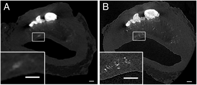
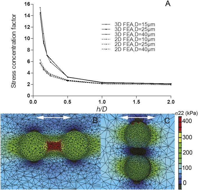
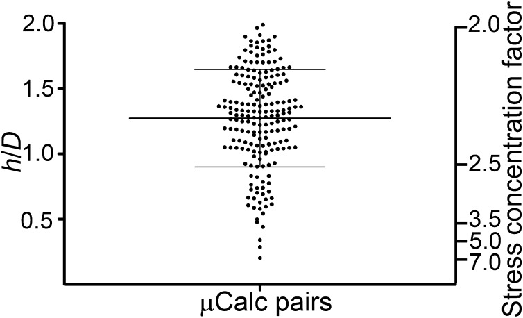
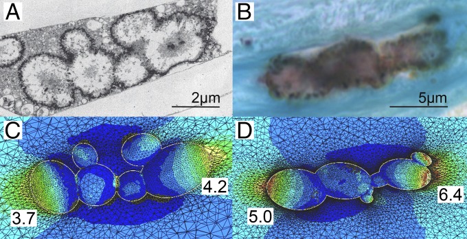
Using 2.1-µm high-resolution microcomputed tomography, we have examined the spatial distribution, clustering, and shape of nearly 35,000 microcalcifications (µCalcs) ≥ 5 µm in the fibrous caps of 22 nonruptured human atherosclerotic plaques. The vast majority of these µCalcs were <15 µm and invisible at the previously used 6.7-µm resolution. A greatly simplified 3D finite element analysis has made it possible to quickly analyze which of these thousands of minute inclusions are potentially dangerous. We show that the enhancement of the local tissue stress caused by particle clustering increases rapidly for gap between particle pairs (h)/particle diameter (D) < 0.4 if particles are oriented along the tensile axis of the cap. Of the thousands of µCalcs observed, there were 193 particle pairs with h/D ≤ 2 (tissue stress factor > 2), but only 3 of these pairs had h/D ≤ 0.4, where the local tissue stress could increase a factor > 5. Using nondecalcified histology, we also show that nearly all caps have µCalcs between 0.5 and 5 µm and that the µCalcs ≥ 5 µm observed in high-resolution microcomputed tomography are agglomerations of smaller calcified matrix vesicles. µCalcs < 5 µm are predicted to be not harmful, because the tiny voids associated with these very small particles will not explosively grow under tensile forces because of their large surface energy. These observations strongly support the hypothesis that nearly all fibrous caps have µCalcs, but only a small subset has the potential for rupture.
Keywords: clustered microcalcifications; finite element analysis of fibrous caps; microcomputed tomography imaging of microcalcifications; vulnerable plaque.
Conflict of interest statement
The authors declare no conflict of interest.
Figures
References
-
- Burke AP, Kolodgie FD, Farb A, Virmani R. Pathogenesis and significance of calcification in coronary aterosclerosis. In: Virmani R, Narula J, Leon M, Willerson JT, editors. The Vulnerable Atherosclerotic Plaque: Strategies for Diagnosis and Management. Oxford: Blackwell; 2007. pp. 77–94.
-
- McCollough CH, et al. Coronary artery calcium: A multi-institutional, multimanufacturer international standard for quantification at cardiac CT. Radiology. 2007;243(2):527–538. - PubMed
-
- Huang H, et al. The impact of calcification on the biomechanical stability of atherosclerotic plaques. Circulation. 2001;103(8):1051–1056. - PubMed
-
- Kume T, et al. Assessment of the coronary calcification by optical coherence tomography. EuroIntervention. 2011;6(6):768–772. - PubMed
Publication types
MeSH terms
Grants and funding
LinkOut - more resources
Full Text Sources
Other Literature Sources
Medical
Miscellaneous
