. 2013 Jun 18;110(25):10306-11.
doi: 10.1073/pnas.1222895110. Epub 2013 Jun 3.

Peripheral elevation of TNF-α leads to early synaptic abnormalities in the mouse somatosensory cortex in experimental autoimmune encephalomyelitis

Affiliations

Peripheral elevation of TNF-α leads to early synaptic abnormalities in the mouse somatosensory cortex in experimental autoimmune encephalomyelitis

Guang Yang et al. Proc Natl Acad Sci U S A. .

Abstract

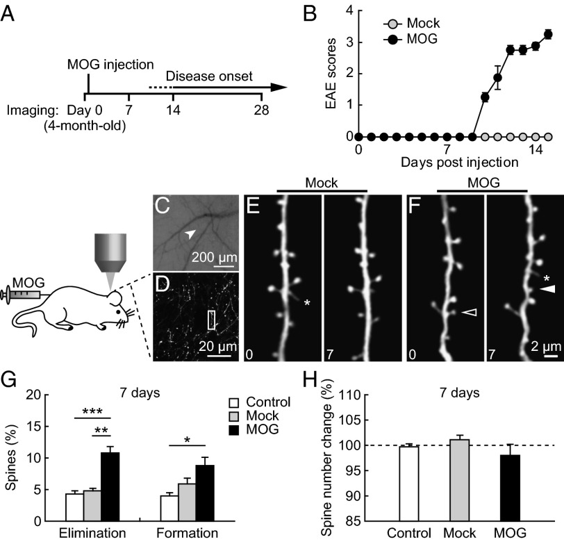
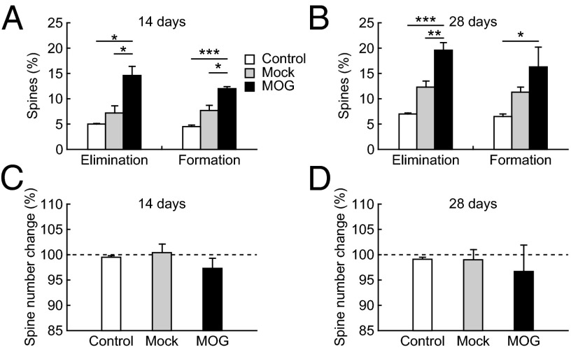
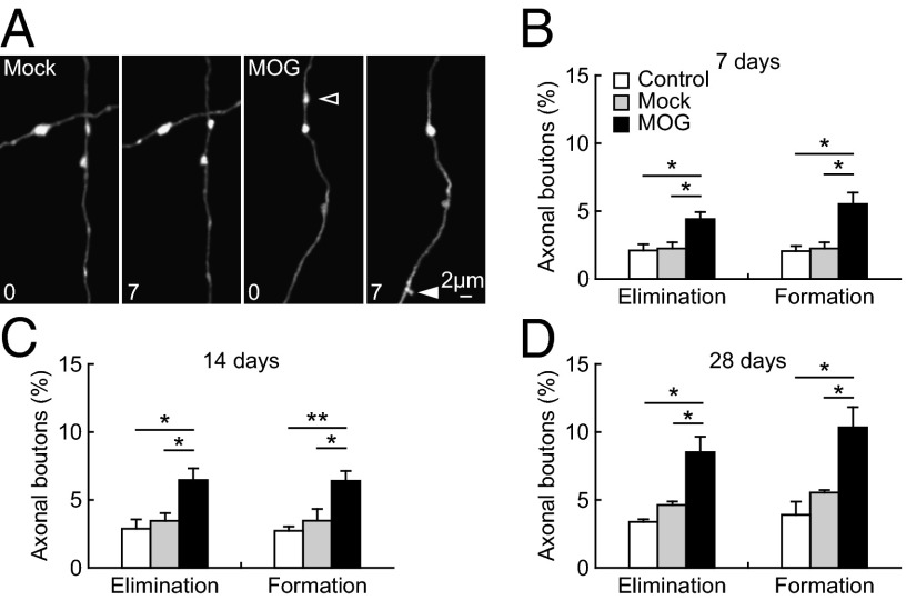
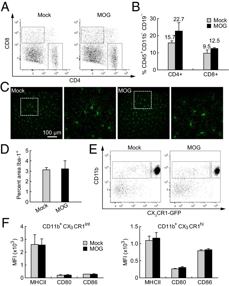
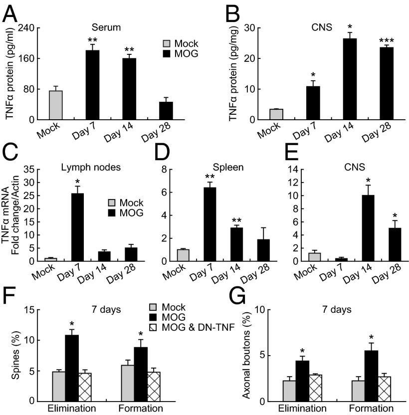
Sensory abnormalities such as numbness and paresthesias are often the earliest symptoms in neuroinflammatory diseases including multiple sclerosis. The increased production of various cytokines occurs in the early stages of neuroinflammation and could have detrimental effects on the central nervous system, thereby contributing to sensory and cognitive deficits. However, it remains unknown whether and when elevation of cytokines causes changes in brain structure and function under inflammatory conditions. To address this question, we used a mouse model for experimental autoimmune encephalomyelitis (EAE) to examine the effect of inflammation and cytokine elevation on synaptic connections in the primary somatosensory cortex. Using in vivo two-photon microscopy, we found that the elimination and formation rates of dendritic spines and axonal boutons increased within 7 d of EAE induction--several days before the onset of paralysis--and continued to rise during the course of the disease. This synaptic instability occurred before T-cell infiltration and microglial activation in the central nervous system and was in conjunction with peripheral, but not central, production of TNF-α. Peripheral administration of a soluble TNF inhibitor prevented abnormal turnover of dendritic spines and axonal boutons in presymptomatic EAE mice. These findings indicate that peripheral production of TNF-α is a key mediator of synaptic instability in the primary somatosensory cortex and may contribute to sensory and cognitive deficits seen in autoimmune diseases.


Conflict of interest statement

The authors declare no conflict of interest.

Figures

Fig. 1.
Fig. 1.
Increased spine turnover before the onset of paralysis in MOG-EAE mice. (A) Schematic diagram of the experiment. (B) Clinical scores of mice with MOG-induced EAE. Four-month-old adult mice were immunized by MOG35–55 on day 0. Clinic symptoms began at days 10–15 postimmunization. No mice showed disease symptoms before day 7. (C) CCD camera view of the vasculature of the somatosensory cortex below the thinned skull. The arrow indicates the region where the two-photon image in D was obtained. (D) Low-magnification image of apical dendrites of layer V pyramidal neurons. A higher-magnification view of the dendritic segment (box in D) is shown in F. (E and F) Repeated imaging of dendritic branches over 7 d in the cortex of Mock- and MOG-immunized animals. Filled and empty arrowheads indicate spines that were formed and eliminated between the two views. Asterisks indicate filopodia. (G) Percentage of spines eliminated and formed over 7 d in no-injection control, Mock-immunized, and MOG-immunized animals. A significant increase in both spine elimination and formation was found in MOG-immunized animals compared with no-injection control and Mock-immunized animals. (H) The total number of spines remains the same over 7 d in all groups of animals. Data are presented as mean ± SEM. *P < 0.05; **P < 0.01; ***P < 0.001.
Fig. 2.
Fig. 2.
Spine turnover in the cortex continually increased during EAE progression. (A and B) Percentage of spines eliminated and formed over 14 d (A) and 28 d (B) in no-injection control, Mock-immunized, and MOG-immunized animals. A significant increase in both spine elimination and formation was found in MOG-immunized animals compared with no-injection control and Mock-immunized animals. (C and D) No significant change in total spine number was observed over 14–28 d in all animal groups. Data are presented as mean ± SEM. *P < 0.05; **P < 0.01; **P < 0.001.
Fig. 3.
Fig. 3.
Increased turnover of axonal boutons after MOG immunization. (A) Repeated imaging of axonal segments in the somatosensory cortex over 7 d in Mock- and MOG-immunized animals (4-mo-old). Filled and empty arrowheads indicate axonal boutons that were formed and eliminated between the two views. (BD) Percentage of axonal boutons eliminated and formed over 7 d (B), 14 d (C), and 28 d (D) in no-injection control, Mock-immunized, and MOG-immunized animals. The turnover of axonal boutons was significantly increased in MOG-immunized animals compared with no-injection and Mock-immunized control in all of the time intervals. Data are presented as mean ± SEM. *P < 0.05; **P < 0.01.
Fig. 4.
Fig. 4.
Absence of peripheral T-cell infiltration and microglia activation in CNS within 7 d after MOG immunization. (A) Flow cytometric data from homogenized brains of Mock- and MOG-immunized mice. The left and right boxes indicate CD8+ and CD4+ populations, respectively. (B) Percentage of CD45+CD11bCD19 cells in the CD4+ and CD8+ gates. There is no significant difference between Mock- and MOG-immunized mice (P > 0.5). (C) Microglia morphology in the cortex was shown by immunolabeling for Iba-1. There is no obvious difference in microglia morphology between Mock- and MOG-immunized animals at day 7 postimmunization. (D) No significant difference in the area covered by Iba-1+ microglia between Mock- and MOG-immunized animals at day 7 (P > 0.9). (E) The nonmicroglial myeloid population (left box: CD11b+ CX3CR1int) and the microglial population (right box: CD11b+ CX3CR1hi) were determined by FACS analysis from the homogenized brain. (F) There was no significant increase in the levels of MHC II, CD80, or CD86 in either the nonmicroglial myeloid population or the microglial population in MOG-immunized animals compared with Mock-immunized controls at day 7 postimmunization (P > 0.3).
Fig. 5.
Fig. 5.
Peripheral TNF-α production contributes to early synaptic instability in the cortex. (A and B) TNF-α protein levels in serum and CNS of Mock- and MOG-immunized animals. TNF-α protein level was significantly increased in both serum and CNS tissues at days 7 and 14 post-MOG immunization, but only in CNS tissues at day 28 post-MOG immunization. (CE) TNF-α mRNA levels in lymph nodes, spleen, and CNS of Mock- and MOG-immunized animals. At day 7 postimmunization, a significant increase of TNF-α mRNA levels was observed in lymph nodes and spleen, but not in CNS of MOG-immunized animals compared with the Mock-immunized control. (F) Spine elimination and formation over 7 d after MOG immunization were significantly reduced in animals treated with DN-TNF. (G) Elimination and formation rates of axonal boutons over 7 d after MOG immunization were significantly reduced in animals treated with DN-TNF. Data are presented as mean ± SEM. *P < 0.05; **P < 0.01; ***P < 0.001.

Similar articles

Cited by

References

    1. Lisak RP. 2007. Neurodegeneration in multiple sclerosis: Defining the problem. Neurology 68(22 Suppl 3):S5–S12; discussion S43–S54.
    1. Magnano I, Aiello I, Piras MR. Cognitive impairment and neurophysiological correlates in MS. J Neurol Sci. 2006;245(1–2):117–122. - PubMed
    1. Mayeux R, Benson DF. Phantom limb and multiple sclerosis. Neurology. 1979;29(5):724–726. - PubMed
    1. Zarei M, Chandran S, Compston A, Hodges J. Cognitive presentation of multiple sclerosis: Evidence for a cortical variant. J Neurol Neurosurg Psychiatry. 2003;74(7):872–877. - PMC - PubMed
    1. Achiron A, Barak Y. Cognitive impairment in probable multiple sclerosis. J Neurol Neurosurg Psychiatry. 2003;74(4):443–446. - PMC - PubMed

Publication types

MeSH terms

Substances

LinkOut - more resources