Case Reports
. 2013 May 21:19:1039-46.
Print 2013.

A novel GUCY2D mutation in a Chinese family with dominant cone dystrophy

Affiliations
Case Reports

A novel GUCY2D mutation in a Chinese family with dominant cone dystrophy

Xin Zhao et al. Mol Vis. .

Abstract

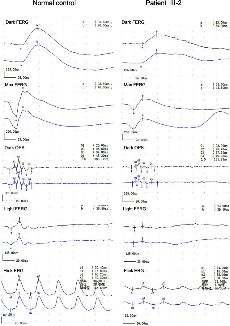
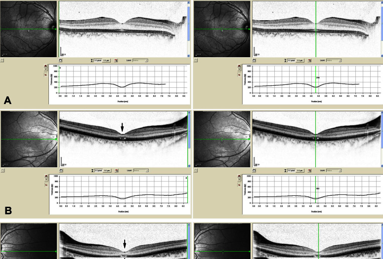
Purpose: To describe the clinical and genetic findings in a Chinese family with autosomal dominant cone dystrophy (adCOD).

Methods: One family was examined clinically, and genomic DNA was extracted from venous blood of all participants. Genotyping and haplotyping analysis was performed on the known genetic loci for adCOD and autosomal dominant cone-rod dystrophies (adCORD) with a panel of polymorphic markers in this family. All coding exons of the AIPL1, PTTPNM3, and GUCY2D gene were directly sequenced. Allele-specific PCR was used to validate a substitution in all available family members and 100 normal controls. Bioinformatics analysis was done using the Garnier-Osguthorpe-Robson method to predict the effect of the variants detected on the secondary structure of the GUCY2D protein.

Results: Clinical examination and pedigree analysis revealed a three-generation family with four members diagnosed with adCOD. Through genotyping, the disease-causing genes were mapped to chromosomes 17p13.1-2 (AIPL1, PITPNM3, and GUCY2D gene). A novel A->G transition at position 2545 (p.T849A) of the cDNA sequence was identified in the GUCY2D gene. No mutation was detected in the AIPL1 and PITPNM3 genes. This missense mutation co-segregated with the disease phenotype of the family but was not found in the 100 normal controls.

Conclusions: A novel missense mutation of the GUCY2D gene was identified in this study. Our results further confirm that the dimerization zone of RetGC-1 is the mutational hot region for COD and CORD.


Figures

Figure 1
Figure 1
Family structure, proband fundus appearance, DNA sequence chromatograms, and co-segregation analysis of the p.T849A mutation with the disease phenotype in a Chinese family with cone dystrophy. A: The pedigree and haplotype analysis of the family with cone dystrophy showed segregation with three microsatellite markers on chromosome 17 listed in rising order from the telomere end. Squares indicate males; circles indicate females; slashed symbols indicate deceased; solid symbols indicate affected; open symbols indicate unaffected; M indicates mutant; and + indicates wild-type. B: Fundus appearance of the proband shows the subtle mottling of the RPE in the macula. C: Heterozygote sequence (sense strand) shows an A/G transition in codon 849 that changed threonine to alanine. D: Allele-specific PCR analysis presents the amplified products of the mutation allele (184 bp) co-segregated with patients in this family. The fragments (325 bp), which are the parts of exon3 of the MYOC gene, were used as the internal control in the allele-specific PCR analysis. E: The sequence alignment portion of the dimerization domain spanning the p.T849 of the GUCD2Y of the human with other species.
Figure 2
Figure 2
Electroretinography of the proband and a normal control. Electroretinography of the proband shows reduced photopic and 30 Hz responses and normal scotopic responses.
Figure 3
Figure 3
Macular optical coherence tomography images from a visually normal subject and the proband of this family with cone dystrophy. A: The macular optical coherence tomography images of the right eye from a normal individual show organization of retinal microstructures with a well defined photoreceptor inner/outer segment layer and normal thickness (214 μm). B and C: The macular optical coherence tomography images of both eyes from the proband exhibit loss of inner/outer segment layer and thinning of the retina in the macular area (151 μm of the right eye, 153 μm of the left eye).

References

    1. Hamel CP. Cone rod dystophies. Orphanet J Rare Dis. 2007;2:7. - PMC - PubMed
    1. Kelsell RE, Gregory-Evans K, Payne A, Perrault I, Kaplan J, Yang RB, Garbers DL, Bird AC, Moore AT, Hunt DM. Mutations in the retinal guanylate cyclase (RETGC-1) gene in dominant cone-rod dystrophy. Hum Mol Genet. 1998;7:1179–84. - PubMed
    1. Gregory-Evans K, Kelsell R, Gregory-Evans C, Downes SM, Fitzke FM, Holder GR, Simunovic M, Mollon JD, Taylor R, Hunt DM, Bird AC, Moore AT. Autosomal dominant cone rod retinal dystrophy (CORD6) from heterozygous mutation of GUCY2D, which encodes retinal guanylate cyclase. Ophthalmology. 2000;107:55–61. - PubMed
    1. Payne AM, Morris AG, Downes SM, Johnson S, Bird AC, Moore AT, Bhattacharya SS, Hunt DM. Clustering and, frequency of mutations in the retinal guanylate cyclase (GUCY2D) gene in patients with dominant cone-rod dystrophies. J Med Genet. 2001;38:611–4. - PMC - PubMed
    1. Kitiratschky VB, Wilke R, Renner AB, Kellner U, Vadala M, Birch DG, Wissinger B, Zrenner E, Kohl S. Mutation analysis identifies GUCY2D as the major gene responsible for autosomal dominant progressive cone degeneration. Invest Ophthalmol Vis Sci. 2008;49:5015–23. - PMC - PubMed

Publication types

MeSH terms

LinkOut - more resources