. 2013 May 21:19:1047-59.
Print 2013.

Retinal O-linked N-acetylglucosamine protein modifications: implications for postnatal retinal vascularization and the pathogenesis of diabetic retinopathy

Affiliations

Retinal O-linked N-acetylglucosamine protein modifications: implications for postnatal retinal vascularization and the pathogenesis of diabetic retinopathy

Zafer Gurel et al. Mol Vis. .

Abstract

Purpose: Hyperglycemia activates several metabolic pathways, including the hexosamine biosynthetic pathway. Uridine diphosphate N-acetylglucosamine (GlcNAc) is the product of the hexosamine biosynthetic pathway and the substrate for O-linked GlcNAc (O-GlcNAc) modification. This modification affects a wide range of proteins by altering their activity, cellular localization, and/or protein interactions. However, the role O-GlcNAcylation may play in normal postnatal retinal vascular development and in the ocular complications of diabetes, including diabetic retinopathy, requires further investigation.

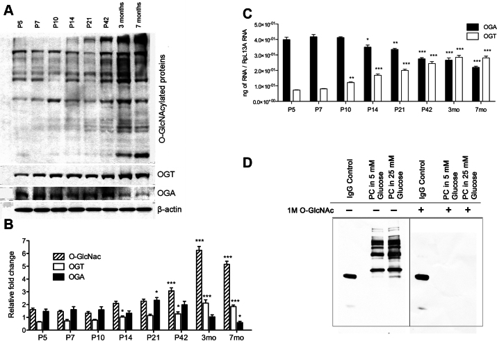
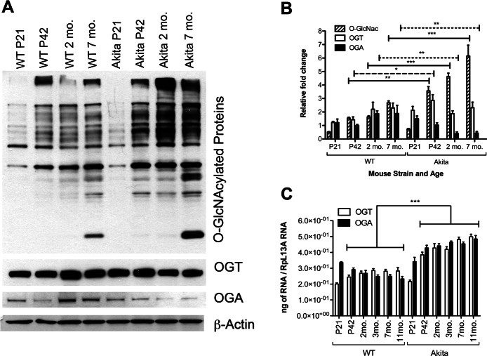
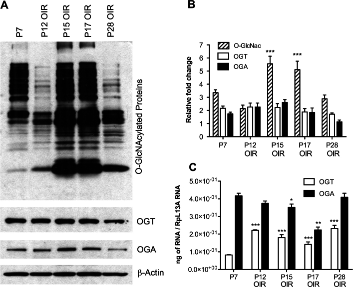
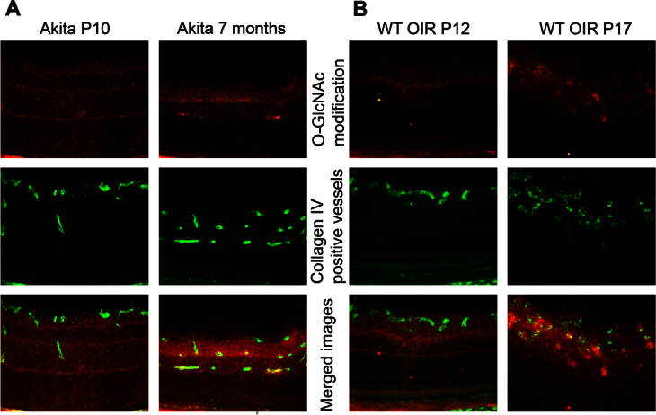
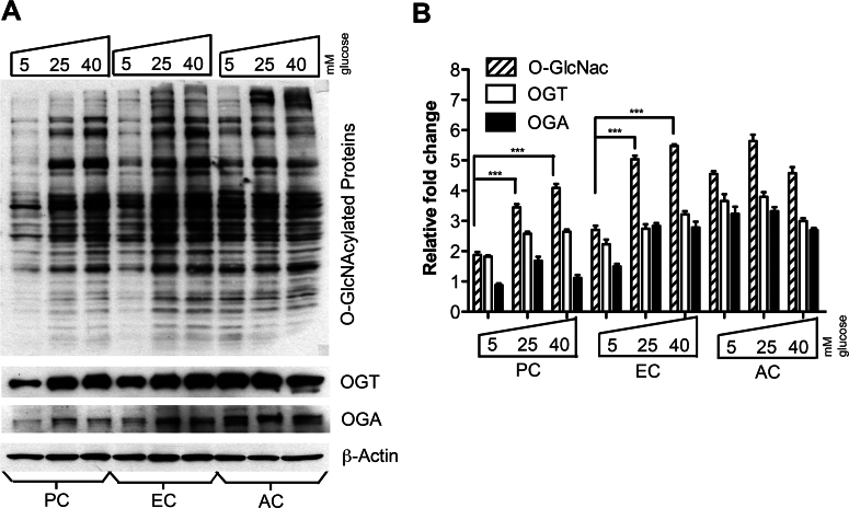
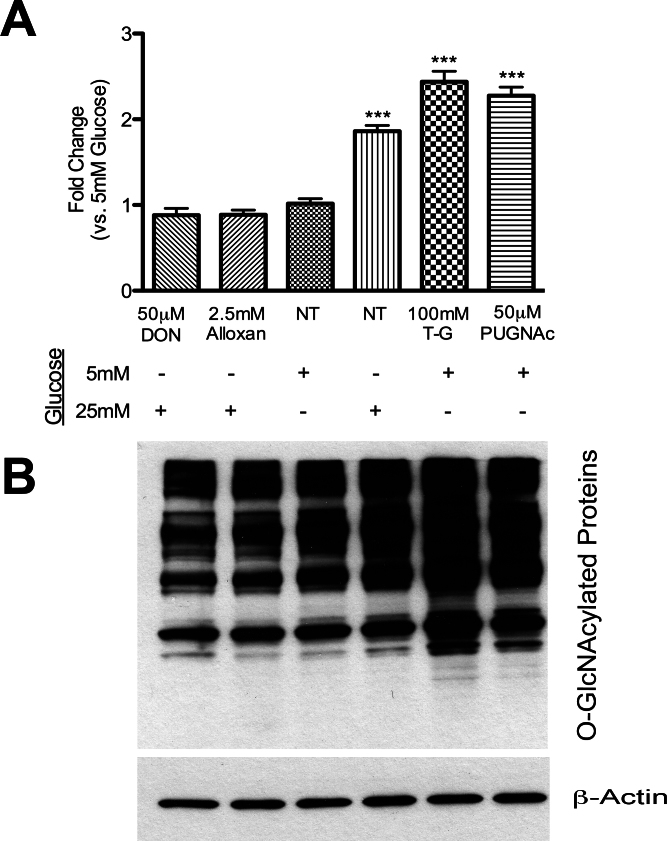
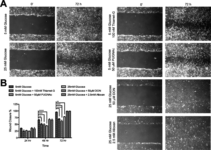
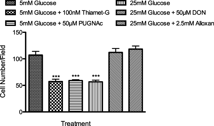
Methods: The total levels of O-GlcNAc-modified proteins were evaluated by western blot analysis of lysates prepared from retinas obtained at different days during postnatal retinal vascularization and oxygen-induced ischemic retinopathy. Similar experiments were performed with retinal lysate prepared from diabetic Ins2(Akita/+) mice with different durations of diabetes and retinal vascular cells cultured under various glucose conditions. The localization of O-GlcNAc-modified proteins in the retinal vasculature was confirmed by immunofluorescence staining. The impact of altered O-GlcNAcylation on the migration of retinal vascular cells was determined using scratch wound and transwell migration assays.

Results: We detected an increase in protein O-GlcNAcylation during mouse postnatal retinal vascularization and aging, in part through the regulation of the enzymes that control this modification. The study of the diabetic Ins2(Akita/+) mouse retina showed an increase in the O-GlcNAc modification of retinal proteins. We also observed an increase in retinal O-GlcNAcylated protein levels during the neovascularization phase of oxygen-induced ischemic retinopathy. Our fluorescence microscopy data confirmed that the alterations in retinal O-GlcNAcylation are similarly represented in the retinal vasculature and in retinal pericytes and endothelial cells. Particularly, the migration of retinal pericytes, but not retinal endothelial cells, was attenuated by increased O-GlcNAc modification.

Conclusions: The O-GlcNAc modification pattern changes during postnatal retinal vascular development and neovascularization, and its dysregulation under hyperglycemia and/or ischemia may contribute to the pathogenesis of the diabetic retinopathy and retinal neovascularization.


Figures

Figure 1
Figure 1
Increased O-linked-N-acetylglucoseamine modification (O-GlcNAcylation) with increased O-GlcNAc transferase (OGT) and decreased O-GlcNAcase (OGA) expression during postnatal retinal vascular development and aging. A: Protein lysates (25 µg) from C57BL/6J mouse retinas were analyzed by western blot analysis for O-GlcNAcylated proteins and the expression of OGT and OGA. B: The β-actin expression was assessed as a loading control and used for normalization and quantification of data, which were obtained after three different runs (B). RNA expression of OGT and OGA were determined by qPCR and normalized by RpL13A RNA expression in samples. The qPCRs were performed with three biologic replicates and in triplicate (C). Validation of O-GlcNAc antibody staining of lysates prepared from pericytes (PC) under various glucose conditions. D: The GlcNAc (1 M) competition during primary antibody incubation was used to validate the specificity of the O-GlcNAc RL2 antibody. IgG control was used to validate the existence of the secondary antibody. Mean±SEM; * (p≤0.05), ** (p≤0.01), and ***(p≤0.001) significantly different from P5.
Figure 2
Figure 2
Increased O-GlcNAcylation and decreased O-GlcNAcase (OGA) expression in the retinas of Ins2Akita/+ mice. O-GlcNAc transferase (OGT) expression did not show any alterations. A: Protein lysates (25 µg) from wild-type C57BL/6J and Ins2Akita/+ mice retina were examined by western blot analysis for O-GlcNAcylated proteins and expression of OGT and OGA. B: The β-actin expression was assessed as a loading control and used for normalization and quantification of data, which were obtained after three different runs. C: Increases in the RNA expression of OGT and OGA were detected in Ins2Akita mice retina by qPCR and normalized to RpL13A RNA expression. The qPCRs were performed with three biologic replicates and in triplicate. Mean±SEM; * (p≤0.05), ** (p≤0.01), and ***(p≤0.001) significantly different from wild-type mice at the same time points of wild-type.
Figure 3
Figure 3
O-GlcNAcylation increases during the neovascularization phase but decreases during the regression phase in an oxygen-induced ischemic retinopathy (OIR) model. O-GlcNAc transferase (OGT) and O-GlcNAcase (OGA) expression did not show any correlation with the alterations in O-GlcNAcylation. A: Retinal lysates (25 µg) from C57BL/6J mice during OIR were analyzed by western blot analysis for O-GlcNAcylated proteins and expression of OGT and OGA. B: The β-actin expression was assessed as a loading control and used for normalization and quantification of data, which were obtained after three different runs. C: RNA expression of OGT and OGA was determined by qPCR and normalized by RpL13A RNA expression in samples. The qPCRs were performed with three biologic replicates and in triplicate. Mean±SEM; * (p≤0.05), ** (p≤0.01), and ***(p≤0.001) significantly different from P7.
Figure 4
Figure 4
O-GlcNAcylated proteins localize to retinal vascular plexus. A: Eye sections from P10 and 7-month-old Ins2Akita/+ mice. B: P12 and P17 oxygen-induced ischemic retinopathy (OIR) wild-type (WT) mice. O-GlcNAcylated proteins labeled with Cy3 (red, first row), vascular plexus labeled with Cy2 (green, second row) and merge images (third row). Please note the high amount of O-GlcNAcylated protein colocalization with the retinal vascular plexus in 7-month-old Ins2Akita/+ and P17 OIR eyes (arrowheads). These images are representative of images evaluated in eyes from at least six mice (original magnification x200).
Figure 5
Figure 5
Increased O-GlcNAcylation in retinal pericytes (PC) and endothelial cells (EC), but not astrocytes (AC), under high glucose conditions. A: Protein lysates (25 µg) from retinal vascular cells were analyzed by western blot analysis for O-GlcNAcylated proteins and expression of O-GlcNAc transferase (OGT) and O-GlcNAcase (OGA). B: The β-actin expression was assessed as a loading control and used for normalization and quantification of data obtained from three different runs (B). Mean±SEM; *** (p≤0.001) significantly different from the 5 mM glucose control.
Figure 6
Figure 6
Alterations in the levels of total O-GlcNAc modified proteins in retinal pericytes (PC) by high glucose and specific inhibitors for glutamine fructose-6-phosphate amidotransferase (GFAT), O-GlcNAc transferase (OGT), or O-GlcNAcase (OGA). A: Protein lysates (50 µg) from retinal vascular cells were analyzed by western blot analysis for O-GlcNAcylated proteins under 5 mM and 25 mM glucose with or without inhibitors. Thiamet-G and PUGNAc are OGA inhibitors. DON is a GFAT inhibitor and Alloxan is an OGT inhibitor. B: The β-actin expression was assessed as a loading control and used for normalization and quantification of data obtained from three different runs. Mean±SEM; *** (p≤0.001) significantly different from 5 mM glucose control.
Figure 7
Figure 7
Increased O-GlcNAc modification by high glucose or OGA inhibitors has a negative effect on retinal pericyte (PC) migration. A: Cell migration was determined by scratch wounding of PC monolayers, and wound closure at 37 °C was monitored by photography. A representative experiment is shown here. B: The quantitative assessment of the wound closure (**p<0.01, ***p<0.001, and ****p<0.0001; n=3).
Figure 8
Figure 8
Transwell migration assay confirmed the migration results observed in the scratch-wound assay. Transwell assays were performed at 33 °C with retinal PC cultured under various conditions. Cells that migrated through the membrane were compared with their control group grown in 5 mM glucose (***p<0.001; n=3).

References

    1. Ockrim Z, Yorston D. Managing diabetic retinopathy. BMJ. 2010;341:c5400. - PubMed
    1. Cheung N, Mitchell P, Wong TY. Diabetic retinopathy. Lancet. 2010;376:124–36. - PubMed
    1. Aiello LP. Angiogenic pathways in diabetic retinopathy. N Engl J Med. 2005;353:839–41. - PubMed
    1. Yang X, Ongusaha PP, Miles PD, Havstad JC, Zhang F, So WV, Kudlow JE, Michell RH, Olefsky JM, Field SJ, Evans RM. Phosphoinositide signalling links O-GlcNAc transferase to insulin resistance. Nature. 2008;451:964–9. - PubMed
    1. McClain DA, Lubas WA, Cooksey RC, Hazel M, Parker GJ, Love DC, Hanover JA. Altered glycan-dependent signaling induces insulin resistance and hyperleptinemia. Proc Natl Acad Sci USA. 2002;99:10695–9. - PMC - PubMed

Publication types

MeSH terms

LinkOut - more resources