

Suprachoroidal delivery in a rabbit ex vivo eye model: influence of drug properties, regional differences in delivery, and comparison with intravitreal and intracameral routes
- PMID: 23734089
- PMCID: PMC3669536
Suprachoroidal delivery in a rabbit ex vivo eye model: influence of drug properties, regional differences in delivery, and comparison with intravitreal and intracameral routes
Retraction in
-
Full retraction: Rajendra S. Kadam, Jason Williams, Puneet Tyagi, Henry F. Edelhauser, Uday B. Kompella. Suprachoroidal delivery in a rabbit ex vivo eye model: influence of drug properties, regional differences in delivery, and comparison with intravitreal and intracameral routes. Mol Vis. 2013; 19:1198-1210.Mol Vis. 2019 Feb 18;25:143. eCollection 2019. Mol Vis. 2019. PMID: 30820149 Free PMC article.
Abstract
Purpose: First, to determine the influence of drug lipophilicity (using eight beta-blockers) and molecular weight (using 4 kDa and 40 kDa fluoroscein isothiocyanate [FITC]-dextrans) on suprachoroidal delivery to the posterior segment of the eye by using a rabbit ex vivo eye model. Second, to determine whether drug distribution differs between the dosed and undosed side of the eye following suprachoroidal delivery. Third, to compare the suprachoroidal delivery of sodium fluorescein (NaF) with the intracameral and intravitreal routes by using noninvasive fluorophotometry.
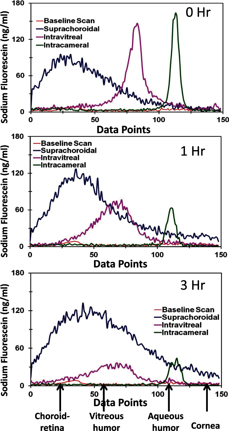
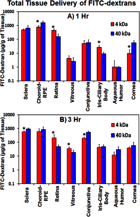
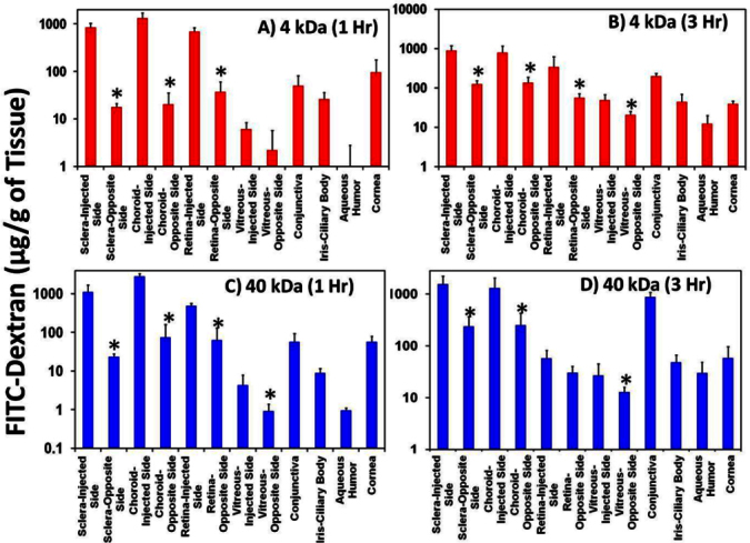
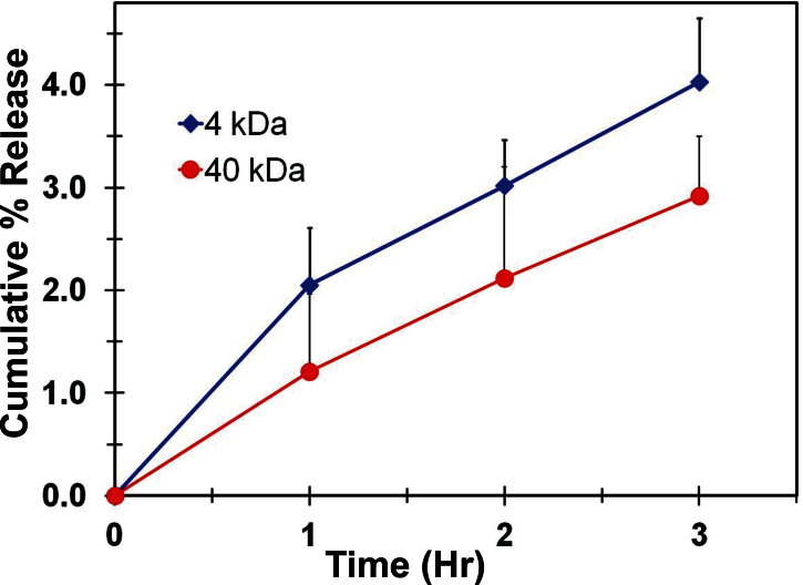
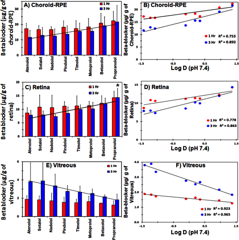
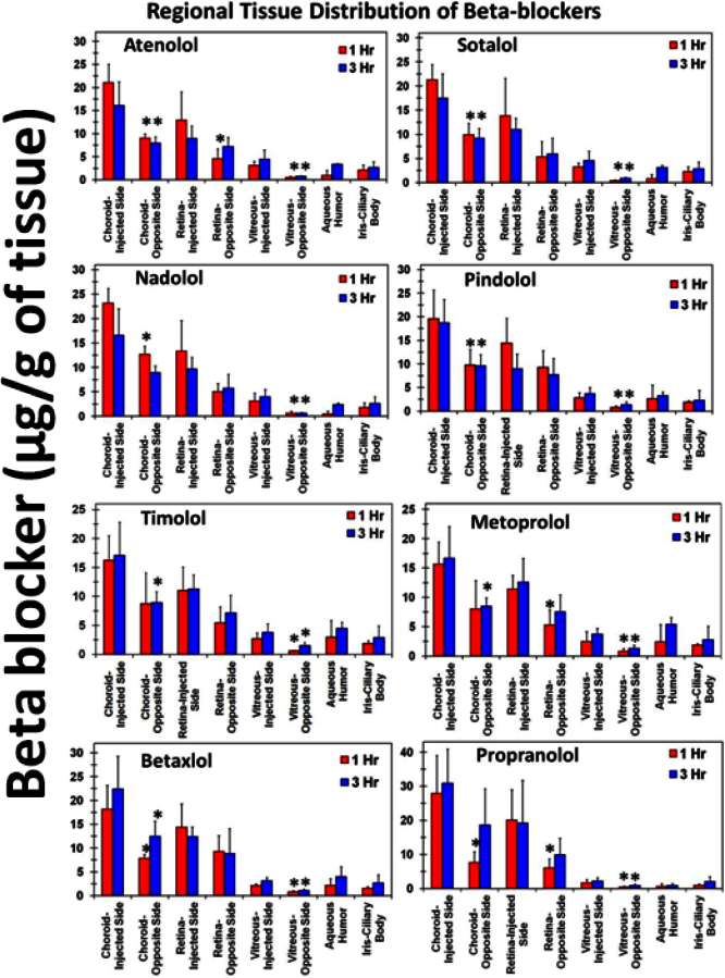
Methods: Using a small hypodermic 26G needle (3/8") with a short bevel (250 µm), location of the suprachoroidal injection in an ex vivo New Zealand white rabbit eye model was confirmed with India ink. Ocular tissue distribution of NaF (25 µl of 1.5 µg/ml) at 37 °C was monitored noninvasively using the Fluorotron Master(TM) at 0, 1, and 3 h following suprachoroidal, intravitreal, or intracameral injections in ex vivo rabbit eyes. For assessing the influence of lipophilicity and molecular size, 25 µl of a mixture of eight beta-blockers (250 µg/ml each) or FITC-dextran (4 kDa and 40 kDa, 30 mg/ml) was injected into the suprachoroidal space of excised rabbit eyes and incubated at 37 °C. Eyes were incubated for 1 and 3 h, and frozen at the end of incubation. Ocular tissues were isolated in frozen condition. Beta-blocker and FITC-dextran levels in excised ocular tissue were measured by liquid chromatography-tandem mass spectrometry and spectrofluorometry, respectively.
Results: Histological sections of India ink-injected albino rabbit eye showed the localization of dye as a black line in the suprachoroidal space. Suprachoroidal injection of NaF showed signal localization to the choroid and retina at 1 and 3 h post injection when compared with intravitreal and intracameral injections. Drug delivery to the vitreous after suprachoroidal injection decreased with an increase in solute lipophilicity and molecular weight. With an increase in drug lipophilicity, drug levels in the choroid-retinal pigment epithelium (RPE) and retina generally increased with some exceptions. Beta-blockers and FITC-dextrans were localized more to the dosed side when compared to the opposite side of the sclera, choroid-RPE, retina, and vitreous. These differences were greater for FITC-dextrans as compared to the beta-blockers.
Conclusions: The suprachoroidal route of injection allows localized delivery to the choroid-RPE and retina for small as well as large molecules. Suprachoroidal drug delivery to the vitreous declines with an increase in drug lipophilicity and molecular weight. Drug delivery differs between the dosed and opposite sides following suprachoroidal injection, at least up to 3 h.
Figures

Comment in
-
Findings of Research Misconduct.Fed Regist. 2018 Dec 6;83(234):62875. Fed Regist. 2018. PMID: 30556543 Free PMC article. No abstract available.
References
-
- Del Amo EM, Urtti A. Current and future ophthalmic drug delivery systems. A shift to the posterior segment. Drug Discov Today. 2008;13:135–43. - PubMed
-
- Wiener JM, Tilly J. Population ageing in the United States of America: implications for public programmes. Int J Epidemiol. 2002;31:776–81. - PubMed
-
- United Nations. Current status of the social situation, wellbeing, participation in development and rights of older persons worldwide;August 2010
-
- Sunkara G, Kompella UB. Membrane transport processes in the eye. In: Ophthalmic drug Delivery Systems. New York: Marcel Dekker Inc.; 2003.
Publication types
MeSH terms
Substances
Grants and funding
LinkOut - more resources
Full Text Sources
Other Literature Sources