. 2013 May 21:7:62.
doi: 10.3389/fncom.2013.00062. eCollection 2013.

Distinct Thalamo-Cortical Controls for Shoulder, Elbow, and Wrist during Locomotion

Affiliations

Distinct Thalamo-Cortical Controls for Shoulder, Elbow, and Wrist during Locomotion

Irina N Beloozerova et al. Front Comput Neurosci. .

Abstract

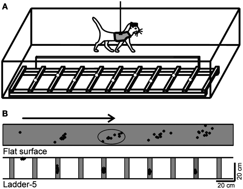
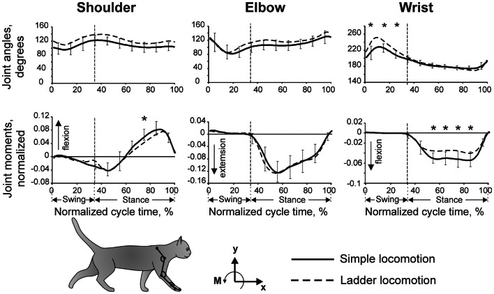
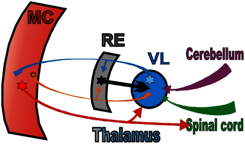
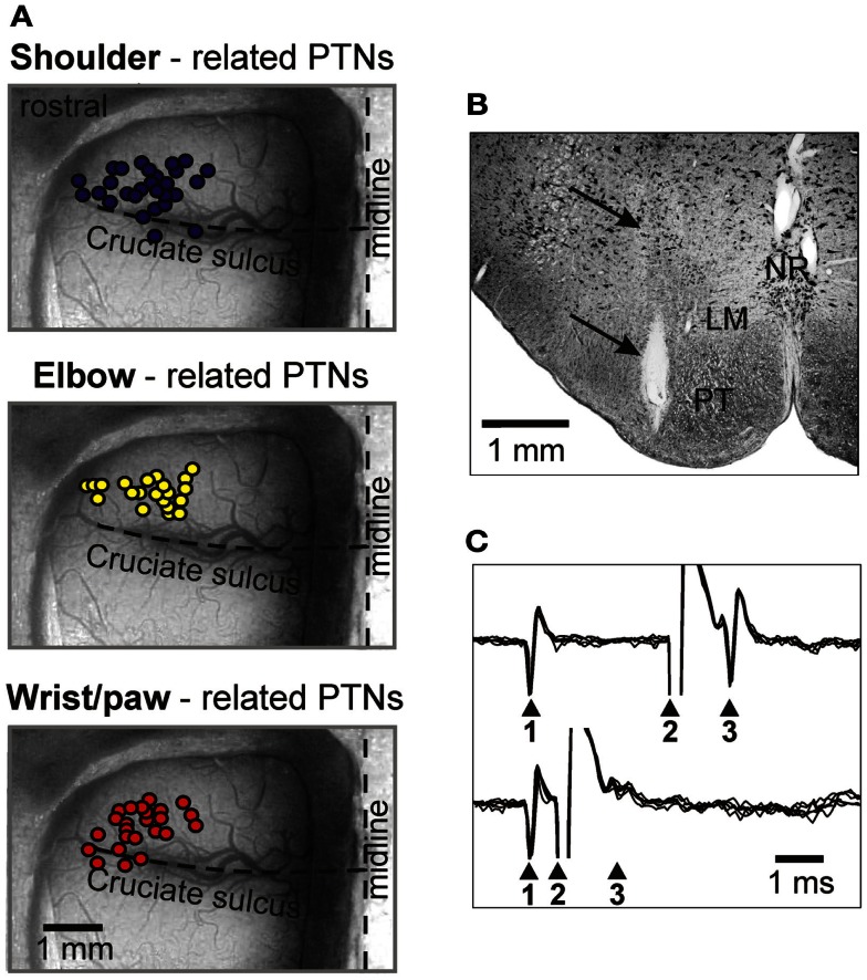
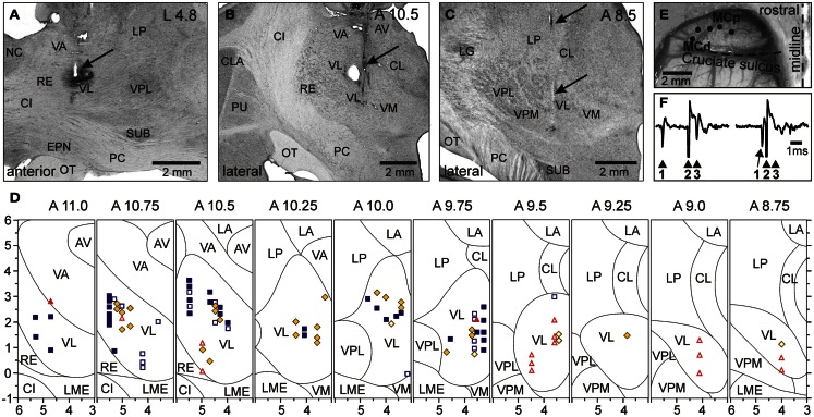
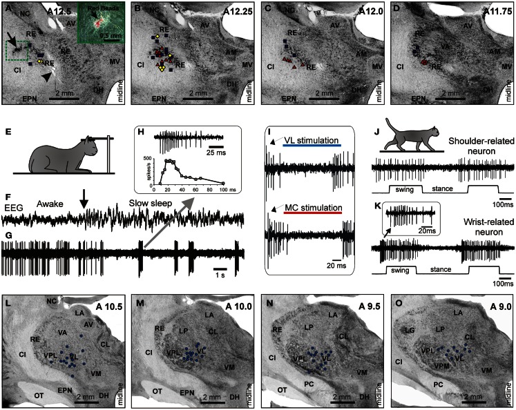
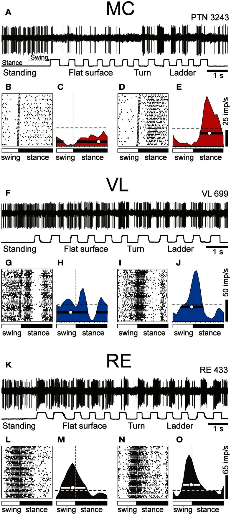
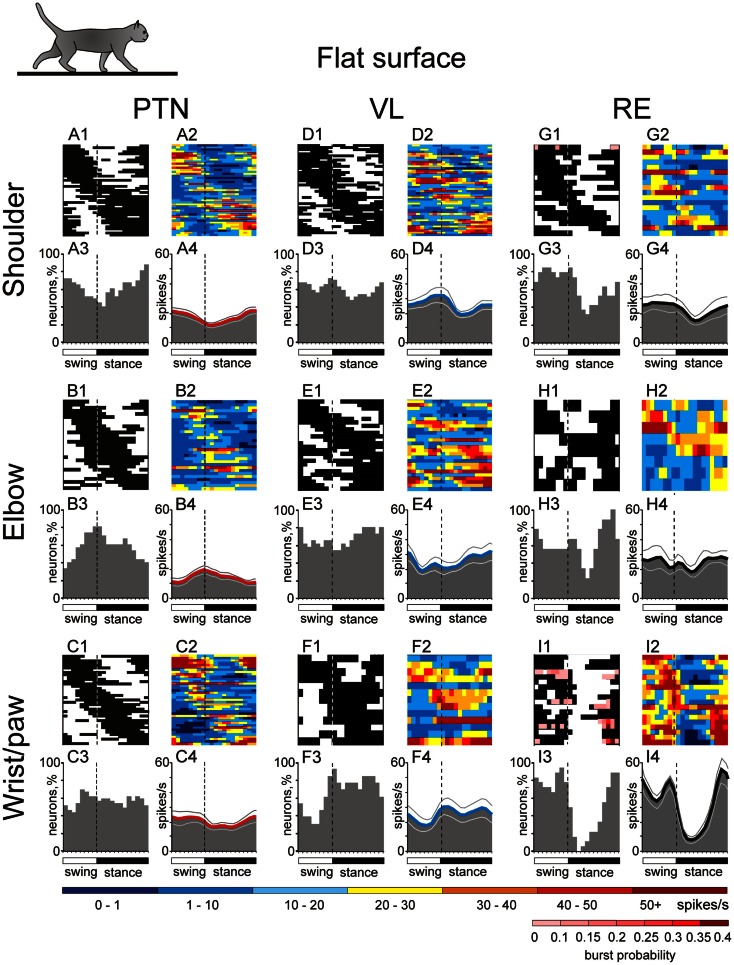
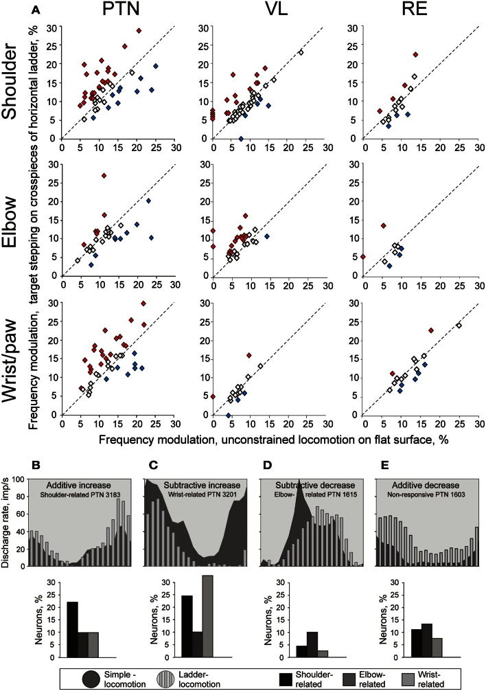
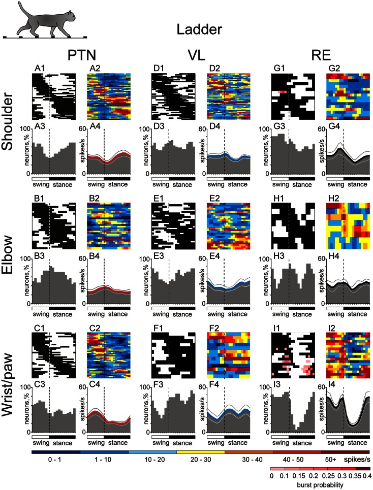
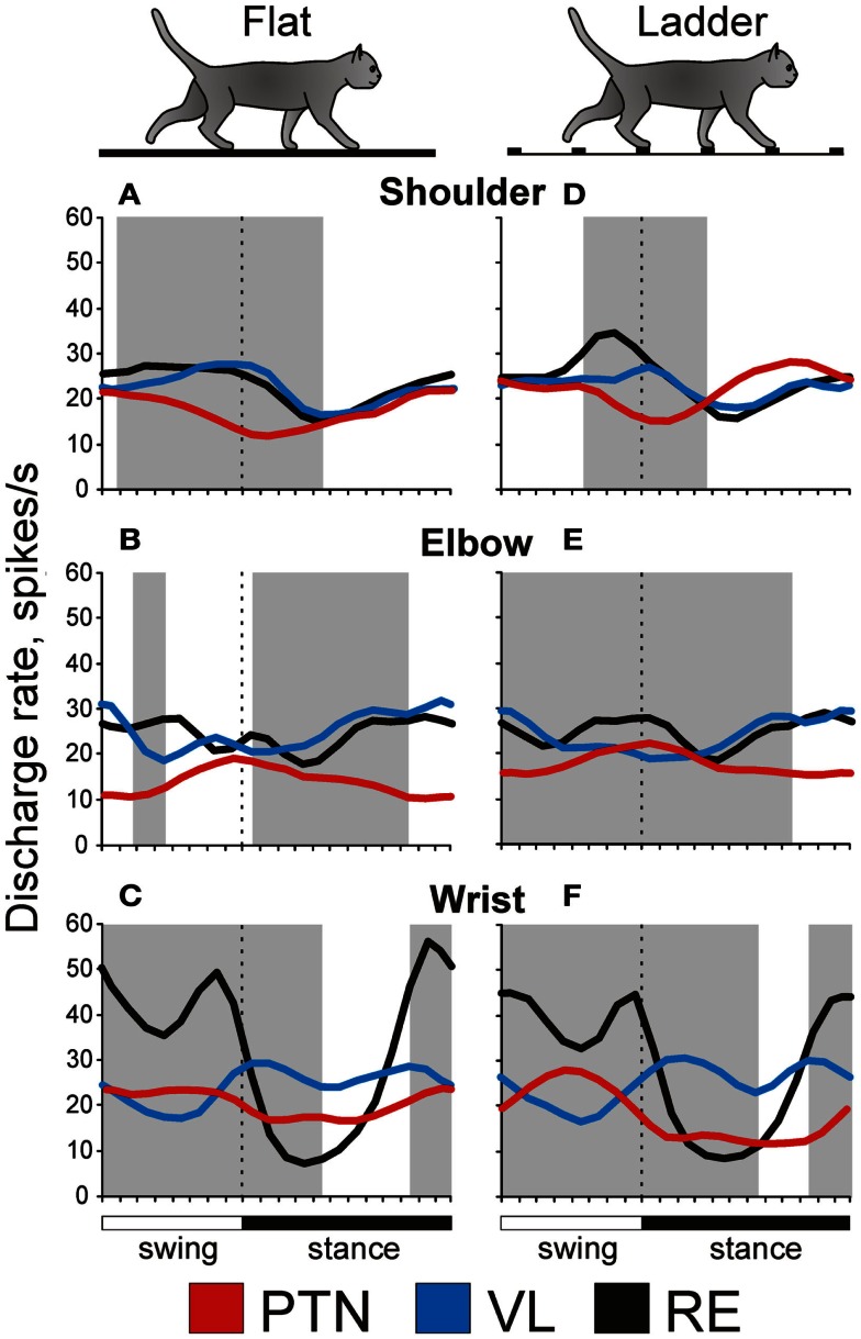
Recent data from this laboratory on differential controls for the shoulder, elbow, and wrist exerted by the thalamo-cortical network during locomotion is presented, based on experiments involving chronically instrumented cats walking on a flat surface and along a horizontal ladder. The activity of the following three groups of neurons is characterized: (1) neurons of the motor cortex that project to the pyramidal tract (PTNs), (2) neurons of the ventrolateral thalamus (VL), many identified as projecting to the motor cortex (thalamo-cortical neurons, TCs), and (3) neurons of the reticular nucleus of thalamus (RE), which inhibit TCs. Neurons were grouped according to their receptive field into shoulder-, elbow-, and wrist/paw-related categories. During simple locomotion, shoulder-related PTNs were most active in the late stance and early swing, and on the ladder, often increased activity and stride-related modulation while reducing discharge duration. Elbow-related PTNs were most active during late swing/early stance and typically remained similar on the ladder. Wrist-related PTNs were most active during swing, and on the ladder often decreased activity and increased modulation while reducing discharge duration. In the VL, shoulder-related neurons were more active during the transition from swing-to-stance. Elbow-related cells tended to be more active during the transition from stance-to-swing and on the ladder often decreased their activity and increased modulation. Wrist-related neurons were more active throughout the stance phase. In the RE, shoulder-related cells had low discharge rates and depths of modulation and long periods of activity distributed evenly across the cycle. In sharp contrast, wrist/paw-related cells discharged synchronously during the end of stance and swing with short periods of high activity, high modulation, and frequent sleep-type bursting. We conclude that thalamo-cortical network processes information related to different segments of the forelimb differently and exerts distinct controls over the shoulder, elbow, and wrist during locomotion.

Keywords: PTN; accuracy; cat; motor cortex; reticular nucleus of thalamus; thalamus; ventro-lateral thalamus; walking.


Figures

Figure 1
Figure 1
Locomotion tasks. (A) Cats walked in an experimental box that was divided into two corridors. In one of the corridors, the floor was flat, while the other corridor contained a horizontal ladder. White circles on the crosspieces of the ladder schematically show placements of cat forelimb paws. This schematic drawing is not to scale. (B) A typical distribution of right forelimb paw prints recorded from one cat during 10 walking passages though each corridor: on a flat surface (simple locomotion) and along the ladder with crosspieces 5 cm wide (complex locomotion). View from above. The direction of the cat’s progression is shown by the arrow on the top. For simple locomotion, paw prints are adjusted to start in the same position. During the ladder task, the first paw placement during ladder locomotion was between the crosspieces. Ellipses enclose approximate areas in which 95% of paw prints were found. (Adapted with modifications from Beloozerova et al., 2010).
Figure 2
Figure 2
Forelimb joint angles and moments during simple and ladder locomotion. Parameters were averaged across five cats. Vertical dashed lines separate the swing and stance phases of the stride. Standard deviations were similar across the two tasks and for clarity are shown only for simple locomotion. Symbol * indicates significant (p < 0.05, post hoc t-test) difference. The cat forelimb model is shown at the bottom. Orientation of each segment was determined as the angle between the negative direction of the vertical axis and the longitudinal segment axis directed from the distal end of the segment to the proximal one. (Adapted with modifications from Beloozerova et al., 2010).
Figure 3
Figure 3
The scheme of the thalamo-cortical network for locomotion. MC, motor cortex; RE, motor compartment of the reticular nucleus of thalamus; VL, ventrolateral nucleus of thalamus. Colored stars and arrows show neurons giving excitatory connections. Black star and arrow shows inhibitory neurons and connection.
Figure 4
Figure 4
Location of MC neurons and identification of PTNs. (A) Area of recording in the forelimb representation of the left motor cortex. Microelectrode entry points into the cortex are combined from eight cats and shown by circles on the photograph of the cortex of one cat. Tracks where PTNs with shoulder-related, elbow-related, and wrist-related receptive fields were recorded are shown by purple, yellow, and red circles, respectively. (B) Reference electrolytic lesion in the left pyramidal tract. Gliosis surrounding the electrode track and the reference lesion mark are indicated by arrows. Abbreviations: LM, lemniscus medialis; NR, nucleus raphes; PT, pyramidal tract. Frontal 50 μm thick section, cresyl violet stain. (C) A collision test determines whether a PTN response was antidromic. Top trace, the PTN spontaneously discharges (arrowhead 1), and the pyramidal tract is stimulated 3 ms later (arrowhead 2). The PTN responds with latency of 1 ms (arrowhead 3). Bottom trace, the PTN spontaneously discharges (arrowhead 1) and the pyramidal tract is stimulated 0.7 ms later (arrowhead 2). PTN does not respond (arrowhead 3) because in 0.7 ms its spontaneous spike was still en route to the site of stimulation in the pyramidal tract, and thus collision/nullification of spontaneous and evoked spikes occurred. (Adapted with modifications from Stout and Beloozerova, 2012).
Figure 5
Figure 5
Location of VL neurons and identification of TCs. (A) The recording site in cat A is shown on a photomicrograph of a parasagittal section of the thalamus. It was located in the rostral VL. The arrow points to the electrolytic lesion mark and the darkened area of tissue filled with WGA-HRP. The site is ∼2 mm caudally to the Nucleus caudatus (NC) of the basal ganglia. (B) The recording site in cat B is shown on a photomicrograph of a coronal section of the thalamus. It was positioned in the middle of the VL. The arrow points to the electrolytic lesion mark and darkened area where fluorescent beads were deposited. The caudal part of putamen (PU), a landmark for the anterior-posterior position of the section, is seen laterally. (C) The recording site in cat C is shown on a photomicrograph of a coronal section of the thalamus. It was positioned in the caudal VL. The arrows point to a track from a reference electrode. The most rostral aspect of the lateral geniculate body (LG), a landmark for the anterior-posterior position of the section, is visible laterally. (A–C) 50 μm thick sections, cresyl violet stain. (D) A photograph of the dorsal surface of the left frontal cortex of cat B. Entrance points of stimulation electrodes into the precruciate sulcus are schematically shown by black dots. Electrodes were placed in the paw (the motor cortex distal forelimb representation, MCd), the elbow and shoulder representations (the motor cortex proximal forelimb representation, MCp) as determined by multiunit recording and micro-stimulation procedures. Cru, cruciate sulcus; Pcd, post-cruciate dimple; mAns, medial ansate sulcus. (E) A collision test determined whether a neuron response was antidromic. Stimulation of the MC evoked a spike in the neuron with a latency of 0.8 ms. To determine whether this spike was elicited antidromically, on a next trial a spontaneous spike of the neuron was used to trigger MC stimulation with 0.4 ms delay. Stimulation delivered with a delay smaller than the time needed for a spontaneous spike to reach the site of stimulation (that is approximately equal to the latent time of an antidromic spike) was not followed by a response. This indicated a collision of ortho- and antidromically conducted spikes and confirmed the antidromic nature of the evoked spike. (F) A reconstruction of positions of individual neurons recorded during locomotion in cats A, B, and C. ■, Purple squares show neurons with somatosensory receptive fields on the shoulder: responding to passive movements in the shoulder joint and/or palpation of muscles on the back or neck; ♦, Yellow diamonds show cells that were activated by movements in the elbow; ▲, Red triangles represent neurons with receptive fields on the wrist or paw. Filled symbols represent neurons with axonal projections to the MC (thalamo-cortical neurons, TCs); open symbols represent neurons whose projections were not identified. Abbreviations: AV, nucleus anterio-ventralis thalami; CI, capsula interna; CL, nucleus centralis lateralis; CLA, claustrum; EPN, nucleus entopeduncularis; LA, nucleus lateralis anterior; LG, lateral geniculate nucleus; LME, lamina medullaris externa thalami; LP, nucleus lateralis posterior; NC, nucleus caudatus; OT, optic tract; PC, pedunculus cerebri; PU, putamen; RE, nucleus reticularis thalami; SUB, nucleus subthalamicus; VA, nucleus ventralis anterior; VL, nucleus ventralis lateralis; VM, nucleus medialis; VPL, nucleus ventralis postero-lateralis; VPM, nucleus ventralis postero-medialis (Adapted with modifications from Marlinski et al., 2012a).
Figure 6
Figure 6
Location and identification of RE neurons. (A–D) Location of RE neurons recorded during locomotion. Estimated locations of neurons are combined from two cats and are shown by various symbols on frontal sections of thalamus of one of them: ■, Purple squares show neurons with somatosensory receptive fields on the shoulder: responding to passive movements in the shoulder joint and/or palpation of muscles on the back or neck; ♦, Yellow diamonds show cells that were activated by movements in the elbow; ▲, Red triangles represent neurons with receptive fields on the wrist or paw. In (A), an arrowhead is pointing to a reference electrolytic lesion and an arrow indicates the site of injection of red fluorescent beads. (A) close-up to the injection site is shown in the insert. Abbreviations: AM, nucleus anterio-medialis; AV, nucleus anterio-ventralis thalami; CI, capsula interna; DH, dorsal hypothalamus; EPN, nucleus entopeduncularis; MV, nucleus medio-ventralis; NC, nucleus caudatus; RE, nucleus reticularis thalami; VA, nucleus ventralis anterior. Frontal 50 μm thick sections, cresyl violet stain. (E–H) Identification of RE neurons by characteristic profile of their bursts during sleep. (E) Cat sleeping with its head restrained. (F,G) An example of activity of a RE neuron while cat is awake and asleep. At the beginning of the record desynchronized activity in EEG indicates that the cat was awake, and the neuron was discharging fairly regularly. The arrow points to the beginning of “spindle waves” in EEG, which are a sign of beginning of slow wave sleep. Shortly thereafter very high frequency irregular bursts separated by long periods of inactivity replaced the regular discharge of the neuron. (H) Close-up on a burst. The first interspike interval in this burst was longer than the second one, and the second interval was longer that the third. Several following interspike intervals were of an approximately similar duration, while the last ones were progressively longer. The lower trace shows change of discharge frequency within the burst. Such a burst with ramping up and then winding down firing rate identifies this neuron as belonging to the RE. (I) Identification of the motor compartment of the RE by responses of neurons to electrical stimulation of the VL (upper trace) and MC (lower trace). In response to either stimulation the cell generates a short latency burst followed by a period of silence and then by another burst. (J) Locomotion-related activity of a representative neuron with shoulder-related receptive field. The activity of this neuron is modulated to strides but does not contain any “sleep-type” busts. (K) Accelerating-decelerating frequency “sleep-type” bursting during locomotion in a wrist/paw-related neuron. A burst is shown in the insert at a fast time scale. Such bursts often appeared at the beginning of the locomotion-related activation of this neuron. (L–O) Thalamic projections to the area of recording in the RE. Neurons in the VL and VL/VPL border zone in one of the cats where red fluorescent beads were injected in the rostro-lateral part of the explored RE area, retrogradely labeled with red fluorescent beads. Neurons are shown on photomicrographs of frontal sections of the left thalamus ipsilateral to the injection site. Each circle represents one labeled neuron. Abbreviations: CL, nucleus centralis lateralis; LA, nucleus lateralis anterior; LG, lateral geniculate nucleus; LP, nucleus lateralis posterior; OT, optic tract; PC, pedunculus cerebri; VL, nucleus ventralis lateralis; VM, nucleus medialis; VPL, nucleus ventralis postero-lateralis; VPM, nucleus ventralis postero-medialis; other abbreviations are as in Figure 5 (Adapted with modifications from Marlinski et al., 2012b).
Figure 7
Figure 7
Example activity of MC, VL, and RE cells during locomotion. (A,F,K) Activity of MC (A), VL (F), and RE (K) cells during standing, simple, and ladder locomotion. The bottom trace shows the stance and swing phases of the step cycle of the right forelimb that is contralateral to the recording site in the cortex and thalamus. (B,C,G,H,L,M) Activities of the same neurons during simple locomotion are presented as rasters of 37–47 step cycles (B,G,L) and as histograms (C,H,M). In the rasters, the duration of step cycles is normalized to 100%, and the rasters are rank-ordered according to the duration of the swing phase. The beginning of the stance phase in each stride is indicated by an open triangle. In the histograms, the horizontal interrupted line shows the level of activity during standing. The horizontal black bar shows the period of elevated firing (PEF) and the circle indicates the preferred phase. (D,E,I,J,N,O) Activities of the same neurons during ladder locomotion are presented as rasters (D,I,N) and as histograms (E,J,O). (Examples of the activity of MC, VL, and RE neurons are adapted with modifications from Beloozerova et al., ; Marlinski et al., ,, respectively).
Figure 8
Figure 8
Activities of the shoulder-, elbow-, and wrist/paw-related cells in the thalamo-cortical network during simple locomotion. (A,D,G) Activity of neurons responsive to movements in the shoulder joint, and/or palpation of back, chest, or neck muscles in the MC (A), VL (D), and RE (G). (A1,D1,G1) Phase distribution of PEFs. (A2,D2,G2) Corresponding phase distribution of discharge frequencies. The average discharge frequency in each 1/20th portion of the cycle is color-coded according to the scale shown at the bottom. (A3,D3,G3) Proportion of active neurons (neurons in their PEFs) in different phases of the step cycle. (A4,D4,G4) The mean discharge rate. Thin lines show SEM. Vertical interrupted lines denote end of swing and beginning of stance phase. (B,E,H) Activity of neurons responsive to passive movement of the elbow joint in the MC (B), VL (E), and RE (H). (C,F,I) Activity of neurons responsive to stimulation of the paw or movement in the wrist joint in the MC (C), VL (F), and RE (I). (Data on the activity of PTNs, VL, and RE neurons are adapted with modifications from Stout and Beloozerova, ; Marlinski et al., ,, respectively).
Figure 9
Figure 9
Change in the depth of frequency modulation upon transition from simple to ladder locomotion. (A) Comparison of depth of modulation in the activity of individual MC, VL, and RE neurons. The abscissa and ordinate of each point show the values of the depth of modulation of a neuron during simple and ladder locomotion, respectively. Neurons whose depths of modulation were statistically significantly different during the two tasks are shown with filled diamonds, the other ones are shown with open diamonds. (B–E) Typical changes in the depth of modulation upon transition from simple to ladder locomotion in PTNs. The area histograms show the activity of typical PTNs during simple locomotion, and the bar histograms show activity of the same PTNs during ladder locomotion. Bar graphs beneath the histograms show the proportion of PTNs from each group exhibiting that type of modulation change. (B): Increase in the depth of modulation by additive mechanism. (C) Increase in the depth of modulation by subtractive mechanism. (D) Decrease in the depth of modulation by subtractive mechanism. (E) Decrease in the depth of modulation by additive mechanism. (Adapted with modifications from Stout and Beloozerova, 2012).
Figure 10
Figure 10
Activities of the shoulder-, elbow-, and wrist/paw-related cells in the thalamo-cortical network during ladder locomotion. (A,D,G) Activity of neurons responsive to movements in the shoulder joint, and/or palpation of back, chest, or neck muscles in the MC (A), VL (D), and RE (G). (A1,D1,G1) Phase distribution of PEFs. (A2,D2,G2) Corresponding phase distribution of discharge frequencies. The average discharge frequency in each 1/20th portion of the cycle is color-coded according to the scale shown at the bottom. (A3,D3,G3) Proportion of active neurons (neurons in their PEFs) in different phases of the step cycle. (A4,D4,G4) The mean discharge rate. Thin lines show SEM. Vertical interrupted lines denote end of swing and beginning of stance phase. (B,E,H) Activity of neurons responsive to passive movement of the elbow joint in the MC (B), VL (E), and RE (H). (C,F,I) Activity of neurons responsive to stimulation of the paw or movement in the wrist joint in the MC (C), VL (F), and RE (I). (Data on the activity of PTNs, VL, and RE neurons are adapted with modifications from Stout and Beloozerova, ; Marlinski et al., ,, respectively).
Figure 11
Figure 11
Distinct thalamo-cortical controls for shoulder, elbow, and wrist during locomotion. Red lines show population activities of shoulder-, elbow-, and wrist/paw-related neurons of the MC, blue lines show those of the VL, and black lines represent the corresponding activities of inhibitory neurons of the RE. Shaded are periods of the step cycle when the activities of the MC and VL are in anti-phase. (Data on the activity of PTNs, VL, and RE neurons are adapted with modifications from Stout and Beloozerova, ; Marlinski et al., ,, respectively).

References

    1. Angaut P. (1979). “The cerebello-thalamic projection in the cat,” in Cerebro-Cerebellar Interactions, eds Massion J., Sasaki K. (North-Holland: Elsevier; ), 1943
    1. Armstrong D. M., Drew T. (1984a). Discharges of pyramidal tract and other motor cortical neurones during locomotion in the cat. J. Physiol. 346, 471–495 - PMC - PubMed
    1. Armstrong D. M., Drew T. (1984b). Locomotor-related neuronal discharges in cat motor cortex compared with peripheral receptive fields and evoked movements. J. Physiol. 346, 497–517 - PMC - PubMed
    1. Armstrong D. M., Drew T. (1985a). Electromyographic responses evoked in muscles of the forelimb by intracortical stimulation in the cat. J. Physiol. 367, 309–326 - PMC - PubMed
    1. Armstrong D. M., Drew T. (1985b). Forelimb electromyographic responses to motor cortex stimulation during locomotion in the cat. J. Physiol. 367, 327–351 - PMC - PubMed

LinkOut - more resources