. 2013 May 29;8(5):e64159.
doi: 10.1371/journal.pone.0064159. Print 2013.

B cells promote tumor progression via STAT3 regulated-angiogenesis

Affiliations

B cells promote tumor progression via STAT3 regulated-angiogenesis

Chunmei Yang et al. PLoS One. .

Abstract

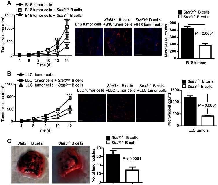
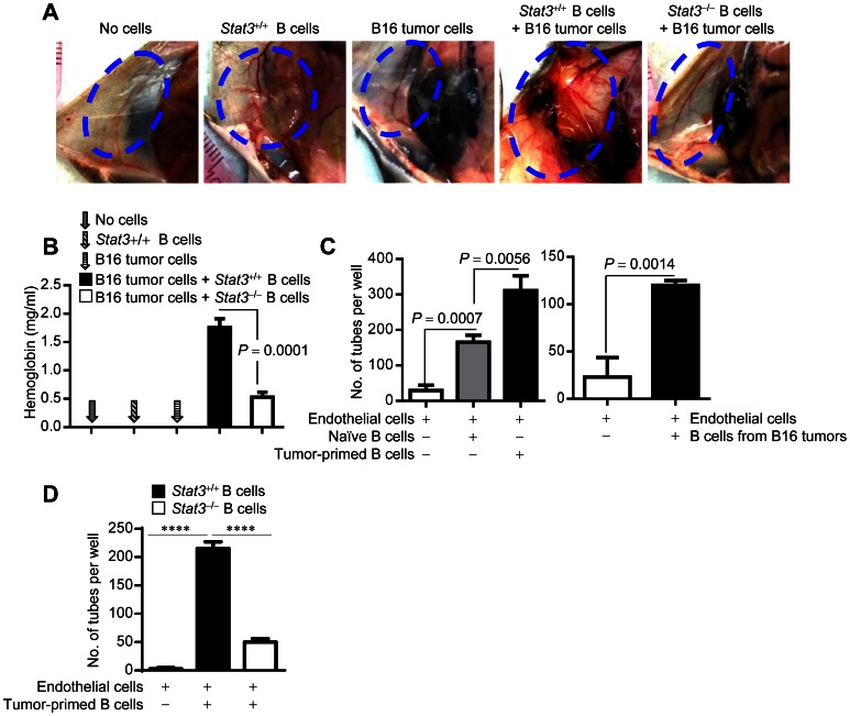
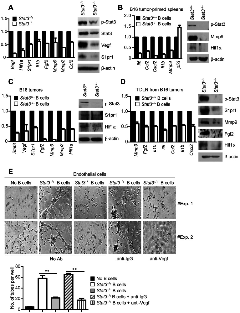
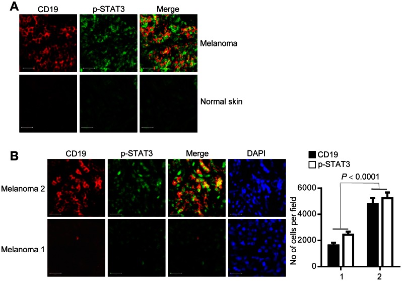
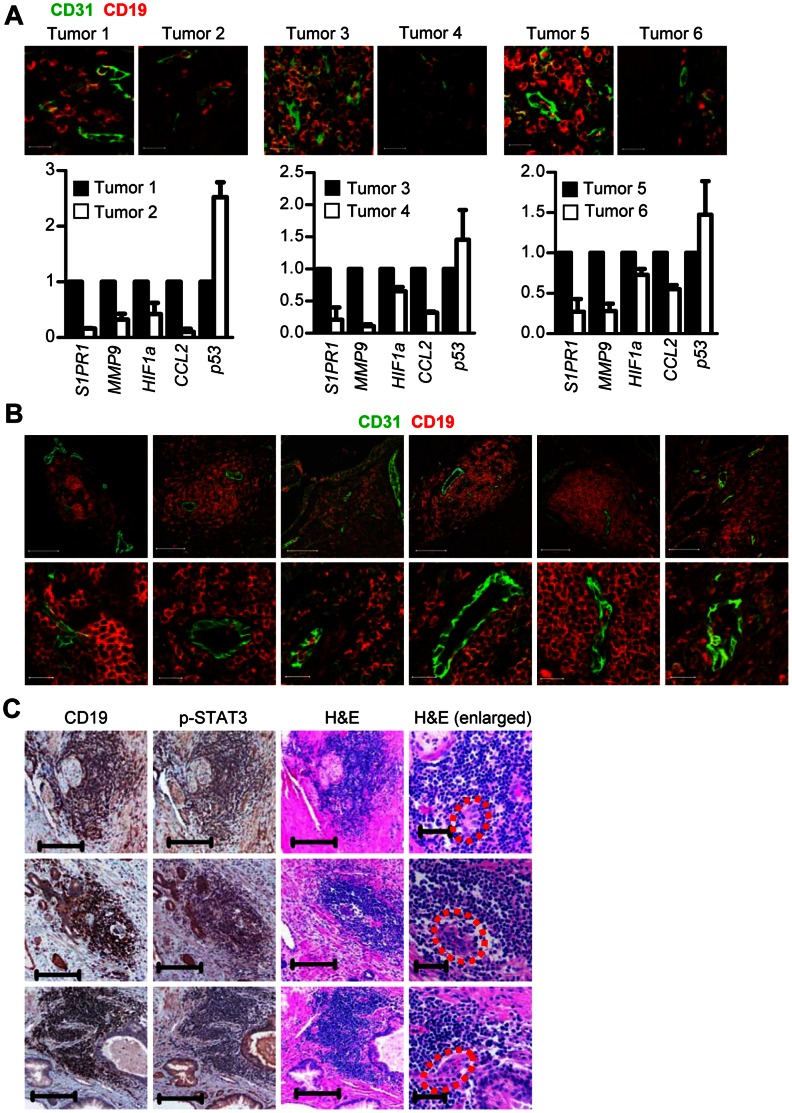
The role of B cells in cancer and the underlying mechanisms remain to be further explored. Here, we show that tumor-associated B cells with activated STAT3 contribute to tumor development by promoting tumor angiogenesis. B cells with or without Stat3 have opposite effects on tumor growth and tumor angiogenesis in both B16 melanoma and Lewis Lung Cancer mouse models. Ex vivo angiogenesis assays show that B cell-mediated tumor angiogenesis is mainly dependent on the induction of pro-angiogenic gene expression, which requires Stat3 signaling in B cells. Furthermore, B cells with activated STAT3 are mainly found in or near tumor vasculature and correlate significantly with overall STAT3 activity in human tumors. Moreover, the density of B cells in human tumor tissues correlates significantly with expression levels of several STAT3-downstream pro-angiogenic genes, as well as the degree of tumor angiogenesis. Together, these findings define a novel role of B cells in promoting tumor progression through angiogenesis and identify STAT3 in B cells as potential therapeutic target for anti-angiogenesis therapy.


Conflict of interest statement

Competing Interests: The authors have declared that no competing interests exist.

Figures

Figure 1
Figure 1. B cells with activated Stat3 accelerate tumor progression and increase blood vessel formation in tumors.
(A and B) Left, Growth curve of B16 (A) or LLC (B) tumors in Rag1−/− mice, without or with Stat3+/+or Stat3 −/− B cells. B cells were enriched from splenocytes of B16 or LLC tumor-bearing mice with or without Stat3 ablated in hematopoietic cells; n = 8; ***, P<0.001. Middle, Immunofluorescent staining of tumors to detect microvessels (anti-CD31, red) and nuclei (Hoechst, blue); scale bars, 200 µm. Right, Graph showing the number of CD31+ vessels in the tumor tissues (3 microscopic fields/section); means ± SD, n = 2. (C) Representative images (left) and a graph (right) showing the number of spontaneous lung metastatic nodule in mice with Stat3+/+ or Stat3−/− B cells receiving B16 tumor cells; means ± SEM, n = 5.
Figure 2
Figure 2. B cells promote tumor angiogenesis by enhancing endothelial cell function in a Stat3-dependent manner.
(A) Representative images of vessel formation in Matrigel plugs implanted in Rag1−/− mice. The Matrigel plugs contain either B16 tumor cell alone, Stat3+/+ B cell alone or B16 tumor cells plus Stat3+/+ or Stat3 −/− B cells, which were isolated from splenocytes of tumor-bearing mice; n = 5; area indicated by blue dot showing level of blood vessel formation. (B) Hemoglobin contents in the pooled Matrigel plugs determined by colorimetric assay; means ± SEM, n = 5. (C) In vitro collagen tube formation assay showing the number of tubes formed by ECs with or without the indicated B cells. Stat3+/+ B cells were enriched from splenocytes of B16 tumor-bearing mice (left) or B16 tumors (right); means ± SEM, n = 3. (D) B cell-mediated endothelial cell tube formation requires Stat3 signaling in B cells. EC tube formation by co-culturing ECs with tumor-primed Stat3+/+ or Stat3−/− splenic B cells. Tumor-primed B cells were enriched from splenocytes of MB49 tumor-bearing mice with Stat3+/+ or Stat3−/− hematopoietic cells; means ± SEM, n = 3; ****, P<0.0001.
Figure 3
Figure 3. Stat3 intrinsic to B cells is crucial for expression of pro-angiogenic genes and endothelial cell tube formation.
(A) Expression levels of mRNA (left) or protein (right) of the indicated genes in B16 tumors. The tumors were formed by implanting B16 tumor cells with the indicated tumor-primed B cells in Rag1−/− mice. (B) mRNA (left) or protein (right) expression levels of the indicated genes in tumor-primed B cells from splenocytes of B16-tumor bearing C57BL/6 mice with Stat3+/+ or Stat3−/− hematopoietic cells. (C and D) mRNA and protein levels of the indicated pro-angiogenic genes in Stat3+/+ or Stat3−/− B cells isolated from either B16 tumors (C) or B16 tumor-draining lymph nodes (TDLN) (D). In all cases, mRNA is determined by real-time RT-PCR. The relative amount of mRNA is compared to that of Stat3+/+ B cells, which is designated as 1 (means ± SEM, n = 3). Western blotting data represent two independent experiments. (E) Representative images of EC tube formation by co-culturing ECs with splenic Stat3+/+ B cells from B16 tumor-bearing mice in the presence of VEGF neutralizing antibodies. IgG antibody at the same concentration is used as control; n = 3 (top). Quantitative graph showing the average number of vessel-like structures formed by ECs upon addition of the indicated B cells; means ± SEM, n = 3; **, P<0.001 (bottom).
Figure 4
Figure 4. B cells with activated STAT3 accumulate in human tumors.
(A) Immunofluorescent staining of human melanoma and normal human skin tissue sections; anti-CD19 (red; B cell marker) and anti-p-STAT3 (green). Scale bars, 20 µm. (B) B cells in primary tumor sites impact overall tumor STAT3 activity. Confocal microscopic images showing primary melanoma tumor tissue staining of B cells and p-STAT3 (left), with quantification of CD19 and p-STAT3 positive cells (right). Scale bars, 20 µm. Total ten microscopic fields (10 X) were examined for each tumor section; n = 2.
Figure 5
Figure 5. B cells with activated STAT3 express pro-angiogenic genes and accumulate around microvessels in human tumors.
(A) B cells are important for expression of pro-angiogenic genes within human prostate tumor tissues. The density of B cells around tumor vasculature in prostate tumor tissue was determined by immunofluorescent staining using anti-CD19 and anti-CD31 antibodies (top); scale bars, 20 µm. Real-time RT-PCR measuring RNA expression levels of pro-angiogenic genes in the consecutive human prostate tumor tissue sections (bottom). The relative amount of mRNA is normalized to 18S and compared to RNA levels in tumor tissues with high p-STAT3, which is designated as 1; means ± SD, n = 2. (B) Immunofluorescent staining of human prostate tumor tissue sections showing B cells and microvessels with CD31+ endothelial cells (green). Scale bars, 100 µm in the original (top) and 20 µm in the enlarged (bottom). (C) B cells around the microvessels display persistently activated STAT3. IHC showing B cells and p-STAT3-positive cells in the same area of human prostate tumor tissues; scale bars, 200 µm. H&E staining of the consecutive tissue sections. Microvessel-like structures are marked by red dots; scale bars, 200 µm in the original and 50 µm in the enlarged.

References

    1. Milne K, Kobel M, Kalloger SE, Barnes RO, Gao D, et al. (2009) Systematic analysis of immune infiltrates in high-grade serous ovarian cancer reveals CD20, FoxP3 and TIA-1 as positive prognostic factors. PLoS One 4: e6412. - PMC - PubMed
    1. Pages F, Berger A, Camus M, Sanchez-Cabo F, Costes A, et al. (2005) Effector memory T cells, early metastasis, and survival in colorectal cancer. N Engl J Med 353: 2654–2666. - PubMed
    1. Galon J, Costes A, Sanchez-Cabo F, Kirilovsky A, Mlecnik B, et al. (2006) Type, density, and location of immune cells within human colorectal tumors predict clinical outcome. Science 313: 1960–1964. - PubMed
    1. Tan TT, Coussens LM (2007) Humoral immunity, inflammation and cancer. Curr Opin Immunol 19: 209–216. - PubMed
    1. Ohri CM, Shikotra A, Green RH, Waller DA, Bradding P (2009) Macrophages within NSCLC tumour islets are predominantly of a cytotoxic M1 phenotype associated with extended survival. Eur Respir J 33: 118–126. - PubMed

Publication types

MeSH terms

Substances