. 2013 Sep;104(9):1154-61.
doi: 10.1111/cas.12213. Epub 2013 Jul 10.

Columnar metaplasia in a surgical mouse model of gastro-esophageal reflux disease is not derived from bone marrow-derived cell

Affiliations

Columnar metaplasia in a surgical mouse model of gastro-esophageal reflux disease is not derived from bone marrow-derived cell

Susumu Aikou et al. Cancer Sci. 2013 Sep.

Abstract

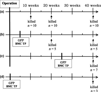
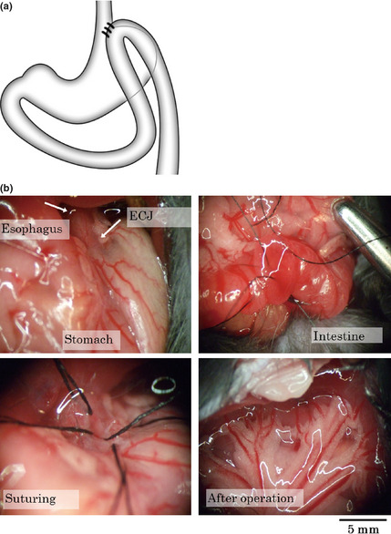
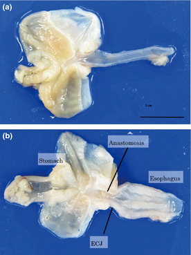
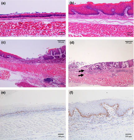
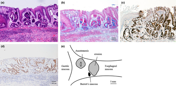
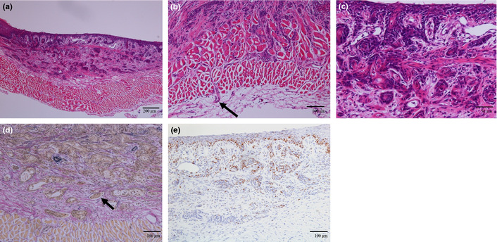
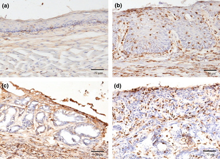
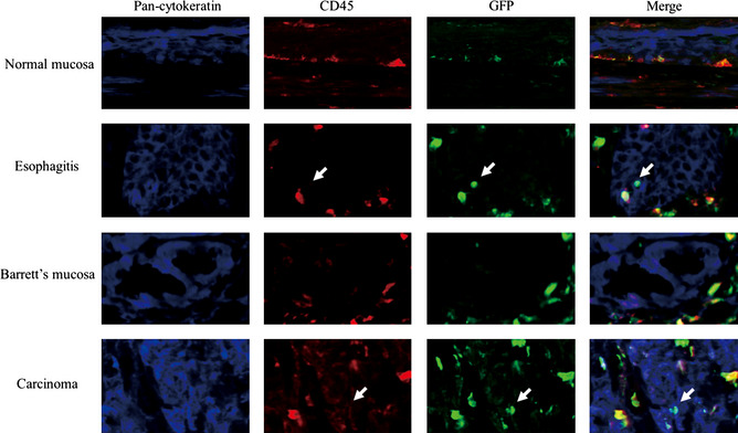
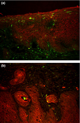
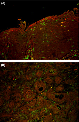
The incidence of esophageal adenocarcinoma has increased in the last 25 years. Columnar metaplasia in Barrett's mucosa is assumed to be a precancerous lesion for esophageal adenocarcinoma. However, the induction process of Barrett's mucosa is still unknown. To analyze the induction of esophageal columnar metaplasia, we established a mouse gastro-esophageal reflux disease (GERD) model with associated development of columnar metaplasia in the esophagus. C57BL/6 mice received side-to-side anastomosis of the esophagogastric junction with the jejunum, and mice were killed 10, 20, and 40 weeks after operation. To analyze the contribution of bone marrow-derived cells to columnar metaplasia in this surgical GERD model, some mice were transplanted with GFP-marked bone marrow after the operation. Seventy-three percent of the mice (16/22) showed thickened mucosa in esophagus and 41% of mice (9/22) developed columnar metaplasia 40 weeks after the operation with a mortality rate of 4%. Bone marrow-derived cells were not detected in columnar metaplastic epithelia. However, scattered epithelial cells in the thickened squamous epithelia in regions of esophagitis did show bone marrow derivation. The results demonstrate that reflux induced by esophago-jejunostomy in mice leads to the development of columnar metaplasia in the esophagus. However, bone marrow-derived cells do not contribute directly to columnar metaplasia in this mouse model.


Figures

Figure 1
Figure 1
Group A underwent only operation to cause the gastroesophageal reflux disease. Group B received bone marrow cell transplantation from green fluorescent protein (GFP) Tg mice 5 weeks after operation. Group C received bone marrow cell transplantation from GFP Tg mice 25 weeks after operation. Group D received only bone marrow cell transplantation from GFP Tg as control group.
Figure 2
Figure 2
(a) Schema of surgical gastroesophageal reflux disease (GERD) model. The jejunum, 1 cm distal to Treitz's ligament, was anastomosed side‐to‐side to the esophago‐gastric junction. (b) The representative procedure of the operation.
Figure 3
Figure 3
Macroscopically, expansion of esophagus diameter and the thickening of the esophageal wall were seen in operated mice.
Figure 4
Figure 4
(a–d) Hematoxylin and eosin staining (a) Normal esophagus mucosa (b) 20 weeks after operation: thickening of the mucous membrane was seen. (c) 10 weeks after operation: inflammatory cell infiltration and erosion were seen. (d) 20 weeks after operation: metaplastic columnar epithelium and double muscularis mucosa were seen (e,f) Immunostaining with anti‐Ki67 antibody. (e) Normal esophageal mucosa. (f) 20 weeks after operation: Proliferating cells stained positively for Ki67 were increased in mucosa of mice after surgery.
Figure 5
Figure 5
Columnar metaplasia. (a) hematoxylin and eosin staining. (b) Alcian blue (pH 2.5) staining. (c) CDX2 immunostaining for columnar metaplasia. (d) Immunostaining with anti‐Ki67 antibody. (e) Schema of distribution of Barrett's mucosa.
Figure 6
Figure 6
Squamous cell carcinoma observed in a mouse 40 weeks after operation. (a–c) Hematoxylin and eosin staining. (b) The tumor invaded to adventitia of esophagus. (c) The tumor is spreading to submucosal layer. (d) Ductal invasion was also seen. The duct is negative for Elastica van Gieson (EVG) staining diagnosed as lymph vessel invasion. (e) Staining with anti‐Ki67 antibody.
Figure 7
Figure 7
Immunohistochemistry against green fluorescent protein (GFP). (a) Normal mucosa of esophagus. (b) Thickening mucosa with esophagitis. (c) There were no GFP positive cell in Barrett's mucosa. (d) There were many GFP positive cells in carcinoma.
Figure 8
Figure 8
Immunofluorescence multiple staining with anti‐CD45, anti‐cytokeratin, anti‐green fluorescent protein (GFP) antibodies. The GFP positive cells in normal esophagus also showed CD45 positivity and identified as leukocyte. A few GFP positive cells did not show CD45 positivity. These cells also positive for cytokeratin and identified as epithelium cells derived from transplanted bone marrow cells. There was no GFP positive, CD45 negative and cytokeratin positive cells in Barrett's mucosa. A few cells in carcinoma showed GFP positivity, CD45 negativity and cytokeratin positivity.
Figure 9
Figure 9
Immunofluorescence double staining with anti‐pan‐cytokeratin and anti‐green fluorescent protein (GFP) antibodies in esophagitis mucosa (a), and in squamous cell carcinoma (b). Pan‐cytokeratin was stained for red and GFP for green. A few cells in esophagitis mucosa and in carcinoma showed GFP positivity and cytokeratin positivity.
Figure 10
Figure 10
Immunofluorescence double staining with anti‐E‐cadherin and anti‐green fluorescent protein (GFP) antibodies in esophagitis mucosa (a), and in squamous cell carcinoma (b). E‐cadherin was stained for red and GFP for green. A few GFP positive cells in esophagitis mucosa and in carcinoma are circumscribed by E‐cadherin.

References

    1. Devesa SS, Blot WJ, Fraumeni JF Jr. Changing patterns in the incidence of esophageal and gastric carcinoma in the United States. Cancer 1998; 83: 2049–53. - PubMed
    1. Pohl H, Welch HG. The role of overdiagnosis and reclassification in the marked increase of esophageal adenocarcinoma incidence. J Natl Cancer Inst 2005; 97: 142–6. - PubMed
    1. Barrett NR. Chronic peptic ulcer of the oesophagus and ‘oesophagitis’. Br J Surg 1950; 38: 175–82. - PubMed
    1. Spechler SJ, Goyal RK. The columnar‐lined esophagus, intestinal metaplasia, and Norman Barrett. Gastroenterology 1996; 110: 614–21. - PubMed
    1. Lagergren J, Bergström R, Lindgren A, Nyren O. Symptomatic gastroesophageal reflux as a risk factor for esophageal adenocarcinoma. N Engl J Med 1999; 340: 825–31. - PubMed

MeSH terms