

Retinal vasculitis in two pediatric patients with systemic lupus erythematosus: a case report
- PMID: 23734963
- PMCID: PMC3682897
- DOI: 10.1186/1546-0096-11-25
Retinal vasculitis in two pediatric patients with systemic lupus erythematosus: a case report
Abstract
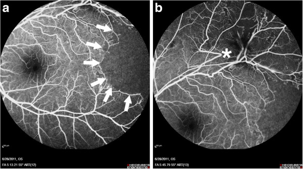
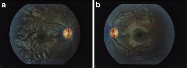
We report two pediatric female patients with systemic lupus erythematosus (SLE) who presented with decreased vision. Both patients were found to have retinal vasculitis and occlusive disease. The first patient also presented with vitreous hemorrhage and later non-arteritic ischemic optic neuropathy. She was treated with panretinal photocoagulation and steroid therapy and later in her disease course was treated with rituximab and cyclophosphamide. Her vision remained decreased. The second patient was treated with rituximab and monthly cyclophosphamide infusions early in her disease course, and her vision improved dramatically. The difference in the presentations and outcomes of these two pediatric patients with SLE highlights the spectrum of severity of SLE retinopathy. We suggest that early recognition of disease and early intervention with B-cell depletion therapy in addition to a traditional cytotoxic agent should be considered in pediatric patients with SLE and occlusive retinopathy.
Figures


References
-
- Ermakova NA, Alekberova ZS, Kosheleva NM, Reshetniak TN. Characteristics of retinal vascular involvement in systemic lupus erythematosus. Vestn Oftalmol. 2001;117(2):21–4. Epub 2001/08/21. Osobennosti porazheniia sosudov setchatki pri sistemnoi krasnoi volchanke. - PubMed
LinkOut - more resources
Full Text Sources
Other Literature Sources
