

Perinatally HIV-infected youth presenting with acute stroke: progression/evolution of ischemic disease on neuroimaging
- PMID: 23735170
- PMCID: PMC3725563
- DOI: 10.1016/j.neurad.2012.08.001
Perinatally HIV-infected youth presenting with acute stroke: progression/evolution of ischemic disease on neuroimaging
Abstract
Background and purpose: Although HIV infection is decreasing in infants and children, there is a steady cohort of perinatally HIV-infected (PHIV) children that are growing older. Increased risk of acute stroke has been reported in PHIV children. Our goal was to evaluate evolution/progression of neuroimaging findings in PHIV youth initially presenting with acute stroke.
Materials and methods: The medical records of PHIV pediatric patients (n = 179) from 1996 to 2010 were reviewed and patients with clinical documentation of acute stroke referred to the neuroradiology service were eligible for the study. Neuroimaging (brain CT, MRI, and MRA) and charts were evaluated; clinical and neuroimaging findings at the initial acute stroke and at the last presentation to the neuroradiology service were documented and analyzed.
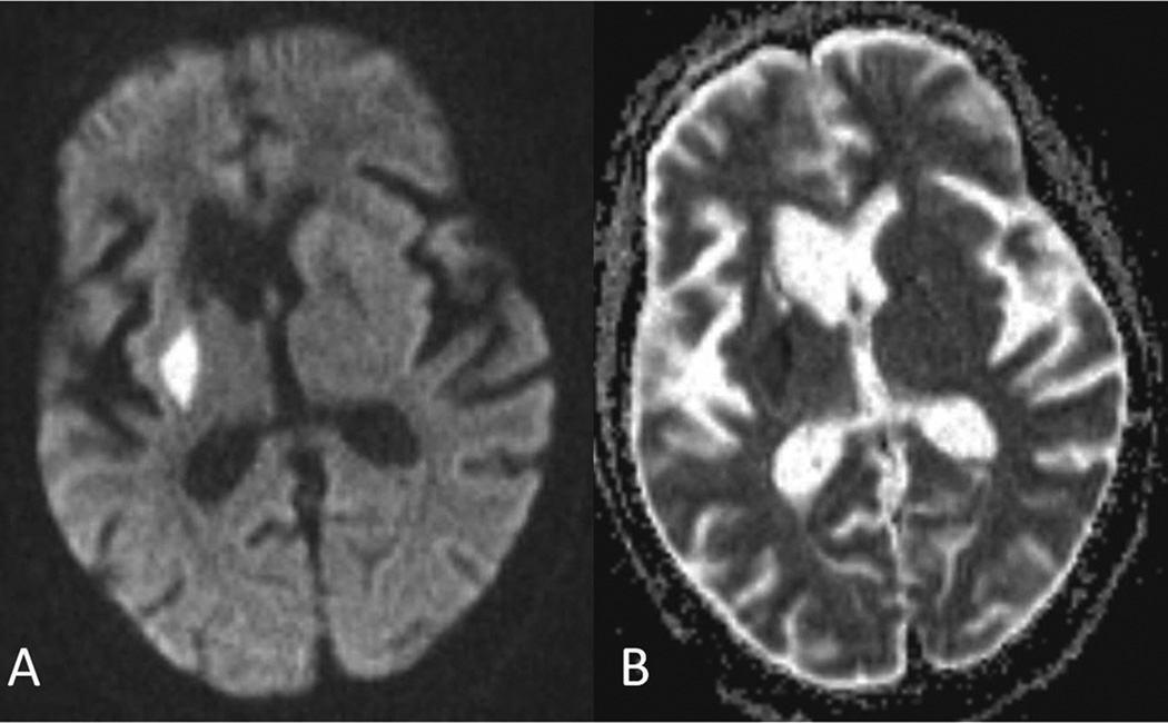
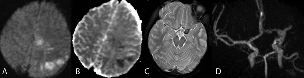
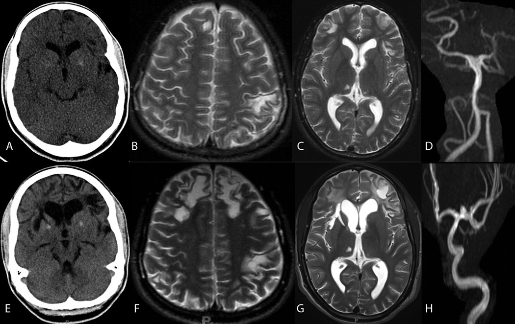
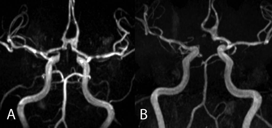
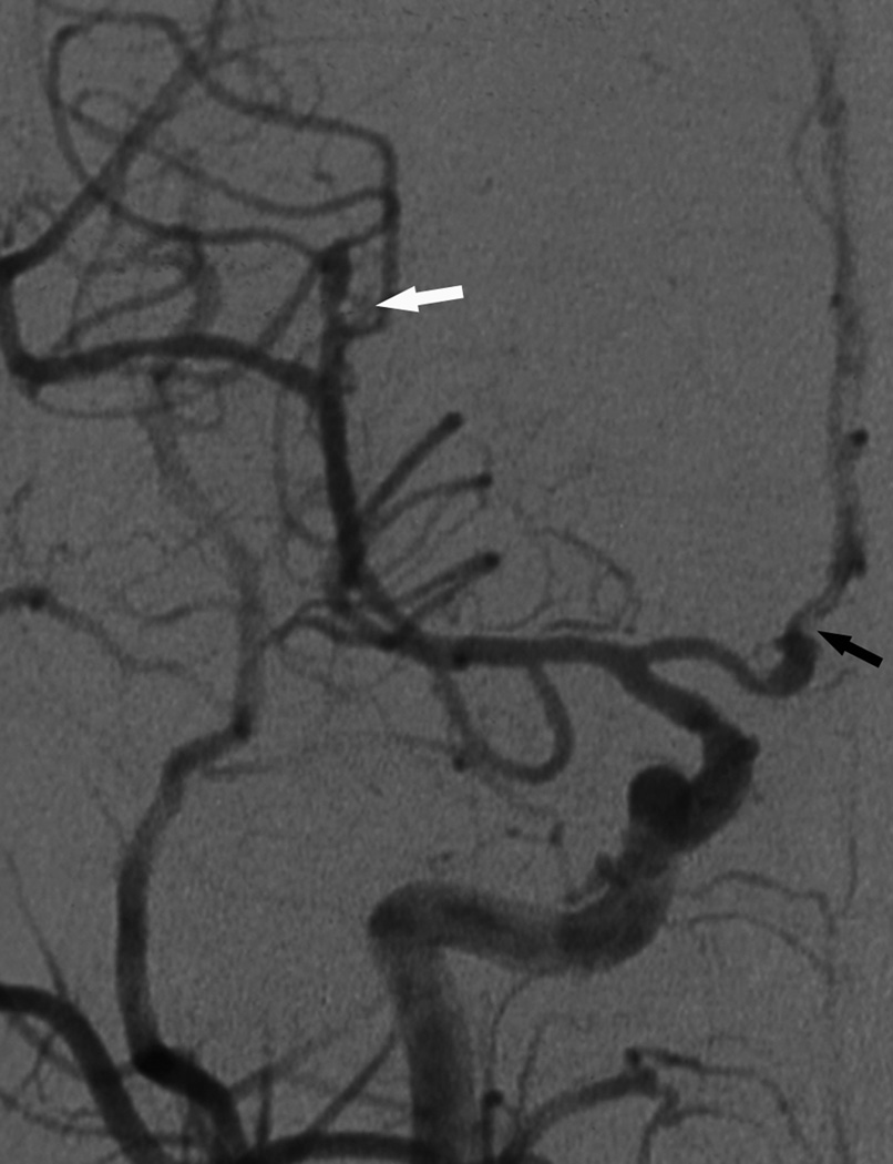
Results: Eight PHIV patients with clinical findings of acute stroke referred to the neuroimaging were identified. CT and MRI findings of infarction were found in all (8/8) patients in their first and/or last neuroimaging study; including basal ganglia-thalami (BGT) infarction (7/8), focal cortical infarction (4/8), and internal capsule infarction (4/8). Imaging depicted cortical atrophy (5/8), BGT calcification (3/8), and posterior reversible encephalopathy syndrome, wallerian degeneration, and periventricular white matter hyperintense T2 signal each in one patient. No tumors or infectious masses, cysts or abscesses were identified. Subsequent available neuroimaging revealed progression of the cerebrovascular disease in seven patients, 5/7 in the absence of new clinical signs or symptoms. Segmental occlusion, narrowing or narrowing/dilatation in the circle of Willis was found in 6/6 patients who underwent MR angiography and fusiform aneurysms were detected in three of them, a saccular aneurysm in one patient.
Conclusion: Asymptomatic progression of cerebrovascular disease was found in PHIV adolescents with prior stroke. These findings may have implications for long-term risk and outcomes for this patient population. There should be a low threshold to evaluate for CNS pathology even with minor symptoms in this population. More studies are necessary to determine if there is a benefit from screening of asymptomatic patients.
Keywords: AIDS; Cerebrovascular accident; HIV; Imaging; Perinatal; Stroke.
Copyright © 2013. Published by Elsevier Masson SAS.
Figures

References
-
- Townsend CL, Willey BA, Cortina-Borja M, et al. Antiretroviral therapy and congenital abnormalities in infants born to HIV-infected women in the UK and Ireland, 1990–2007. AIDS. 2009;23:519–524. - PubMed
-
- Hazra R, Siberry GK, Mofenson LM. Growing up with HIV: children, adolescents, and young adults with perinatally acquired HIV infection. Annu Rev Med. 2010;61:169–185. - PubMed
-
- Gutierrez J, Ortiz G. HIV/AIDS patients with HIV vasculopathy and VZV vasculitis: a case series. Clin Neuroradiol. 2011 Sep;21(3):145–151. - PubMed
MeSH terms
Grants and funding
LinkOut - more resources
Full Text Sources
Other Literature Sources
Medical
