. 2013 Sep;23(9):1075-83.
doi: 10.1093/glycob/cwt044. Epub 2013 Jun 4.

The structure of the Mycobacterium smegmatis trehalose synthase reveals an unusual active site configuration and acarbose-binding mode

Affiliations

The structure of the Mycobacterium smegmatis trehalose synthase reveals an unusual active site configuration and acarbose-binding mode

Sami Caner et al. Glycobiology. 2013 Sep.

Abstract

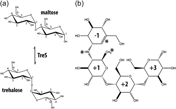
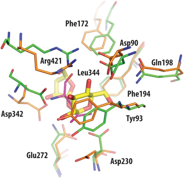
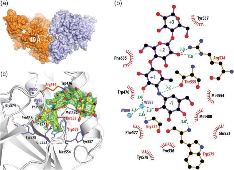
Trehalose synthase (TreS) catalyzes the reversible conversion of maltose into trehalose in mycobacteria as one of three biosynthetic pathways to this nonreducing disaccharide. Given the importance of trehalose to survival of mycobacteria, there has been considerable interest in understanding the enzymes involved in its production; indeed the structures of the key enzymes in the other two pathways have already been determined. Herein, we present the first structure of TreS from Mycobacterium smegmatis, thereby providing insights into the catalytic machinery involved in this intriguing intramolecular reaction. This structure, which is of interest both mechanistically and as a potential pharmaceutical target, reveals a narrow and enclosed active site pocket within which intramolecular substrate rearrangements can occur. We also present the structure of a complex of TreS with acarbose, revealing a hitherto unsuspected oligosaccharide-binding site within the C-terminal domain. This may well provide an anchor point for the association of TreS with glycogen, thereby enhancing its role in glycogen biosynthesis and degradation.

Keywords: GH13 glycoside hydrolase; drug design; enzyme inhibition; trehalose synthase; tuberculosis.


Figures

Fig. 1.
Fig. 1.
(a) Interconversion of α-1-4 linked maltose and α-1-1 linked trehalose catalyzed by TreS. Numbers indicate carbon atom positions in the sugar ring. (b) Chemical structure of the inhibitor acarbose, a pseudo-tetrasaccharide. Asterisks indicate features that distinguish acarbose from the related substrate maltotetraose. These features include an unsaturated ring, a methyl group in place of a hydroxymethyl group and an N-linked “glycosidic” bond. Ring components have been numbered with respect to the N-linkage of this inhibitor.
Fig. 2.
Fig. 2.
Sequence alignment between TreS of M. smegmatis (GenBank accession ID: YP_006571064) and M. tuberculosis (GenBank accession ID: EFI32604). Colored boxes highlight the domain organization found in M. smegmatis (Figure 3). Green indicates domain A, yellow indicates domain B, red indicates domain C and blue indicates the extended active site loop found in domain A. Catalytic residues are shown with red letters in black boxes. Leu344 is highlighted in green and is boxed as well.
Fig. 3.
Fig. 3.
Illustration of the domain architecture of our TreS structure and representative GH13 family members. Protein surfaces of domains A, B and C are shown in green, yellow and red, respectively. Additional domains are shown in blue. A white star indicates the position of the catalytic center. (a) human pancreatic α-amylase (PDB ID: 1hny); (b) malto-oligosyltrehalose synthase TreY (PDB ID: 1iv8); (c) trehalulose synthase MutB (PDB ID: 2pwe); (d) the asymmetric unit dimer of TreS from our studies of the M. smegmatis enzyme (PDB ID: 3zo9). A schematic of the expected tetrameric assembly by two TreS homodimers is shown in the lower right of frame (D).
Fig. 4.
Fig. 4.
Ion binding in the structure of TreS. Domains are indicated by large capital letters and follow the same coloring scheme as Figure 2. For clarity, loops have been smoothed and only one enzyme molecule of the asymmetric unit is shown. Ion positions are indicated by colored spheres (red = Cl, green = Ca2+, magenta = Mg2+). Chloride (i) is located in a homologous position compared with the allosterically activating chloride ion of the human α-amylases (Brayer et al. 1995; Maurus et al. 2005). Chloride (ii) and (iii) are located in loops L7 (residues 338–384) and L1 (residues 43–77), respectively, which surround the active site. Chloride (iv) binding would appear to be due to crystal packing interactions and is not present in the second molecule of this enzyme in the asymmetric unit. The sidechains of the catalytic triad composed of Asp230 (nucleophile), Glu272 (acid/base catalyst), and Asp 342 (substrate binding), along with the conserved active site residue Arg228, are shown using orange stick representations.
Fig. 5.
Fig. 5.
(a) Superposition of active site residues and loop L7 of TreS (orange), human pancreatic α-amylase (blue), trehalulose synthase MutB (green) and malto-oligosyltrehalose synthase TreY (cyan), with residue numbering according to the TreS structure. With the exception of the nucleophilic Asp230 in TreS, the catalytic residues for the other α-glucosidase enzymes are in comparable positions within the superposed structures (arrow indicates conformational change in TreS). The active site conformation of the bound inhibitor acarbose (gray sticks) in the active site of human α-amylase structure (blue) is shown (Maurus et al. 2005) in an overlay to evaluate the ligand-binding pocket in comparison with that of TreS. Importantly, the active site loop L7 of TreS (orange) protrudes into the catalytic center, suggesting that the structure we have determined is that of an inactive form of the enzyme. In addition, the side chain of Leu344 at the extremity of this loop projects into the active site to a position that would be <2 Å from the nucleophilic aspartate in the amylase structure. This feature presumably accounts for the displacement of the Asp230 catalytic nucleophile of TreS. (b) A cross sectional surface representation of TreS in the vicinity of the active site pocket. The narrow entrance to the active site from the surface of TreS is indicated by a dashed arrow and is delimited in this structure by the conformation of loop L7 and the positioning of the side chain of Leu344 in particular. A further consequence of the placement of Leu344 is a steric conflict with the side chain of the catalytic nucleophile Asp230, which as a consequence adopts a buried conformation, therefore becoming solvent and substrate inaccessible.
Fig. 6.
Fig. 6.
Superposition of active site residues of TreS (orange) and trehalulose synthase MutB (green), with residue numbering according to the TreS structure. With the exception of the side chain of the nucleophilic Asp230 in TreS, other catalytic residues are in comparable positions between the superimposed structures. Also drawn in yellow is the disaccharide substrate sucrose of MutB, as found bound in the active site of this enzyme. Leu344 of TreS (shown in magenta) overlaps with the sucrose molecule of MutB, hence prohibiting ligand access to the catalytic Asp230.
Fig. 7.
Fig. 7.
(a) Structure of the TreS/acarbose complex. The acarbose-binding site (within the white circle) is found in a crevice at the interface between two monomers of TreS (only a dimer is shown) and is located ∼40 Å from the active site of this enzyme (white asterisk). (b) The detailed structure of the observed acarbose-binding site and the overall fit of this inhibitor to a FobsFobs electron density map (green mesh) contoured at the 3σ level. (c) LIGPLOT+ (Laskowski and Swindells 2011) representation of the interactions formed to acarbose in its binding site. Green dashed lines represent hydrogen bonds with residues (red letters) and water molecules (blue spheres). Residues involved in hydrophobic interactions are shown as red semicircles.

References

    1. Adams PD, Afonine PV, Bunkoczi G, Chen VB, Davis IW, Echols N, Headd JJ, Hung LW, Kapral GJ, Grosse-Kunstleve RW, et al. PHENIX: A comprehensive Python-based system for macromolecular structure solution. Acta Crystallogr D Biol Crystallogr. 2010;66:213–221. doi:10.1107/S0907444909052925. - DOI - PMC - PubMed
    1. Banner DW, Bloomer AC, Petsko GA, Phillips DC, Pogson CI, Wilson IA, Corran PH, Furth AJ, Milman JD, Offord RE, et al. Structure of chicken muscle triose phosphate isomerase determined crystallographically at 2.5 angstrom resolution using amino acid sequence data. Nature. 1975;255:609–614. doi:10.1038/255609a0. - DOI - PubMed
    1. Barry CE, 3rd, Lee RE, Mdluli K, Sampson AE, Schroeder BG, Slayden RA, Yuan Y. Mycolic acids: Structure, biosynthesis and physiological functions. Prog Lipid Res. 1998;37:143–179. doi:10.1016/S0163-7827(98)00008-3. - DOI - PubMed
    1. Barry CE, III, Mdluli K. Drug sensitivity and environmental adaptation of mycobacterial cell wall components. Trends Microbiol. 1996;4:275–281. doi:10.1016/0966-842X(96)10031-7. - DOI - PubMed
    1. Boraston AB, Bolam DN, Gilbert HJ, Davies GJ. Carbohydrate-binding modules: Fine-tuning polysaccharide recognition. Biochem J. 2004;382:769–781. doi:10.1042/BJ20040892. - DOI - PMC - PubMed

Publication types

MeSH terms

LinkOut - more resources