. 2013 Jun;19(6):916-24.
doi: 10.3201/eid1906.121641.

Transmission potential of Rift Valley fever virus over the course of the 2010 epidemic in South Africa

Affiliations

Transmission potential of Rift Valley fever virus over the course of the 2010 epidemic in South Africa

Raphaëlle Métras et al. Emerg Infect Dis. 2013 Jun.

Abstract

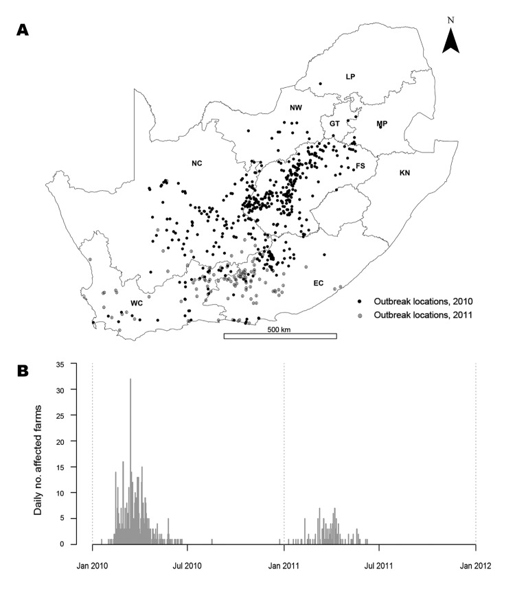
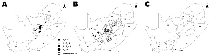
A Rift Valley fever (RVF) epidemic affecting animals on domestic livestock farms was reported in South Africa during January-August 2010. The first cases occurred after heavy rainfall, and the virus subsequently spread countrywide. To determine the possible effect of environmental conditions and vaccination on RVF virus transmissibility, we estimated the effective reproduction number (Re) for the virus over the course of the epidemic by extending the Wallinga and Teunis algorithm with spatial information. Re reached its highest value in mid-February and fell below unity around mid-March, when vaccination coverage was 7.5%-45.7% and vector-suitable environmental conditions were maintained. The epidemic fade-out likely resulted first from the immunization of animals following natural infection or vaccination. The decline in vector-suitable environmental conditions from April onwards and further vaccination helped maintain Re below unity. Increased availability of vaccine use data would enable evaluation of the effect of RVF vaccination campaigns.

Keywords: Rift Valley fever; Rift Valley fever virus; South Africa; epidemic; likelihood functions; transmission; viruses; zoonoses.


Figures

Figure 1
Figure 1
Rift Valley fever epidemic, South Africa, 2010–2011. A) Location of cases. Unmarked area in center right is Lesotho (no data). B) Epidemic curve for the 2 years. NC, Northern Cape; NW, North West; LP, Limpopo; GT, Gauteng; MP, Mpumalanga; FS, Free State; KN, KwaZulu-Natal; EC, Eastern Cape; WC, Western Cape.
Figure 2
Figure 2
Schematic representation of the Wallinga-Teunis algorithm extended with spatial information. Farm i could get infection from Farm j, but it also could get infection from Farms k1, k2, and k3. In this example, the most likely time difference between onset of symptoms is 4 days (based on the serial interval distribution, given below the x-axis), and the most likely distance between farms is short (<1 km). Therefore, Farm j is the most likely farm to have infected Farm i (this scenario maximizes the probability in both dimensions). See the online Technical Appendix (wwwnc.cdc.gov/EID/article/19/6/12-1641-Techapp1.pdf) for details.
Figure 3
Figure 3
Distribution of D0 by time and distance [D0(s,t)]. D0(s,t) is a measure of spatiotemporal interaction between cases that was estimated by using the space–time K-function (19,20); the distribution is indicated by the pink dashed line. The green, yellow, and blue lines are the smoothed distributions, which were obtained with bandwidth values of 1, 3, and 5, respectively. A) Plot of D0(s,t) values by distance on day 1. B) D0(s,t) values by time at distance of 5 km. C) Plot of D0(s,t) values by distance on day 5. D) D0(s,t) values by time at distance of 15 km.
Figure 4
Figure 4
Rift Valley fever incidence (bars), daily effective reproduction number (Re; red dashed line), and smoothed mean of Re (solid red line) over the course of 2010 epidemic in Free State Province, South Africa. Blue dots, estimates of concurrent total monthly rainfall; dashed green line, average daily temperature. Vaccination coverage (VC) by March 31, 2010, and May 31, 2010, for Scenarios A–C (descriptions follow) are indicated at the top of the graph. Scenarios: Scenario A assumed that vaccination coverage was applied throughout South Africa in proportion to the livestock population; Scenario B assumed that the number of vaccines used in a province over a specific period was proportional to the number of cases reported in that province over that same period; Scenario C assumed that all vaccines were used in Free State Province during Period 2 (January 19–March 31, 2010) and Period 3 (April 1–May 31, 2010) and that no vaccine had been used before the epidemic (Period 1, April 1, 2009–January 18, 2010). The horizontal dashed line represents the threshold value Re = 1.
Figure 5
Figure 5
Effective reproduction number (Re) per affected farm, by province, over the 2010 Rift Valley fever epidemic, South Africa. A) January and February. B) March and April. C) May and June. July and August are not displayed because no cases were reported in July, and Re was 0 for the only farm reported in August. NC, Northern Cape; NW, North West; LP, Limpopo; GT, Gauteng; MP, Mpumalanga; FS, Free State; KN, KwaZulu-Natal; EC, Eastern Cape; WC, Western Cape. The unmarked area to the right of center is Lesotho (no data).
Figure 6
Figure 6
Mean effective daily reproduction number (Re) during Rift Valley fever epidemic, South Africa, 2010. Re was estimated by using D0(s,t) values (dashed black line) and D0(s,t) smoothed surfaces obtained with bandwidth values of 1 (dark gray), 3 (medium gray), and 5 (light gray). D0(s,t) values were estimated by using the space–time K-function (19,20) and are a measure of the spatiotemporal proximity between cases. The horizontal dashed line represents the threshold value Re = 1.
Figure 7
Figure 7
Daily temperature and total monthly rainfall during January–August 2010, Free State and Northern Cape Provinces, South Africa.

References

    1. Pepin M, Bouloy M, Bird BH, Kemp A, Paweska J. Rift Valley fever virus (Bunyaviridae: Phlebovirus): an update on pathogenesis, molecular epidemiology, vectors, diagnostics and prevention. Vet Res. 2010;41:61 and. 10.1051/vetres/2010033 - DOI - PMC - PubMed
    1. Bird BH, Ksiazek TG, Nichol ST, Maclachlan NJ. Rift Valley fever virus. J Am Vet Med Assoc. 2009;234:883–93 and. 10.2460/javma.234.7.883 - DOI - PubMed
    1. Linthicum KJ, Anyamba A, Tucker CJ, Kelley PW, Myers MF, Peters CJ. Climate and satellite indicators to forecast Rift Valley fever epidemics in Kenya. Science. 1999;285:397–400 and. 10.1126/science.285.5426.397 - DOI - PubMed
    1. Anyamba A, Chretien JP, Small J, Tucker CJ, Formenty PB, Richardson JH, et al. Prediction of a Rift Valley fever outbreak. Proc Natl Acad Sci U S A. 2009;106:955–9 and. 10.1073/pnas.0806490106 - DOI - PMC - PubMed
    1. Logan TM, Linthicum KJ, Thande PC, Wagateh JN, Nelson GO, Roberts CR. Egg hatching of Aedes mosquitoes during successive floodings in a Rift Valley fever endemic area in Kenya. J Am Mosq Control Assoc. 1991;7:109–12 . - PubMed

Publication types

LinkOut - more resources