. 2013 Jul 17;32(14):2056-72.
doi: 10.1038/emboj.2013.131. Epub 2013 Jun 4.

The ALS8 protein VAPB interacts with the ER-Golgi recycling protein YIF1A and regulates membrane delivery into dendrites

Affiliations

The ALS8 protein VAPB interacts with the ER-Golgi recycling protein YIF1A and regulates membrane delivery into dendrites

Marijn Kuijpers et al. EMBO J. .

Abstract

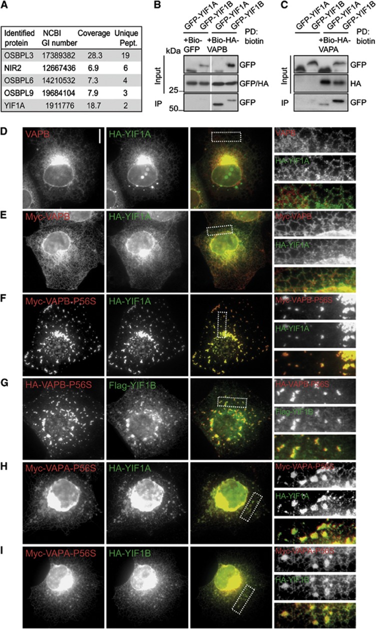
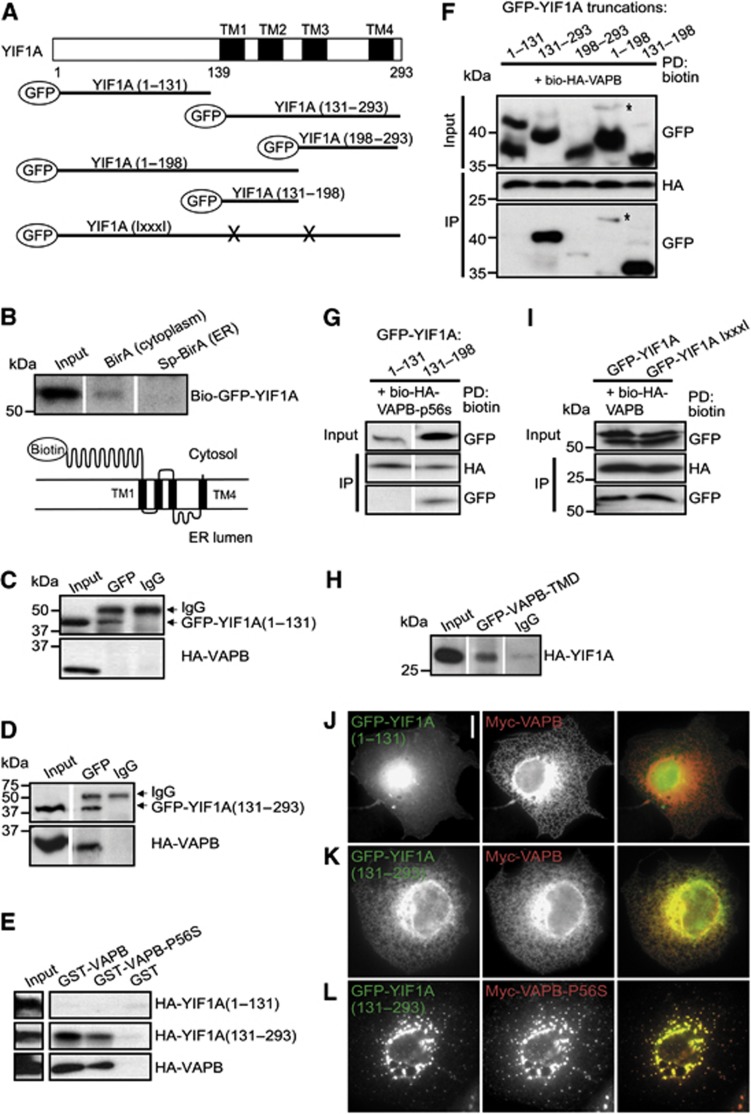
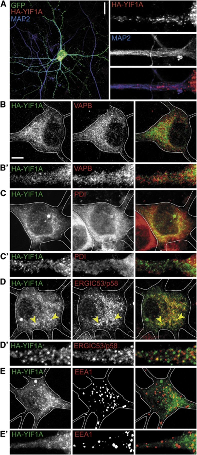
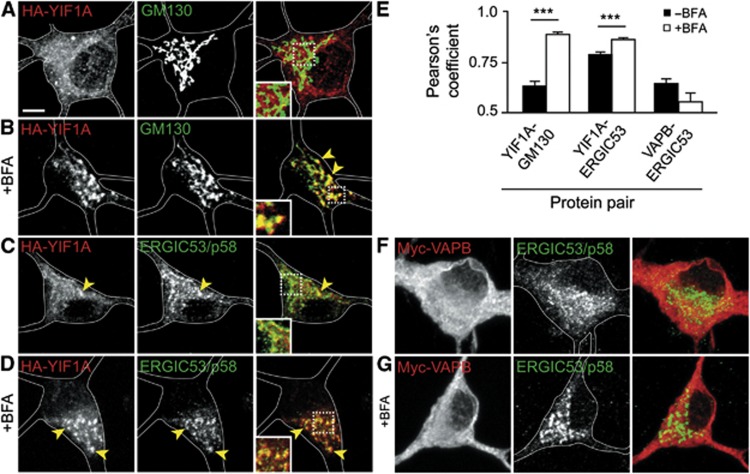
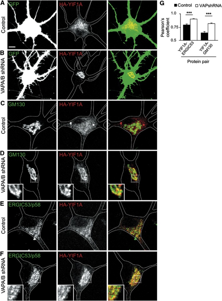
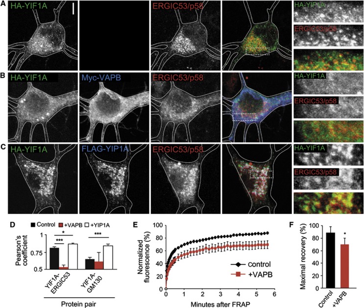
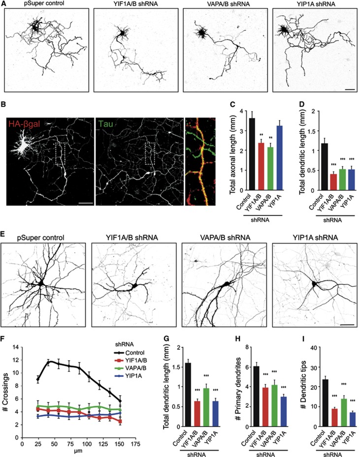
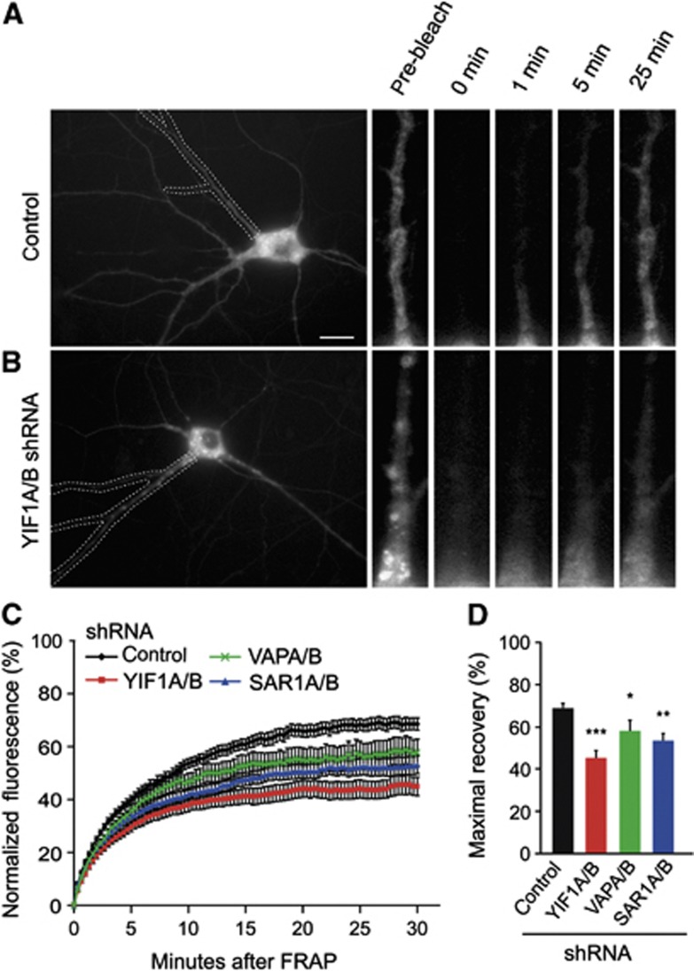
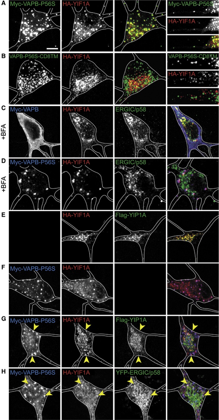
The vesicle-associated membrane protein (VAMP) associated protein B (VAPB) is an integral membrane protein localized to the endoplasmic reticulum (ER). The P56S mutation in VAPB has been linked to motor neuron degeneration in amyotrophic lateral sclerosis type 8 (ALS8) and forms ER-like inclusions in various model systems. However, the role of wild-type and mutant VAPB in neurons is poorly understood. Here, we identified Yip1-interacting factor homologue A (YIF1A) as a new VAPB binding partner and important component in the early secretory pathway. YIF1A interacts with VAPB via its transmembrane regions, recycles between the ER and Golgi and is mainly localized to the ER-Golgi intermediate compartments (ERGICs) in rat hippocampal neurons. VAPB strongly affects the distribution of YIF1A and is required for intracellular membrane trafficking into dendrites and normal dendritic morphology. When VAPB-P56S is present, YIF1A is recruited to the VAPB-P56S clusters and loses its ERGIC localization. These data suggest that both VAPB and YIF1A are important for ER-to-Golgi transport and that missorting of YIF1A may contribute to VAPB-associated motor neuron disease.


Conflict of interest statement

The authors declare that they have no conflict of interest.

Figures

Figure 1
Figure 1
Interaction of YIF1A with wild-type and mutant VAPB. (A) Identification of wild-type VAPB binding partners by mass spectrometry in HeLa cell extract. The table shows proteins identified with a significant Mascot score in the pull-down with streptavidin beads from an extract of HeLa cells co-expressing Bio-GFP-VAPB and biotin ligase BirA. The list is corrected for background proteins, which were identified in a control pull-down from HeLa cells expressing bio-GFP. Abbreviations used in the table to indicate the identified proteins: OSBPL, oxysterol binding protein-like; NIR, N-terminal domain-interacting receptor. (B) Biotin pull-downs (PD) from HEK293T extract transfected with Bio-HA-VAPB and GFP-YIF1A, GFP-YIF1B or control bio-GFP and probed for GFP and HA. (C) Biotin pull-downs from HEK293T extract transfected with Bio-HA-VAPA and GFP-YIF1A or GFP-YIF1B and probed for GFP and HA. The ratio input/pellet is 2–5% for all pull-down and immunoprecipitation experiments. (D) COS-7 cells transfected with HA-YIF1A and stained with anti-HA (green) and anti-VAPB (red) antibodies. (E, F) COS-7 cells double transfected with HA-YIF1A and myc-VAPB (D) or myc-VAPB-P56S (E) stained with anti-HA (green) and anti-myc (red) antibodies. (G) COS-7 cells double transfected with HA-VAPB-P56S and Flag-YIF1B, fixed and stained with anti-HA (green) and anti-Flag (red) antibodies. (H, I) COS-7 cells double transfected with myc-VAPA-P56S and HA-YIF1A (H) or HA-YIF1B (I) stained with anti-HA (green) and anti-myc (red) antibodies. Panels on the right side show enlargements of the boxed regions. Scale bar, 10 μm.
Figure 2
Figure 2
The transmembrane domain of YIF1A interacts with VAPB. (A) YIF1A deletion constructs were made containing amino acids 1–131 of YIF1A, amino acids 131–293, 198–293, 1–198 and amino acids 131–198. GxxxG motifs in transmembrane domain one and three were mutated by replacing the glycine residues with isoleucine. The predicted transmembrane domains are labelled with TM. (B) Biotin pull-down to determine the topology of YIF1A using HEK293T extracts transfected with bio-GFP-YIF1A and BirA (cytoplasm) or SP-BirA (ER lumen). Bio-GFP-YIF1A binds to streptavidin beads in the presence of cytoplasmic BirA but not in the presence of luminal BirA. Samples were immunoblotted using anti-GFP antibodies. (C, D) Analysis of YIF1A binding domain by co-immunoprecipitation. HEK293T cells co-transfected with (C) GFP-YIF1A(1–131) or (D) GFP-YIF1A(131-293) and HA-VAPB were immunoprecipitated with anti-GFP or IgG (control) antibodies. (E) Binding domain analysis by GST pull-down assay using lysates of HEK293T cells expressing HA-YIF1A truncated constructs and GST-VAPB or GST-VAPB-P56S. Samples were immunoblotted using anti-HA antibodies. (F) Biotin pull-downs (PD) from HEK293T extracts transfected with GFP-YIF1A truncated constructs and bio-HA-VAPB. Probed for GFP and HA. The asterisk denotes a band corresponding to the YIF1A(1–198) protein. (G) Biotin pull-down (PD) from HEK293T extracts transfected with GFP-YIF1A truncated constructs and bio-HA-VAPB-P56S and probed for GFP and HA. (H) Immunoprecipitation from extract of HEK293T cells co-expressing GFP-VAPB-TMD and HA-YIF1A. Immunoblot is probed for HA. (I) Biotin pull-down from HEK293T extracts transfected with GFP-YIF1A IxxxI and bio-HA-VAPB and probed for GFP and HA. The ratio input/pellet is 2–5% for all pull-down and immunoprecipitation experiments. (JL) COS-7 cells double transfected with myc-VAPB (J, K) or myc-VAPB-P56S (L) and GFP-YIF1A truncation constructs, fixed and stained with anti-myc (red) antibodies. Scale bar, 10 μm.
Figure 3
Figure 3
YIF1A localization in cultured hippocampal neurons.(A) Representative images of rat hippocampal neurons (DIV16) co-transfected with HA-YIF1A and GFP to visualize morphology and labelled with anti-HA (red) and anti-MAP2 (blue) antibodies. Scale bar, 20 μm. In the right panel, a dendritic segment is enlarged to show the presence of HA-YIF1A in proximal dendrites. (BE) Representative images of rat hippocampal neurons transfected with HA-YIF1A and labelled with anti-HA (green) and anti-VAPB (red in B), anti-PDI (red in C), anti-ERGIC53/p58 (red in D) or anti-EEA1 (red in E). Solid lines indicate the cell edge and arrows showco-localization. Scale bars represent 20 μm in (A) and 5 μm in (B). (B’–E’) Enlargement of dendritic segments to show localization of endogenous proteins and overexpressed YIF1A.
Figure 4
Figure 4
YIF1A localizes to the ER–Golgi intermediate compartment (ERGIC). (A) Image of the cell body of a hippocampal neuron transfected with HA-YIF1A and stained with anti-HA (red) and anti-GM130 (green) antibodies. (B) Redistribution of HA-YIF1A by BFA treatment. Neurons were transfected with HA-YIF1A, treated with BFA (5 μg/ml) for 15 min fixed and labelled with anti-HA (red) and anti-GM130 (green).(C) Representative image of the cell body of a hippocampal neuron transfected with HA-YIF1A and labelled with anti-HA (red) and anti-ERGIC (green). Redistribution of HA-YIF1A after BFA treatment is shown in (D). (E) Summary of co-localization experiments. Pearson’s coefficient (rp) for YIF1A versus ERGIC53/p58, YIF1A versus GM130 and VAPB versus ERGIC53/p58 in control (black bars) and BFA-treated cells (white bars). Twenty-three to twenty-five ROIs were analysed for each condition. Error bars indicate s.e.m., ***P<0.001. (F, G) Images of cell bodies of hippocampal neurons transfected with myc-VAPB and labelled with anti-myc (red) and anti-ERGIC53/p58 (green). BFA treatment has no effect on myc-VAPB distribution (G). Solid lines indicate the cell edges; the insets show magnifications of boxed areas and arrows indicateco-localization. Scale bar, 5 μm.
Figure 5
Figure 5
Effect of VAP knockdown on YIF1A localization in cultured hippocampal neurons. (A, B) Representative images of cell bodies of hippocampal neurons co-transfected at DIV16 for 4 days with HA-YIF1A (red), GFP and pSuper control vector (A) or pSuper-VAPA and pSuper-VAPB shRNAs (B). VAP knockdown results in relocalization of HA-YIF1A. (CF) Cell bodies of neurons co-transfected with HA-YIF1A (red) and pSuper control vector (C, E) or pSuper-VAPA and pSuper-VAPB shRNAs (D, F). Neurons were stained for either GM130 (C, D) or ERGIC53/p58 (E, F). Solid lines indicate the cell edges. Scale bar, 5 μm. (G) Summary of co-localization experiments. Pearson’s coefficient (rp) for YIF1A versus ERGIC53/p58 and GM130 in control (black bars) and VAP knockdown neurons (white bars). Seventeen to twenty-three ROIs were analysed for each condition. Error bars indicate s.e.m., ***P<0.001.
Figure 6
Figure 6
VAPB overexpression relocalizes YIF1A. (AC) Hippocampal neurons transfected with HA-YIF1A and stained with anti-HA (green) and anti-ERGIC53/p58 (blue). Co-transfection with either myc-VAPB (B) or FLAG-YIP1A (C) results in relocalization of HA-YIF1A. Solid lines indicate the cell edges and panels on the right side show enlargements of the boxed regions. Scale bar, 5 μm. (D) Summary of co-localization experiments. Pearson’s coefficient (rp) for YIF1A versus ERGIC53/p58 and GM130 in control (black bars), VAPB overexpressing (red bars) and YIP1A overexpressing neurons (white bars). Thirteen to twenty-three ROIs were analysed for each condition. (E) Fluorescent recovery plots showing the rates of GFP-YIF1A recovery in cell bodies of control neurons and neurons overexpressing VAPB. Fluorescent intensity was normalized to intensity before bleaching. P=0.008; repeated measures ANOVA. (F) Histogram representing the maximal recovery of fluorescence (estimated mobile fraction) in hippocampal neurons expressing GFP-YIF1A with (n=11) or without VAPB overexpression (n=10). Data are presented as means±s.e.m., *P<0.05, ***P<0.001.
Figure 7
Figure 7
YIF1 and VAP are required for normal dendrite morphology. (A) Hippocampal neurons co-transfected at DIV1 with indicated constructs and β-galactosidase to visualize morphology. (B) Representative image of a hippocampal neurons (DIV5) co-transfected with empty pSuper and β-galactosidase and co-stained with Tau (green) to highlight the axon. (C, D) Quantification of total axonal length and dendritic length after 4 days overexpression of knockdown constructs or empty pSuper as control (14–16 cells were analysed for each condition). (E) Hippocampal neurons co-transfected at DIV15 with indicated constructs and β-galactosidase to visualize morphology. (F) Sholl analysis and quantification of the total dendritic length (G), number of primary dendrites (H) and dendritic tips (I) after 4 days overexpression of knockdown constructs or empty pSuper as control (15–17 cells were analysed for each condition). Error bars indicate s.e.m., **P<0.01, ***P<0.001. Scale bars represent 50 μm.
Figure 8
Figure 8
YIF1 and VAP play a role in membrane trafficking in primary hippocampal neurons. (A, B) Representative images of membrane-bound GFP (CD8-GFP) moving from soma into dendrites in hippocampal neurons (DIV16–19) co-expressing CD8-GFP with either empty pSuper control vector (A) or YIF1A and YIF1B shRNAs (B). Boxed area indicates photobleached dendritic region. In the right panel, proximal part of photobleached dendrite is shown. Scale bar, 20 μm. (C) Fluorescent recovery plots showing the rates of CD8-GFP recovery in photobleached dendrites of control and knockdown neurons. Fluorescent intensity was normalized to intensity before bleaching. P<0.001 for YIF1A/B versus control, P=0.033 for VAPA/B versus control and P=0.008 for SAR1A/B versus control (repeated measures ANOVA followed by Tukey’s post hoc test). (D) Maximal fluorescence recovery. Nine to fifteen cells were analysed for each condition. Data are presented as means±s.e.m., *P<0.05, **P<0.01, ***P<0.001; one-way ANOVA, Tukey’s post hoc test.
Figure 9
Figure 9
Mutant VAPB expression results in recruitment of YIF1A to clusters. (A) Co-transfection of hippocampal neurons with HA-YIF1A (red) and myc-VAPB-P56S (green) shows that YIF1A is recruited to the VAPB-P56S clusters. The far right panel shows enlargement of a dendrite. (B) Hippocampal neuron co-transfected with VAPB-P56S-CD8TM (green) and HA-YIF1A (red). (C, D) Images show that in the presence of overexpressed wild-type VAPB, HA-YIF1A localizes to ERGIC after BFA treatment. However, when VAPBP-P56S is expressed YIF1A loses its localization to the ERGIC. (EG) Neurons co-transfected with HA-YIF1A (green) and Flag-YIP1A (D), myc-VAPB-P56S (E) or both (F). (H) Hippocampal neuron co-transfected with myc-VAPB-P56S (blue), YFP-ERGIC (green) and HA-YIF1A (red). Scale bar, 5 μm.

References

    1. Al Awabdh S, Miserey-Lenkei S, Bouceba T, Masson J, Kano F, Marinach-Patrice C, Hamon M, Emerit MB, Darmon M (2012) A new vesicular scaffolding complex mediates the G-protein-coupled 5-HT1A receptor targeting to neuronal dendrites. J Neurosci 32: 14227–14241 - PMC - PubMed
    1. Altschul SF, Madden TL, Schaffer AA, Zhang J, Zhang Z, Miller W, Lipman DJ (1997) Gapped BLAST and PSI-BLAST: a new generation of protein database search programs. Nucleic Acids Res 25: 3389–3402 - PMC - PubMed
    1. Amarilio R, Ramachandran S, Sabanay H, Lev S (2005) Differential regulation of endoplasmic reticulum structure through VAP-Nir protein interaction. J Biol Chem 280: 5934–5944 - PubMed
    1. Anagnostou G, Akbar MT, Paul P, Angelinetta C, Steiner TJ, de Belleroche J (2010) Vesicle associated membrane protein B (VAPB) is decreased in ALS spinal cord. Neurobiol Aging 31: 969–985 - PubMed
    1. Appenzeller-Herzog C, Hauri HP (2006) The ER-Golgi intermediate compartment (ERGIC): in search of its identity and function. J Cell Sci 119: 2173–2183 - PubMed

Publication types

MeSH terms

Supplementary concepts