. 2013 Oct 15;22(20):4084-101.
doi: 10.1093/hmg/ddt258. Epub 2013 Jun 4.

The DcpS inhibitor RG3039 improves survival, function and motor unit pathologies in two SMA mouse models

Affiliations

The DcpS inhibitor RG3039 improves survival, function and motor unit pathologies in two SMA mouse models

Rocky G Gogliotti et al. Hum Mol Genet. .

Abstract

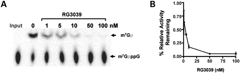
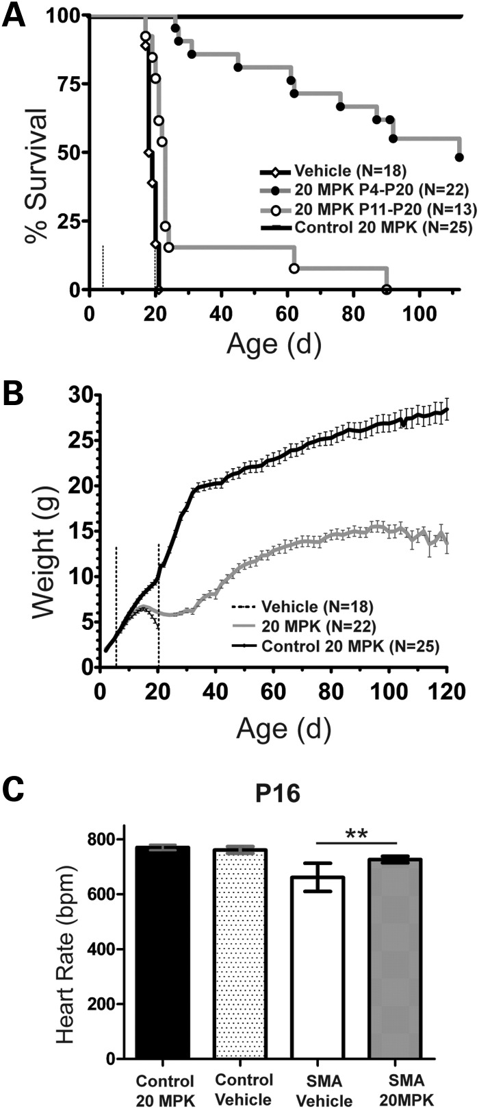
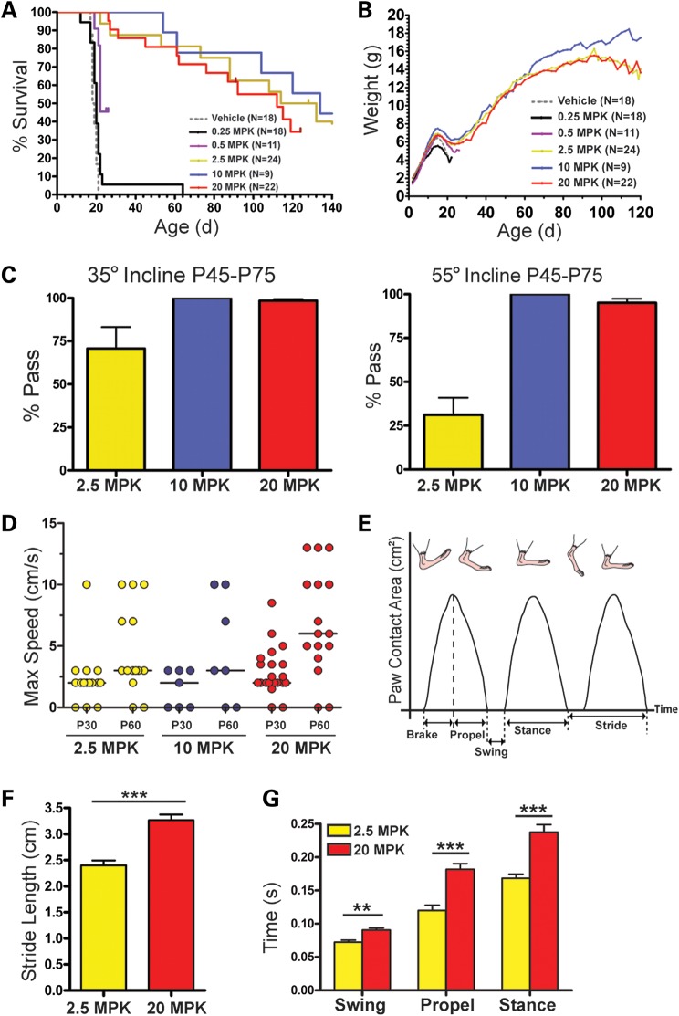
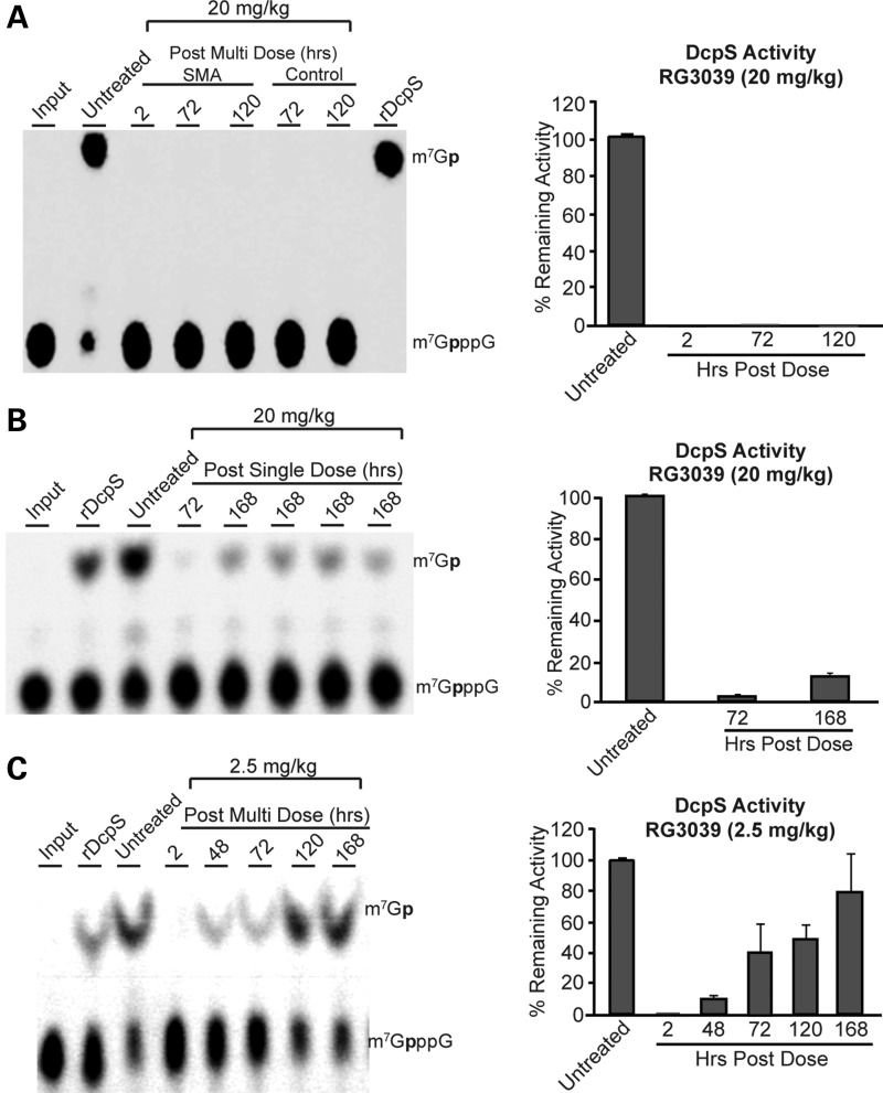
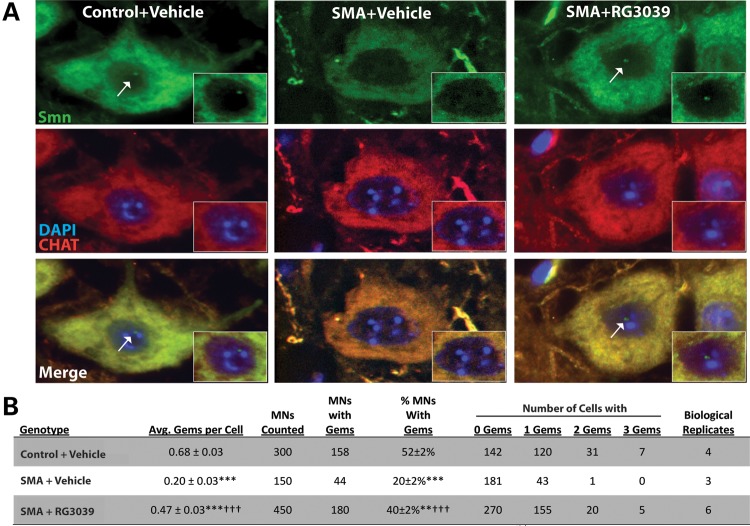
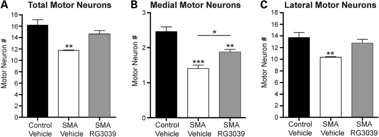
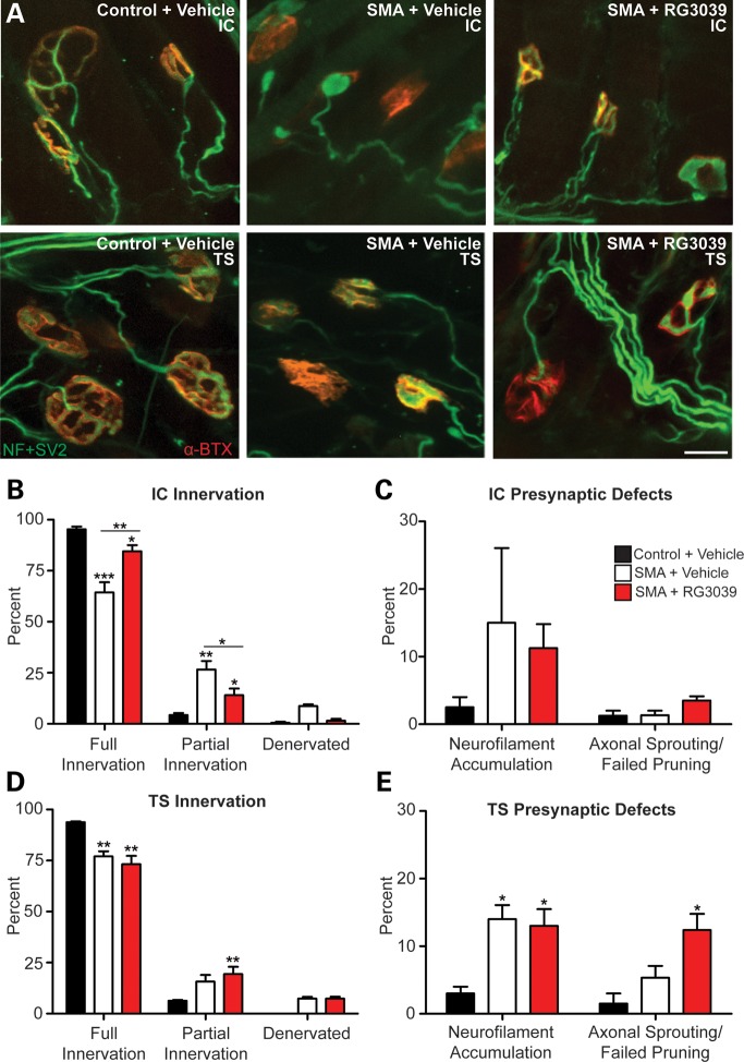
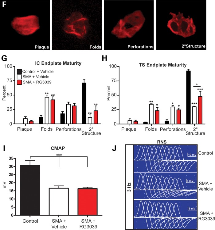
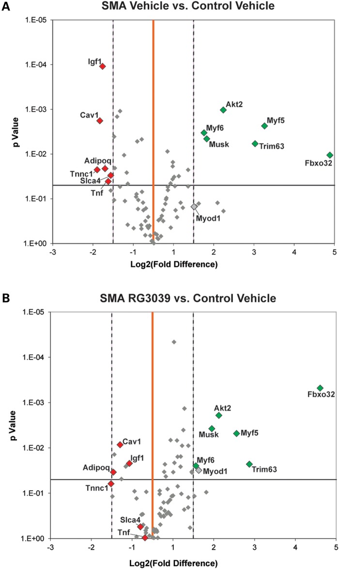
Spinal muscular atrophy (SMA) is caused by insufficient levels of the survival motor neuron (SMN) protein due to the functional loss of the SMN1 gene and the inability of its paralog, SMN2, to fully compensate due to reduced exon 7 splicing efficiency. Since SMA patients have at least one copy of SMN2, drug discovery campaigns have sought to identify SMN2 inducers. C5-substituted quinazolines increase SMN2 promoter activity in cell-based assays and a derivative, RG3039, has progressed to clinical testing. It is orally bioavailable, brain-penetrant and has been shown to be an inhibitor of the mRNA decapping enzyme, DcpS. Our pharmacological characterization of RG3039, reported here, demonstrates that RG3039 can extend survival and improve function in two SMA mouse models of varying disease severity (Taiwanese 5058 Hemi and 2B/- SMA mice), and positively impacts neuromuscular pathologies. In 2B/- SMA mice, RG3039 provided a >600% survival benefit (median 18 days to >112 days) when dosing began at P4, highlighting the importance of early intervention. We determined the minimum effective dose and the associated pharmacokinetic (PK) and exposure relationship of RG3039 and DcpS inhibition ex vivo. These data support the long PK half-life with extended pharmacodynamic outcome of RG3039 in 2B/- SMA mice. In motor neurons, RG3039 significantly increased both the average number of cells with gems and average number of gems per cell, which is used as an indirect measure of SMN levels. These studies contribute to dose selection and exposure estimates for the first studies with RG3039 in human subjects.


Figures

Figure 1.
Figure 1.
RG3039 inhibits the decapping enzyme DcpS. (A) RG3039 inhibits hDcpS enzyme activity in a dose-dependent manner in vitro (n = 3 experimental replicates) and is graphically shown in (B). The IC50 is 4.2 ± 0.13 nm and IC90 is 40 nm.
Figure 2.
Figure 2.
Oral delivery of RG3039 increases survival, weight and heart rate in 2B/− SMA mice. (A) The Kaplan–Meier survival curve comparing RG3039 control (2B/+, n = 25), vehicle 2B/− SMA (n = 18) and RG3039 2B/− SMA (n = 22) all dosed P4–P20; also included is P11–P20 RG3039 2B/− SMA (n = 13). RG3039 was dosed at 20 mg/kg (MPK). The median survival for each group, 20 MPK P4–P20 (112 days) and 20 MPK P11–P20 (23 days), was significant when compared with vehicle-treated 2B/− SMA mice (18.5 days), log-rank test, P < 0.0001. The median survival of pairwise comparison between P4–P20 and P11–P20 SMA RG3039-treated mice was significantly different, by log-rank test, P < 0.0001. Dashed lines in panels (A) and (B) represent the treatment window, P4–P20. (B) Weight curves from P2 to P120 of RG3039-treated control (black), P4–P20 2B/− SMA vehicle (dashed) and P4–P120 RG3039-treated 2B/− SMA mice (20 MPK) (gray). Note the max weight achieved in both SMA treatment groups is P15 with a subsequent decline. After P25, RG3039-treated 2B/− SMA mice parallel the weight gain seen in control mice until ∼P105. Weight is shown as the average ± SEM. (C) Heart rates from ECG recording at P16 were obtained from conscious mice and used to assist in unblinding the drug study. Control 20 MPK (n = 6), control vehicle (n = 7), SMA 20 MPK (n = 9) and SMA vehicle (n = 10). Heart rate is shown as the average ± SEM. *P < 0.05, **P < 0.01, Student's t-test.
Figure 3.
Figure 3.
RG3039 treatment improves survival and function of 2B/− SMA mice in a dose-dependent manner. (A) Kaplan–Meier survival curves of 2B/− SMA pups treated with varying doses (0.25–20 mg/kg) (po) daily from P4 to P20 and followed for survival and function. The median survival of drug-treated mice with a significant survival benefit compared with vehicle-treated SMA mice was 22–134 days [0.5 mg/kg (n = 11), 2.5 mg/kg (n = 24), 10 mg/kg (n = 9) and 20 mg/kg (n = 22), by log-rank test, P < 0.0001]. The lowest dose 0.25 mg/kg (n = 18) did not differ significantly from vehicle-treated SMA mice (n = 18). (B) Body weight graphs (average ± SEM) of the mice shown in (A). (CG) Functional assays post-weaning across the surviving treatment cohorts of inclined planes (C) and gait dynamics (D–G). (D) The maximum ambulatory speed (cm/s) of each RG3039-treated 2B/− SMA mouse alive at that time point. (E) Schematic representation of the basic components of stride as a function of paw contact area. (F) P60 stride length at 10 cm/s and (G) examples of hind-limb gait differences at 10 cm/s. The P-values between different treatment cohorts (C and D) were determined by one-way ANOVA and Bonferroni multiple post hoc comparisons. Statistical comparisons in (F) were done by Student's t-test with normally distributed data comparing P60 SMA mice at 10 cm/s that had been previously dosed at 2.5 and 20 mg/kg. *P ≤ 0.05, **P ≤ 0.01, ***P ≤ 0.001.
Figure 4.
Figure 4.
RG3039 inhibits DcpS activity. The inhibition of DcpS decapping activity in brain homogenates following po dosing of RG3039 to 2B/− SMA mice was analyzed at the indicated time points following last dose from (A) 20 mg/kg/day P4–P20 dose group, (B) a single po dose (20 mg/kg) administered at P11 or (C) 2.5 mg/kg/day P4–P20 dose group. The ex vivo DcpS inhibition results are shown as thin layer chromatographic analyses and quantified in the corresponding bar graphs. For DcpS ex vivo assay, the purified recombinant human DcpS and the radiolabeled m7Gp*ppG substrate were used as described previously (23). The authentic radiolabeled product (m7Gp*), substrate (m7Gp*ppG) marked ‘input’ as positive controls for DcpS inhibition (cap structure) and a negative vehicle (DMSO) control are shown in lanes 1, 8 and 2, respectively, in (A) and in lanes 1, 2 and 3 in (B) and (C) (p* = 32P).
Figure 5.
Figure 5.
RG3039 increases Smn gem number in 2B/− SMA motor neurons. (A) Immunofluorescence of ChAT (red) positive motor neurons stained for Smn (green) at P16 from pups treated with vehicle or RG3039 (20 mg/kg) from P4 to P16. Nuclear gems (white arrows) are lost in 2B/− SMA motor neurons and are restored following treatment with RG3039. (B) Table quantifying gem number in motor neurons from vehicle-treated at B/+control and 2B/− SMA as well as RG3039-treated 2B/− SMA pups. Data are presented as mean ± SEM. The P-values between different treatment cohorts were determined by one-way ANOVA and Bonferroni multiple post hoc comparisons. **P ≤ 0.01, ***P ≤ 0.001 relative to control; †††P ≤ 0.001 relative to 2B/− SMA vehicle.
Figure 6.
Figure 6.
RG3039 attenuates motor neuron loss in 2B/− SMA mice. Motor neuron number was analyzed in cryosections of lumbar spinal cord segments L2–L5 from P16 vehicle-treated control (2B/+) pups (black bars, n = 4), vehicle-treated 2B/− SMA mice (white bars, n = 3) and RG3039-treated 2B/− SMA pups (gray bars, n = 6), which were fluorescently labeled with anti-ChAT antibody. Only ChAT+ neurons with visible nuclei were analyzed. Asterisks show statistical significance (*P < 0.05, **P < 0.01, ***P < 0.001) as determined by one-way ANOVA followed by Bonferroni multiple post hoc comparisons. Asterisks above the error bar show significance compared with control, 2B/+ pups (black bars), while asterisks above the line show significance between 2B/− SMA vehicle and RG3039 SMA pups.
Figure 7.
Figure 7.
RG3039 improves NMJ pathology and function. 2B/− SMA pups and controls were treated with RG3039 (20 mg/kg) or vehicle from P4 to P18 (n = 5/group and n > 50 NMJs/pup). (A) Whole mount NMJ analysis of IC and TS muscles stained with neurofilament + synaptic vesicle 2 antibodies (green) and α-Bungarotoxin (red) to mark the pre- and post-synaptic junctions. The merged image of the two (yellow) shows the innervation of the muscle and nerve at the motor endplate. Quantification of innervation status of IC (B) and TS (D) muscles and presynaptic defects in the IC (C) and TS (E). Post-synaptic endplate maturity as defined in (F) was used to quantify vehicle and RG3039-treated IC (G) and TS (H) NMJ maturity. (I) CMAP at P18 measured from the gastrocnemius muscle after nerve stimulation at the sciatic notch from vehicle-treated control (2B/+) pups (black bars, n = 4), vehicle-treated 2B/− SMA mice (white bars, n = 5) and RG3039-treated 2B/− SMA pups (red bars, n = 5). (J) CMAPs recorded during RNS showing stable (top, control) decremental response >10% (middle, vehicle SMA) and borderline decremental response of 11% (lower, RG3039 SMA). Untreated/vehicle-treated 2B/− SMA mice (n = 6), and RG3039-treated 2B/− SMA pups (n = 5). Note asterisks show statistical significance (*P < 0.05, **P < 0.01, ***P < 0.001) as determined by two-way ANOVA (A, D, G and H), one-way ANOVA (C, E and I) followed by Bonferroni multiple post hoc comparisons. Asterisks above the error bar show significance compared with control, 2B/+ pups (black bars), while asterisks above the line show significance between 2B/− SMA vehicle and RG3039 2B/− SMA pups.
Figure 7.
Figure 7.
RG3039 improves NMJ pathology and function. 2B/− SMA pups and controls were treated with RG3039 (20 mg/kg) or vehicle from P4 to P18 (n = 5/group and n > 50 NMJs/pup). (A) Whole mount NMJ analysis of IC and TS muscles stained with neurofilament + synaptic vesicle 2 antibodies (green) and α-Bungarotoxin (red) to mark the pre- and post-synaptic junctions. The merged image of the two (yellow) shows the innervation of the muscle and nerve at the motor endplate. Quantification of innervation status of IC (B) and TS (D) muscles and presynaptic defects in the IC (C) and TS (E). Post-synaptic endplate maturity as defined in (F) was used to quantify vehicle and RG3039-treated IC (G) and TS (H) NMJ maturity. (I) CMAP at P18 measured from the gastrocnemius muscle after nerve stimulation at the sciatic notch from vehicle-treated control (2B/+) pups (black bars, n = 4), vehicle-treated 2B/− SMA mice (white bars, n = 5) and RG3039-treated 2B/− SMA pups (red bars, n = 5). (J) CMAPs recorded during RNS showing stable (top, control) decremental response >10% (middle, vehicle SMA) and borderline decremental response of 11% (lower, RG3039 SMA). Untreated/vehicle-treated 2B/− SMA mice (n = 6), and RG3039-treated 2B/− SMA pups (n = 5). Note asterisks show statistical significance (*P < 0.05, **P < 0.01, ***P < 0.001) as determined by two-way ANOVA (A, D, G and H), one-way ANOVA (C, E and I) followed by Bonferroni multiple post hoc comparisons. Asterisks above the error bar show significance compared with control, 2B/+ pups (black bars), while asterisks above the line show significance between 2B/− SMA vehicle and RG3039 2B/− SMA pups.
Figure 8.
Figure 8.
Volcano plots comparing skeletal muscle regeneration and atrophy gene expression fold changes between (A) vehicle-treated 2B/− SMA and vehicle-treated control (2B/+) and (B) RG3039-treated 2B/− SMA and vehicle-treated control (2B/+). The X-axis represents fold change (log2) and the Y-axis t-test P-value (−log10). The central vertical line (orange) represents a fold value of zero. Each dot represents the expression of a single gene. Dots to the right or left of the dashed vertical lines have a 2-fold increase or decrease, respectively. Dots above the horizontal line indicate significant difference (P < 0.05). Those dots shown in color (red; upregulated and green; downregulated) are both significant and expressed ± >2-fold in (A). Vehicle control (n = 4); vehicle SMA (n = 5); RG3039 (n = 5).

References

    1. Crawford T.O., Pardo C.A. The neurobiology of childhood spinal muscular atrophy. Neurobiol. Dis. 1996;3:97–110. - PubMed
    1. Pearn J. Incidence, prevalence, and gene frequency studies of chronic childhood spinal muscular atrophy. J. Med. Genet. 1978;15:409–413. - PMC - PubMed
    1. Feldkotter M., Schwarzer V., Wirth R., Wienker T.F., Wirth B. Quantitative analyses of SMN1 and SMN2 based on real-time lightCycler PCR: fast and highly reliable carrier testing and prediction of severity of spinal muscular atrophy. Am. J. Hum. Genet. 2002;70:358–368. - PMC - PubMed
    1. Munsat T.L., Davies K.E. International SMA consortium meeting (26–28 June 1992, Bonn, Germany) Neuromuscul. Disord. 1992;2:423–428. - PubMed
    1. Zerres K., Rudnik-Schoneborn S. Natural history in proximal spinal muscular atrophy. Clinical analysis of 445 patients and suggestions for a modification of existing classifications. Arch. Neurol. 1995;52:518–523. - PubMed

Publication types

MeSH terms

Substances