Review
. 2011 Apr;1(2):971-93.
doi: 10.1002/cphy.c100053.

The kallikrein-kinin system as a regulator of cardiovascular and renal function

Affiliations
Review

The kallikrein-kinin system as a regulator of cardiovascular and renal function

Nour-Eddine Rhaleb et al. Compr Physiol. 2011 Apr.

Abstract

Autocrine, paracrine, endocrine, and neuroendocrine hormonal systems help regulate cardio-vascular and renal function. Any change in the balance among these systems may result in hypertension and target organ damage, whether the cause is genetic, environmental or a combination of the two. Endocrine and neuroendocrine vasopressor hormones such as the renin-angiotensin system (RAS), aldosterone, and catecholamines are important for regulation of blood pressure and pathogenesis of hypertension and target organ damage. While the role of vasodepressor autacoids such as kinins is not as well defined, there is increasing evidence that they are not only critical to blood pressure and renal function but may also oppose remodeling of the cardiovascular system. Here we will primarily be concerned with kinins, which are oligopeptides containing the aminoacid sequence of bradykinin. They are generated from precursors known as kininogens by enzymes such as tissue (glandular) and plasma kallikrein. Some of the effects of kinins are mediated via autacoids such as eicosanoids, nitric oxide (NO), endothelium-derived hyperpolarizing factor (EDHF), and/or tissue plasminogen activator (tPA). Kinins help protect against cardiac ischemia and play an important part in preconditioning as well as the cardiovascular and renal protective effects of angiotensin-converting enzyme (ACE) and angiotensin type 1 receptor blockers (ARB). But the role of kinins in the pathogenesis of hypertension remains controversial. A study of Utah families revealed that a dominant kallikrein gene expressed as high urinary kallikrein excretion was associated with a decreased risk of essential hypertension. Moreover, researchers have identified a restriction fragment length polymorphism (RFLP) that distinguishes the kallikrein gene family found in one strain of spontaneously hypertensive rats (SHR) from a homologous gene in normotensive Brown Norway rats, and in recombinant inbred substrains derived from these SHR and Brown Norway rats this RFLP cosegregated with an increase in blood pressure. However, humans, rats and mice with a deficiency in one or more components of the kallikrein-kinin-system (KKS) or chronic KKS blockade do not have hypertension. In the kidney, kinins are essential for proper regulation of papillary blood flow and water and sodium excretion. B2-KO mice appear to be more sensitive to the hypertensinogenic effect of salt. Kinins are involved in the acute antihypertensive effects of ACE inhibitors but not their chronic effects (save for mineralocorticoid-salt-induced hypertension). Kinins appear to play a role in the pathogenesis of inflammatory diseases such as arthritis and skin inflammation; they act on innate immunity as mediators of inflammation by promoting maturation of dendritic cells, which activate the body's adaptive immune system and thereby stimulate mechanisms that promote inflammation. On the other hand, kinins acting via NO contribute to the vascular protective effect of ACE inhibitors during neointima formation. In myocardial infarction produced by ischemia/reperfusion, kinins help reduce infarct size following preconditioning or treatment with ACE inhibitors. In heart failure secondary to infarction, the therapeutic effects of ACE inhibitors are partially mediated by kinins via release of NO, while drugs that activate the angiotensin type 2 receptor act in part via kinins and NO. Thus kinins play an important role in regulation of cardiovascular and renal function as well as many of the beneficial effects of ACE inhibitors and ARBs on target organ damage in hypertension.


Figures

Figure 1
Figure 1
Site of kininogen cleavage (solid arrows) by the main kininogenases (glandular and plasma kallikrein). The broken arrows indicate sites of kinin cleavage by kininases (kininase I, kininase II, neutral endopeptidases 24.11 and 24.1 5, and aminopeptidases). [Modified after Carretero and Scicli (49).]
Figure 2
Figure 2
Kinins act via the B2 and B1 receptors. Most of the known effects of kinins are mediated by the B2 receptor that in terms act by stimulating the release of various intermediaries: eicosanoids, endothelial derive hyperpolarizing factor (EDRF), nitric oxide (NO), tissue plasminogen activator (†PA), glucose transporter (GLU-1 and -2 [modified from (42)].
Figure 3
Figure 3
Kinins participate in the activation of the innate/adaptive immunity in a variety of acute and/or chronic inflammatory diseases. Upon tissue injury, kinins via activation of dendritic cells (DCs) B2 receptors triggered mechanism related to maturation of DCs, including migration to sites of inflammation and release of proinflammatory cytokines and chemokines such as IL-12, which promote differentiation of naive CD4+ to Th1 and CD8+ cytotoxic effector T cells (CETCs) producing IFN-γ and other proinflammatory cytokines, reaching infected or injured peripheral tissues such as lung, kidneys, heart or brain (244). [Modified from Monteiro et al. (175).] Copyright 2009. The American Association of Immunologists, Inc.
Figure 4
Figure 4
Localization of the kallikrein-kinin system along the nephron. Kallikrein is produced in the convoluted connecting tubule, kininogens in the cortical portion of the collecting duct and kinins along the collecting duct. Filtered blood kinins are mainly destroyed by kininases located along the proximal tubule. Activation of kinin receptors in the nephron leads to production of prostaglandin E2 (PGE2) along the medullary portion of the collecting duct. Drawing of the nephron was kindly provided by Dr. William H. Beierwaltes.
Figure 5
Figure 5
Diurnal and nocturnal mean blood pressure of bradykinin B2 receptor knockout (−/−) and wild type (+/+) mice. Blood pressure was measured 24 h by a telemetric system (Rhaleb N-E and Carretero O.A. unpublished data).
Figure 6
Figure 6
ACE has multiple substrates, and inhibition of their hydrolysis may explain the cardioprotective effect of ACE inhibitors.
Figure 7
Figure 7
Antihypertensive effect of ACE inhibitor in bradykinin B2 receptor knockout (−/−) mice. Mice with 2K-1C hypertension were given plain water (vehicle) or water mixed with ACE inhibitor, ramipril, (4 mg/kg/day) to drink 5 weeks after blood pressure was increased. ACE inhibitor normalized blood pressure in B2−/− hypertensive mice. *P < 0.001, 2K-1C versus sham; **P < 0.001, 2K-1 C versus 2K-1C + ramipril. (Rhaleb N-E and Carretero O.A. unpublished data.)
Figure 8
Figure 8
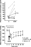
Role of kinins in the acute antihypertensive effects an ACE inhibitor (enalaprilat) in rats with severe hypertension. Top: Blood kinin concentrations before (C) and after administration of the ACE inhibitor. Bottom: Mean blood pressure before and after ACE inhibition, open and closed circles represent rats pretreated with a kinin antagonist or vehicle, respectively. Values are mean ± SEM (bottom). [Reprinted from Carbonell et al. (40).]
Figure 9
Figure 9
Two-dimensional M-mode echocardiographs of B2−/− mice and B2+/+ mice with sham coronary ligation (sham) or heart failure induced by coronary artery ligation. ACE inhibitor ramipril (2.5 mg/kg/day) reduced LV chamber size and thickeness in B2+/+, but only marginally in B2−/−, indicating an important role of kinin in the cardoprotective effects of ACE inibition in heart failure model. IS indicates interventricular septum; DD, left ventricular (LV) diastolic dimension; and PW, LV posterior wall. (Reprinted from Yang et al. (311).]
Figure 10
Figure 10
Effect of prolylcarboxypeptidase (PRCP) blockade by a siRNA on bradykinin (BK) release in Ad-AT2R-transfected mouse coronary endothelial cells (ECs). (A) Representative Western blot showing PRCP protein expression (top) and quantitative analysis of PRCP protein (bottom); PRCP SiRNA, but not scarmbled-SiRNA (S-siRNA) significantly blocked PRCP expression. (B) Fold change in bradykinin release relative to Ad-GFP-transfected cells; BK release was further increased when Ad-AT2R-transfected ECs were treated with CGP42112A, a selective AT2 agonist (CGP; 0.1 μmol/l). SiRNA, but not S-siRNA significantly blunted Ad-AT2R-induced bradykinin release, n = 4. [Reprinted from Zhu, Carretero, Yang et al., (318).]
Figure 11
Figure 11
The renin-angiotensin and kallikrein-kinin systems. In both systems, a substrate is cleaved by an enzyme of restricted specificity, releasing a peptide that is either already active (lysbradykinin, bradykinin) or inactive (angiotensin I). Upon further processing by a specific peptidase, angiotensin I is converted to a vasoactive peptide (angiotensin II). In turn, vasoactive peptides are inactivated by peptidases. Angiotensin-Converting enzyme is common to both systems but has different roles: it processes angiotensin I to angiotensin II and is the main kinin-inactivating peptidase. [Reprinted from Liu et al. (143).]

Similar articles

Cited by

References

    1. Farmer SG, editor. The Kinin System (The Handbook of lmmunopharmacology) San Diego, CA: Academic Press Inc; 1997. pp. 1–323.
    1. Abadir PM, Carey RM, Siragy HM. Angiotensin AT2 receptors directly stimulate renal nitric oxide in bradykinin B2-receptor-null mice. Hypertension. 2003;42:600–604. - PubMed
    1. Abadir PM, Periasamy A, Carey RM, Siragy HM. Angiotensin II type 2 receptor-bradykinin B2 receptor functional heterodimerization. Hypertension. 2006;48:1–7. - PubMed
    1. Abbas A, Gorelik G, Carbini LA, Scicli AG. Angiotensin-(1-7) induces bradykinin-mediated hypotensive responses in anesthetized rats. Hypertension. 1997;30:217–221. - PubMed
    1. AbdAlla S, Abdel-Baset A, Lother H, el Massiery A, Quitterer U. Mesangial AT1/B2 receptor heterodimers contribute to angiotensin II hyperresponsiveness in experimental hypertension. J Mol Neurosci. 2005;26:185–192. - PubMed

Publication types

MeSH terms

Substances