. 2013 Aug;52(8):775-84.
doi: 10.1002/gcc.22073. Epub 2013 Jun 5.

Novel YAP1-TFE3 fusion defines a distinct subset of epithelioid hemangioendothelioma

Affiliations

Novel YAP1-TFE3 fusion defines a distinct subset of epithelioid hemangioendothelioma

Cristina R Antonescu et al. Genes Chromosomes Cancer. 2013 Aug.

Abstract

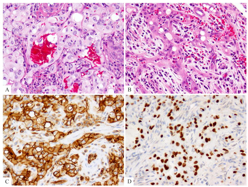
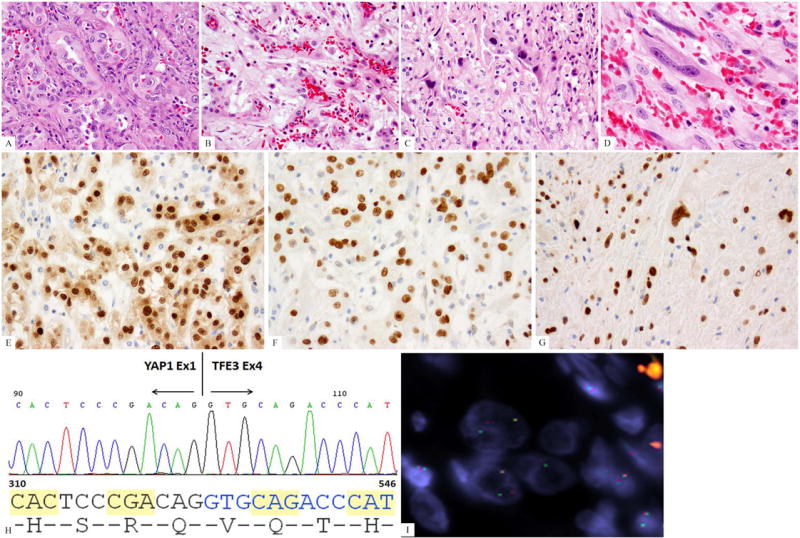
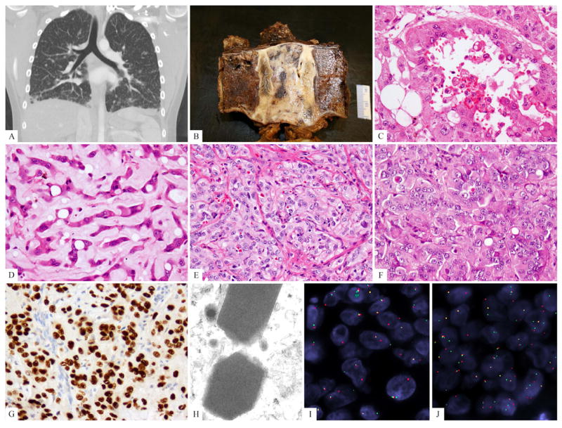
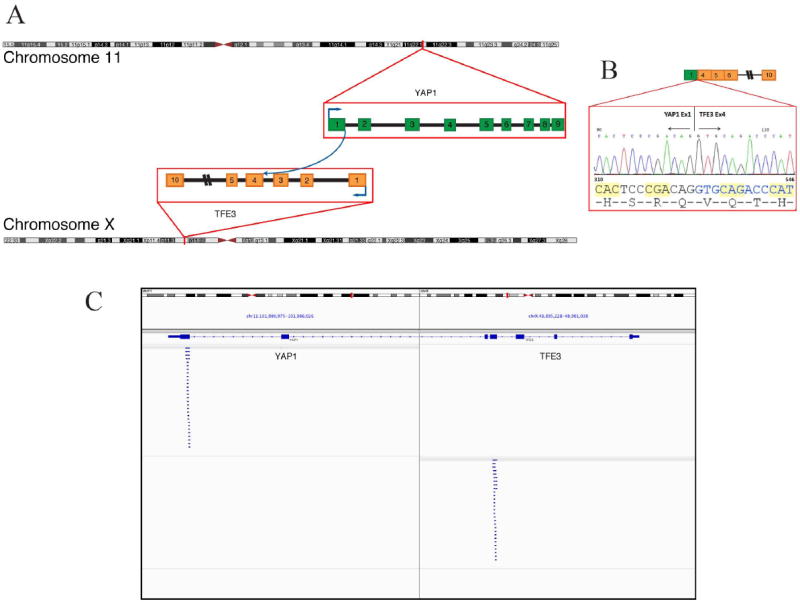
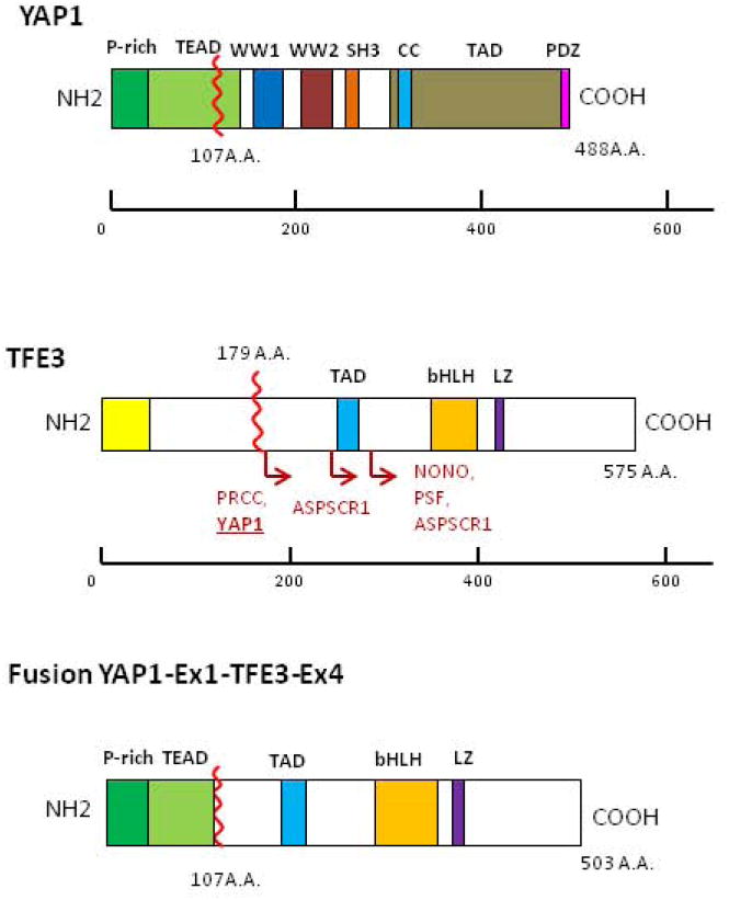
Conventional epithelioid hemangioendotheliomas (EHE) have a distinctive morphologic appearance and are characterized by a recurrent t(1;3) translocation, resulting in a WWTR1-CAMTA1 fusion gene. We have recently encountered a fusion-negative subset characterized by a somewhat different morphology, including focally well-formed vasoformative features, which was further investigated for recurrent genetic abnormalities. Based on a case showing strong transcription factor E3 (TFE3) immunoreactivity, fluorescence in situ hybridization (FISH) analysis for TFE3 gene rearrangement was applied to the index case as well as to nine additional cases, selected through negative WWTR1-CAMTA1 screening. A control group, including 18 epithelioid hemangiomas, nine pseudomyogenic HE, and three epithelioid angiosarcomas, was also tested. TFE3 gene rearrangement was identified in 10 patients, with equal gender distribution and a mean age of 30 years old. The lesions were located in somatic soft tissue in six cases, lung in three and one in bone. One case with available frozen tissue was tested by RNA sequencing and FusionSeq data analysis to detect novel fusions. A YAP1-TFE3 fusion was thus detected, which was further validated by FISH and reverse transcription polymerase chain reaction (RT-PCR). YAP1 gene rearrangements were then confirmed in seven of the remaining nine TFE3-rearranged EHEs by FISH. No TFE3 structural abnormalities were detected in any of the controls. The TFE3-rearranged EHEs showed similar morphologic features with at least focally, well-formed vascular channels, in addition to a variably solid architecture. All tumors expressed endothelial markers, as well as strong nuclear TFE3. In summary, we are reporting a novel subset of EHE occurring in young adults, showing a distinct phenotype and YAP1-TFE3 fusions.


Conflict of interest statement

Conflict of interest: none

Figures

Fig. 1
Fig. 1
Index case displaying a distinctive pseudo-alveolar pattern was tested for TFE3 immunohistochemistry and gene rearrangements (EHE2). (A, B) Morphologic appearance showing mature vessel lumen formation lined by epithelioid endothelial cells with moderate amount of densely to lightly eosinophilic cytoplasm, focal intra-cytoplasmic vacuoles and stromal inflammation including eosinophils. (C) CD31 and (D) TFE3 strong reactivity.
Fig. 2
Fig. 2
Morphologic progression of EHE after multiple local recurrences (EHE1). (A) Histologic appearance of the primary tumor showing well-formed vasoformative features, with epithelioid cells with moderate nuclear pleomorphism, nuclear indentations and pseudo-inclusions; (B-D). Histologic appearance of the latest recurrence 20 years after the initial diagnosis showing a spectrum of well-differentiated to solid to frankly malignant and spindle cell areas; (E) ERG immunostaining showing strong nuclear reactivity; (F, G) TFE3 strong reactivity in both primary as well as latest sarcomatous recurrence; (H) ABI sequence from the RT-PCR product showing YAP1 exon1 being fused to TFE3 exon 4. (I) YAP1 break-apart by FISH (green, telomeric; red signals, centromeric).
Fig. 3
Fig. 3
Clinical and pathologic spectrum of TFE3-rearranged EHE. (A) CT scan showing bilateral ground-glass opacities suggestive of interstitial lung disease, most predominantly in the lower lobes but diffusely present (EHE9); (B) Gross appearance of the T2 vertebral en-bloc resection showing an ill-defined white-gray lesion (EHE3). (C, D). Biphasic morphologic appearance showing a pseudo-alveolar component with abundant, densely eosinophilic cytoplasm and the other resembling classis EHE, with cord-like arrangement and myxoid stroma (EHE3); (E) foamy cytoplasm (histiocytoid), mild nuclear pleomorphism (EHE7); (F) predominantly solid and nested growth pattern, showing densely eosinophilic cytoplasm, rare vacuoles, and lack of significant intervening stroma (EHE4); (G) strong TFE3 immunostaining; (H) ultrastructural study showing distinctive rhomboid crystals with a periodicity ranging from 9.05-11.63, mean of 10.34 nm, (46,000 magnification, EHE10). (I) TFE3 and (J) YAP1 break-apart signals by FISH (EHE 7) (green, telomeric; red, centromeric).
Fig. 4
Fig. 4
YAP1-TFE3 gene fusion. (A) Schematic representation of the YAP1-TFE3 fusion indicating the loci that are fused together. (B) Experimental validation of the fusion shows the sequence of the junction between exon 1 of YAP1 and exon 4 or TFE3. (C) Integrative Genome Viewer (IGV) snapshot of the reads supporting the fusion candidate as determined by RNA-seq.
Fig 5
Fig 5
Protein domains of YAP1, TFE3 and projected YAP1-TFE3 fusion protein. Schematic representations of YAP1 showing prolin-rich domain (P-rich), TEAD binding domain (TEAD), WW1/WW2 domains, SRC homology 3 domain (SH3), coiled-coil domain (CC), transactivation domain (TAD), PSD-95/DLG1/ZO-1 (PDZ) domain and for TFE3 showing common domains: glutamine-rich domain (Gln-rich); activation domain (AD); basic, a positively charged domain; helix-loop-helix (HLH) and domain; leucine zipper (LZ) domain. For TFE3, locations of fusions in some cancer types are shown. ASPSCR1, alveolar soft part sarcoma chromosome region, candidate 1; NONO, non-POU domain containing, octamer-binding; PRCC, papillary renal cell carcinoma; PSF, splicing factor proline/glutamine-rich.

References

    1. Antonescu CR, Zhang L, Chang NE, Pawel BR, Travis W, Katabi N, Edelman M, Rosenberg AE, Nielsen GP, Dal Cin P, Fletcher CD. EWSR1-POU5F1 fusion in soft tissue myoepithelial tumors. A molecular analysis of sixty-six cases, including soft tissue, bone, and visceral lesions, showing common involvement of the EWSR1 gene. Genes Chromosomes Cancer. 2010;49:1114–1124. - PMC - PubMed
    1. Argani P, Lui MY, Couturier J, Bouvier R, Fournet JC, Ladanyi M. A novel CLTC-TFE3 gene fusion in pediatric renal adenocarcinoma with t(X;17)(p11.2;q23) Oncogene. 2003;22:5374–5378. - PubMed
    1. Clark J, Lu YJ, Sidhar SK, Parker C, Gill S, Smedley D, Hamoudi R, Linehan WM, Shipley J, Cooper CS. Fusion of splicing factor genes PSF and NonO (p54nrb) to the TFE3 gene in papillary renal cell carcinoma. Oncogene. 1997;15:2233–2239. - PubMed
    1. Dong J, Feldmann G, Huang J, Wu S, Zhang N, Comerford SA, Gayyed MF, Anders RA, Maitra A, Pan D. Elucidation of a universal size-control mechanism in Drosophila and mammals. Cell. 2007;130:1120–1133. - PMC - PubMed
    1. Errani C, Zhang L, Panicek DM, Healey JH, Antonescu CR. Epithelioid hemangioma of bone and soft tissue: a reappraisal of a controversial entity. Clin Orthop Relat Res. 2012;470:1498–1506. - PMC - PubMed

Publication types

MeSH terms

Substances