Review
. 2013 Aug;242(8):909-22.
doi: 10.1002/dvdy.23992. Epub 2013 Jun 27.

Angiogenesis and intramembranous osteogenesis

Affiliations
Review

Angiogenesis and intramembranous osteogenesis

Christopher J Percival et al. Dev Dyn. 2013 Aug.

Abstract

Background: Angiogenesis is likely critical for the process of intramembranous osteogenesis; however, the developmental relationship between blood vessels and bone mineralization is not well studied within intramembranous bones. Given its importance, changes in angiogenesis regulation are likely to contribute to evolutionarily and medically relevant craniofacial variation.

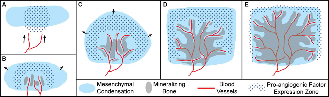
Results: We summarize what is known about the association between angiogenesis and intramembranous osteogenesis, supplementing with information from the better-studied processes of endochondral ossification and distraction osteogenesis. Based on this review, we introduce a model of angiogenesis during early intramembranous osteogenesis as well as a series of null hypotheses to be tested.

Conclusions: This model can serve as a basis of future research on the spatio-temporal association and regulatory interactions of mesenchymal, vascular, and bone cells, which will be required to illuminate the potential effects of angiogenesis dysregulation on craniofacial skeletal phenotypes.

Keywords: craniofacial development; intramembranous ossification; vascular invasion.


Figures

Figure 1
Figure 1
Ossification type and cellular origin of postnatal day eight (P8) mouse craniofacial bones, as shown on (A) a right lateral view, (B) a superior interior view lacking calvaria, nasals, and a mandible, and (C) an interior view lacking a mandibles. Red: endochondral ossification; Blue: intramembranous ossification; Diagonal lines: neural crest derived cellular origin; Dots: mesoderm derived cellular origin. The stars identify small portions of medosderm derived bone within the mostly neural crest derived presphenoid. Ossification identification from (Depew et al., 2002). Cellular origin of cranial base from (McBratney-Owen et al., 2008). Calvarial cellular origin from (Jiang et al., 2002). Other cellular origin from (Noden and Trainor, 2005).
Figure 2
Figure 2
High resolution images of perinatal mouse frontal and parietal bones produced by multiple imaging modalities. Note the lattice-like pattern at the edges of ossification, with bone filling in the gaps in older areas of bone. All images are an oblique dorsal view for which caudal is towards the bottom of the image and medial is to the left. A) Surface reconstruction around the medial coronal suture of a newborn mouse from an image produced at the High-Resolution X-ray Computed Tomography Facility at the University of Texas at Austin. The hard edges of the image represent the extent of the region of the bone that was imaged. B) Lightfield microscope image of the medial coronal suture of a whole-mount Ailzarin red/Alacian blue clear and stained E18.5 mouse. Image courtesy of Mizuho Kawasaki and Kazuhiko Kawasaki. C) Two photon laser scanning microscopy image of the lateral coronal suture of an E19.5 C57BL/6 mouse. The bones have been marked with calcein. Image produced by Kevin Flaherty and Patrick Drew at Penn State.
Figure 3
Figure 3
Schematic of the epiphyseal ossification of endochondral long bones, with emphasis on the process of capillary growth into calcified epiphyseal cartilage and subsequent trabecular ossification. A) Chondrocytes differentiate from proliferating prechondrocytes within the growth plate. The chondrocytes are pushed toward the diaphysis by this continuous process and then enlarge under hypoxia, leading to mineralization of surrounding cartilage and the attraction of blood vessels required for bone formation. B) A magnified view of bracketed zone from ‘A’ showing capillaries, in association with chondroclasts, growing towards hypertrophic chondrocytes as a precursor to osteoblast activity and bone growth at the epiphysis. Figure adapted from (Streeten and Brandi, 1990; Lewinson and Silbermann, 1992; Bloom and Fawcett, 1994; Kronenberg, 2003).
Figure 4
Figure 4
Schematic of the cellular zones of distraction osteogenesis. The fibrous interzone (FIZ) forms first and is composed of a variety of cells, including osteoblasts that deposit osteoid along parallel collagen bundles. Zones of microcolumn formation (MCF) form on either side of the insult and include invading vascular sinuses and vessels originating from the cut bone. These vessels and microcolumns of mineralizing bone are parallel to tension applied during distraction. Between the FIZ and MCF is the primary mineralization front (PMF), which is a thin zone of high cellular proliferation. The MCF continues expanding as the portions of the original bone are pulled apart, while the FIZ remains a constant width.
Figure 5
Figure 5
Hypothetical schematic of the association between mesenchymal precursors, pro-angiogenic factor (e.g. VEGF, HIF) expression, invading blood vessels, and bone formation during the initial period of intramembranous ossification. A) Blood vessels (red line) are drawn (arrows) to the border of the avascular mesenchymal condensation (light blue solid) by pro-angiogenic factors (blue dots). B) As mesenchymal cells migrate outwards (arrows), blood vessels invade the condensation near the center of ossification at or around the time of initial ossification (grey solid), which occurs in proximity to invading vasculature. C,D) Migration of cells derived from the original condensation continues outward (arrows) until they receive some signal to stop, often at sutures that form between the advancing mesenchymal fronts of two bones. Vessels continue to extend outward through the mesenchymal condensation towards regions of pro-angiogenic factor expression. New bone mineralization occurs proximate to sprouting vessels, as previous sites of bone formation begin to merge and mature. E) After the end of rapid mesenchymal cell expansion from the condensation, vessels and associated regions of ossification approach the edge, forming an ossification front at the suture margin that will allow for continued calvarial growth.
Figure 6
Figure 6
Radiographic image of the two parietal bones of a human fetal skull after vascular perfusion with radioactive Thorotrast (white). The vasculature within the developing parietal bones can be seen radiating outward from their centers. Image reproduced with kind permission of Springer Science+Business Media (p65, Brookes and Revell, 1998).

References

    1. Abzhanov A, Rodda SJ, McMahon AP, Tabin CJ. Regulation of skeletogenic differentiation in cranial dermal bone. Development. 2007;134:3133–3144. - PubMed
    1. Al-Aql ZS, Alagl AS, Graves DT, Gerstenfeld LC, Einhorn TA. Molecular mechanisms controlling bone formation during fracture healing and distraction osteogenesis. Journal of Dental Research. 2008;87:107–118. - PMC - PubMed
    1. Amizuka N, Hasegawa T, Oda K, Luiz de Freitas P, Hoshi K, Li M, Ozawa H. Histology of epiphyseal cartilage calcification and endochondral ossification. Front Biosci. 2012;4:2085–2100. - PubMed
    1. Aronson J, Good B, Stewart C, Harrison B, Harp J. Preliminary studies of mineralization during distraction osteogenesis. Clinical Orthopaedics and Related Research. 1990;250:43–49. - PubMed
    1. Aronson J. Temporal and spatial increases in blood flow during distraction osteogenesis. Clinical Orthopaedics and Related Research. 1994;301:124–131. - PubMed

Publication types

MeSH terms

LinkOut - more resources