

Elevation of conjunctival epithelial CD45INTCD11b⁺CD16⁺CD14⁻ neutrophils in ocular Stevens-Johnson syndrome and toxic epidermal necrolysis
- PMID: 23737478
- PMCID: PMC3711386
- DOI: 10.1167/iovs.13-11859
Elevation of conjunctival epithelial CD45INTCD11b⁺CD16⁺CD14⁻ neutrophils in ocular Stevens-Johnson syndrome and toxic epidermal necrolysis
Abstract
Purpose: Ocular complications related to Stevens-Johnson Syndrome (SJS)-Toxic Epidermal Necrolysis (TEN) may persist and progress after resolution of systemic disease. This is thought to be related in part to persistent ocular innate-immune signaling. In this study, our aim was to characterize infiltrative conjunctival cellular profiles during acute (<12 months) and chronic (>12 months) disease.
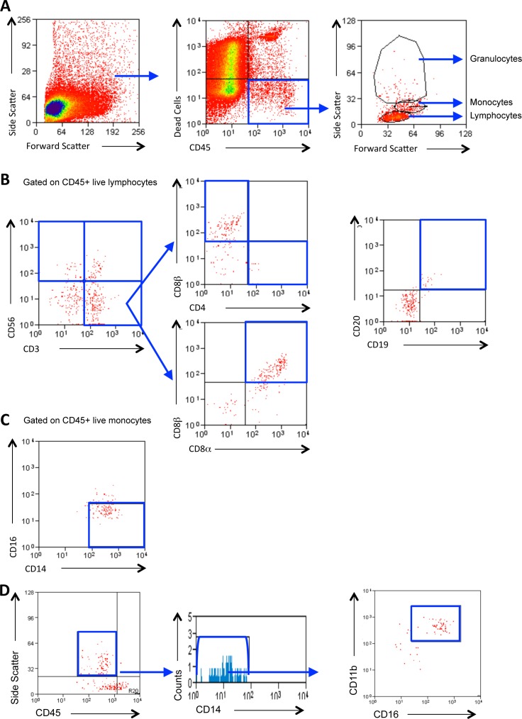
Methods: Consecutive patients presenting with SJS-TEN over a 12-month period were followed for 1 year. Detailed clinical examination and conjunctival impression cell recovery was analyzed by flow cytometry for the presence of intraepithelial leukocytes and compared with healthy controls (n = 21).
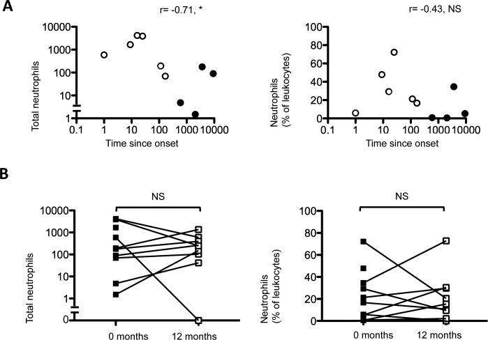
Results: Ten patients were recruited of whom six had acute disease and five were classified as TEN (SCORTEN = 1, n = 4). Conjunctival inflammation was graded as absent/mild in a total of nine patients; but despite this, evidence of fornix shrinkage was observed in nine subjects. This inversely correlated with disease duration (P < 0.05). A reduction in percentage of CD8αβ(+) T cells compared with controls (80% vs. 57%; P < 0.01) was associated with a corresponding increase in the number/percentage of CD45(INT)CD11b(+)CD16(+)CD14(-) neutrophils (186 vs. 3.4, P < 0.01, 31% vs. 0.8%, P < 0.001). Neutrophils inversely correlated with disease duration (r = -0.71, P = 0.03), yet there was no absolute change in the CD8αβ(+) or neutrophil populations during the study period (P = 1.0).
Conclusions: These data highlight that a neutrophilic infiltrate is present in mildly inflamed or clinically quiescent conjunctival mucosa in patients with ocular SJS-TEN, where neutrophil numbers inversely correlate with disease duration. Neutrophil persistence endorses the hypothesis of an unresolved innate-inflammatory process that might account for disease progression.
Keywords: SJS; TEN; innate immunity; neutrophils.
Figures

Similar articles
-
Neutrophil-driven and interleukin-36γ-associated ocular surface inflammation in chronic Stevens-Johnson syndrome.Allergy. 2024 Aug;79(8):2128-2143. doi: 10.1111/all.16126. Epub 2024 Apr 29. Allergy. 2024. PMID: 38682250
-
A cross-sectional comparative study on chronic ocular manifestations of Stevens-Johnson syndrome and toxic epidermal necrolysis in Chinese eyes: a 15-year case series.Int Ophthalmol. 2018 Jun;38(3):1155-1160. doi: 10.1007/s10792-017-0576-5. Epub 2017 May 25. Int Ophthalmol. 2018. PMID: 28547534
-
Multistep Grading System for Evaluation of Chronic Ocular Sequelae in Patients With Stevens-Johnson Syndrome.Am J Ophthalmol. 2019 Jul;203:69-77. doi: 10.1016/j.ajo.2019.01.028. Epub 2019 Feb 4. Am J Ophthalmol. 2019. PMID: 30731084
-
Ocular surface inflammation mediated by innate immunity.Eye Contact Lens. 2010 Sep;36(5):269-81. doi: 10.1097/ICL.0b013e3181ee8971. Eye Contact Lens. 2010. PMID: 20703156 Review.
-
Immunologic Mediators in Stevens-Johnson Syndrome and Toxic Epidermal Necrolysis.Semin Ophthalmol. 2016;31(1-2):85-90. doi: 10.3109/08820538.2015.1115255. Semin Ophthalmol. 2016. PMID: 26959133 Review.
Cited by
-
Molecular mechanisms and treatments for ocular symblephara.Surv Ophthalmol. 2022 Jan-Feb;67(1):19-30. doi: 10.1016/j.survophthal.2021.04.008. Epub 2021 Apr 29. Surv Ophthalmol. 2022. PMID: 33932469 Free PMC article. Review.
-
Vulvovaginal and ocular involvement and treatment in female patients with Stevens-Johnson syndrome and toxic epidermal necrolysis: A review.Int J Womens Dermatol. 2021 Sep 2;7(5Part A):520-528. doi: 10.1016/j.ijwd.2021.08.012. eCollection 2021 Dec. Int J Womens Dermatol. 2021. PMID: 35024409 Free PMC article. Review.
-
Leukocyte Distribution in the Open Eye Tears of Normal and Dry Eye Subjects.Curr Eye Res. 2018 Oct;43(10):1253-1259. doi: 10.1080/02713683.2018.1500611. Epub 2018 Aug 9. Curr Eye Res. 2018. PMID: 30005585 Free PMC article.
-
Age-related Defects in Ocular and Nasal Mucosal Immune System and the Immunopathology of Dry Eye Disease.Ocul Immunol Inflamm. 2016 Jun;24(3):327-47. doi: 10.3109/09273948.2014.986581. Epub 2014 Dec 23. Ocul Immunol Inflamm. 2016. PMID: 25535823 Free PMC article. Review.
-
Update on the role of impression cytology in ocular surface disease.Taiwan J Ophthalmol. 2019 Sep 12;9(3):141-149. doi: 10.4103/tjo.tjo_57_19. eCollection 2019 Jul-Sep. Taiwan J Ophthalmol. 2019. PMID: 31572650 Free PMC article. Review.
References
-
- Wojnarowska FT, Briggaman RA. Management of Blistering Diseases. London: Chapman and Hall Medical; 1990.
-
- Saw VP, Dart JK. Ocular mucous membrane pemphigoid: diagnosis and management strategies. Ocul Surf. 2008; 6: 128–142 - PubMed
-
- Bastuji-Garin S, Fouchard N, Bertocchi M, Roujeau JC, Revuz J, Wolkenstein P. SCORTEN: a severity-of-illness score for toxic epidermal necrolysis. J Invest Dermatol. 2000; 115: 149–153 - PubMed
-
- Schopf E, Stuhmer A, Rzany B, Victor N, Zentgraf R, Kapp JF. Toxic epidermal necrolysis and Stevens-Johnson syndrome. An epidemiologic study from West Germany. Arch Dermatol. 1991; 127: 839–842 - PubMed
-
- Bastuji-Garin S, Rzany B, Stern RS, Shear NH, Naldi L, Roujeau JC. Clinical classification of cases of toxic epidermal necrolysis, Stevens-Johnson syndrome, and erythema multiforme. Arch Dermatol. 1993; 129: 92–96 - PubMed
Publication types
MeSH terms
Substances
Grants and funding
LinkOut - more resources
Full Text Sources
Other Literature Sources
Research Materials
Miscellaneous
