

A renewable tissue resource of phenotypically stable, biologically and ethnically diverse, patient-derived human breast cancer xenograft models
- PMID: 23737486
- PMCID: PMC3732575
- DOI: 10.1158/0008-5472.CAN-12-4081
A renewable tissue resource of phenotypically stable, biologically and ethnically diverse, patient-derived human breast cancer xenograft models
Abstract
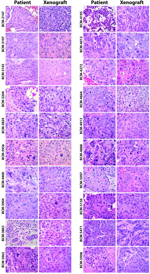
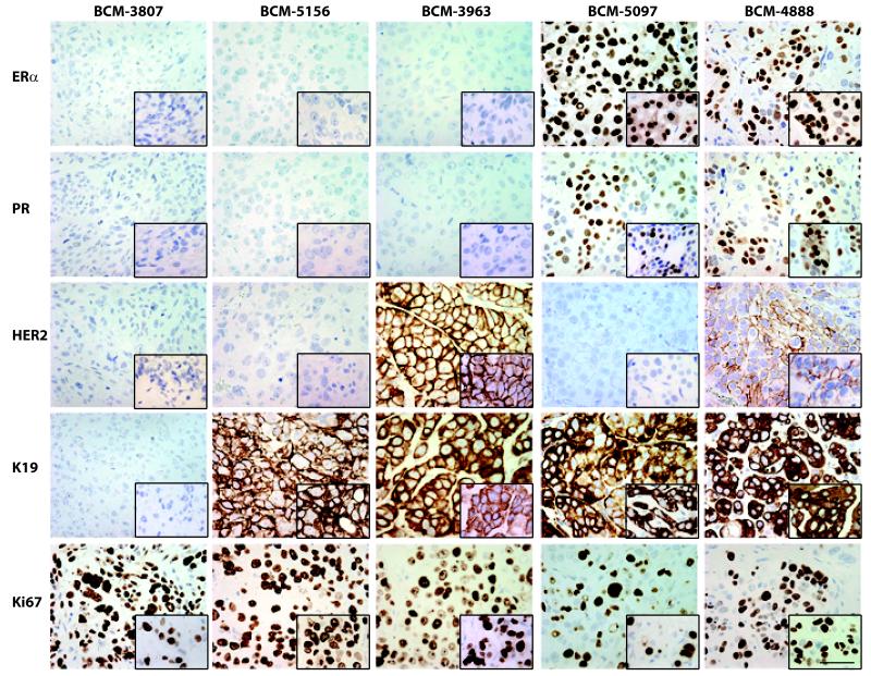
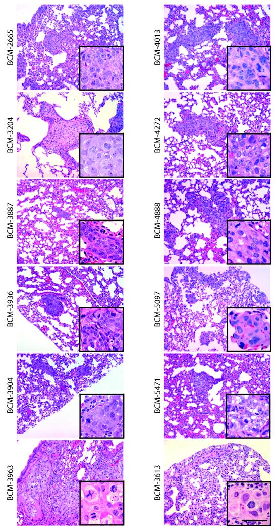
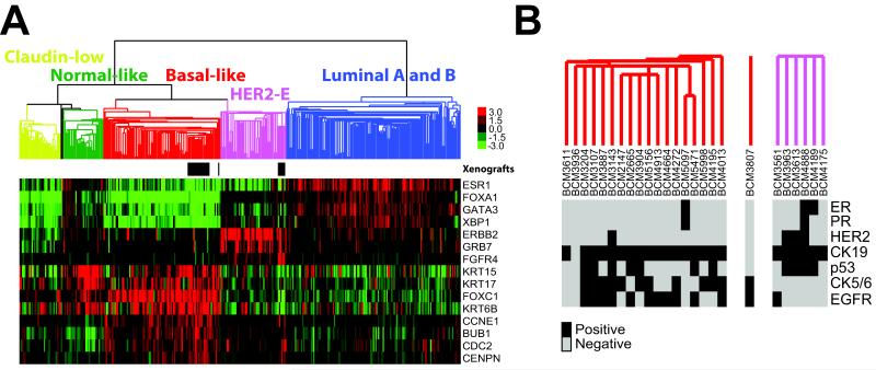
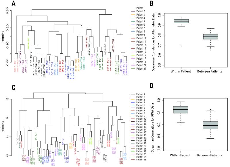
Breast cancer research is hampered by difficulties in obtaining and studying primary human breast tissue, and by the lack of in vivo preclinical models that reflect patient tumor biology accurately. To overcome these limitations, we propagated a cohort of human breast tumors grown in the epithelium-free mammary fat pad of severe combined immunodeficient (SCID)/Beige and nonobese diabetic (NOD)/SCID/IL-2γ-receptor null (NSG) mice under a series of transplant conditions. Both models yielded stably transplantable xenografts at comparably high rates (∼21% and ∼19%, respectively). Of the conditions tested, xenograft take rate was highest in the presence of a low-dose estradiol pellet. Overall, 32 stably transplantable xenograft lines were established, representing 25 unique patients. Most tumors yielding xenografts were "triple-negative" [estrogen receptor (ER)-progesterone receptor (PR)-HER2+; n = 19]. However, we established lines from 3 ER-PR-HER2+ tumors, one ER+PR-HER2-, one ER+PR+HER2-, and one "triple-positive" (ER+PR+HER2+) tumor. Serially passaged xenografts show biologic consistency with the tumor of origin, are phenotypically stable across multiple transplant generations at the histologic, transcriptomic, proteomic, and genomic levels, and show comparable treatment responses as those observed clinically. Xenografts representing 12 patients, including 2 ER+ lines, showed metastasis to the mouse lung. These models thus serve as a renewable, quality-controlled tissue resource for preclinical studies investigating treatment response and metastasis.
©2013 AACR.
Figures
Comment in
-
Breast cancer: Stable breast cancer xenograft models.Nat Rev Clin Oncol. 2013 Aug;10(8):426. doi: 10.1038/nrclinonc.2013.111. Epub 2013 Jun 25. Nat Rev Clin Oncol. 2013. PMID: 23799372 No abstract available.
References
-
- Rae-Venter B, Reid LM. Growth of human breast carcinomas in nude mice and subsequent establishment in tissue culture. Cancer Res. 1980;40(1):95–100. - PubMed
-
- Sebesteny A, Taylor-Papadimitriou J, Ceriani R, Millis R, Schmitt C, Trevan D. Primary human breast carcinomas transplantable in the nude mouse. J Natl Cancer Inst. 1979;63(6):1331–1337. - PubMed
-
- Noel A, Borcy V, Bracke M, Gilles C, Bernard J, Birembaut P, Mareel M, Foidart JM. Heterotransplantation of primary and established human tumour cells in nude mice. Anticancer Res. 1995;15(1):1–7. - PubMed
Publication types
MeSH terms
Grants and funding
- P30 CA016672/CA/NCI NIH HHS/United States
- R01 CA112305/CA/NCI NIH HHS/United States
- CA16672/CA/NCI NIH HHS/United States
- P50 CA50183/CA/NCI NIH HHS/United States
- P30 CA125123/CA/NCI NIH HHS/United States
- P01 CA030195/CA/NCI NIH HHS/United States
- U54 CA149196/CA/NCI NIH HHS/United States
- P30-CA125123/CA/NCI NIH HHS/United States
- P01 CA30195/CA/NCI NIH HHS/United States
- P50 CA098258/CA/NCI NIH HHS/United States
- R01 CA127857/CA/NCI NIH HHS/United States
- P50 CA058223/CA/NCI NIH HHS/United States
- P50-CA58223/CA/NCI NIH HHS/United States
LinkOut - more resources
Full Text Sources
Other Literature Sources
Medical
Molecular Biology Databases
Research Materials
Miscellaneous
