. 2013 Jun 18;110(25):10201-6.
doi: 10.1073/pnas.1300016110. Epub 2013 Jun 4.

Andrographolide derivatives inhibit guanine nucleotide exchange and abrogate oncogenic Ras function

Affiliations

Andrographolide derivatives inhibit guanine nucleotide exchange and abrogate oncogenic Ras function

Harrison J Hocker et al. Proc Natl Acad Sci U S A. .

Abstract

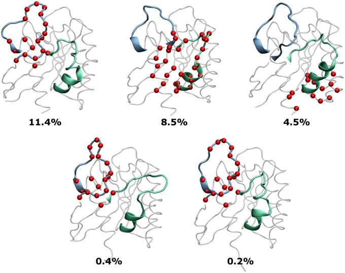
Aberrant signaling by oncogenic mutant rat sarcoma (Ras) proteins occurs in ∼15% of all human tumors, yet direct inhibition of Ras by small molecules has remained elusive. Recently, several small-molecule ligands have been discovered that directly bind Ras and inhibit its function by interfering with exchange factor binding. However, it is unclear whether, or how, these ligands could lead to drugs that act against constitutively active oncogenic mutant Ras. Using a dynamics-based pocket identification scheme, ensemble docking, and innovative cell-based assays, here we show that andrographolide (AGP)--a bicyclic diterpenoid lactone isolated from Andrographis paniculata--and its benzylidene derivatives bind to transient pockets on Kirsten-Ras (K-Ras) and inhibit GDP-GTP exchange. As expected for inhibitors of exchange factor binding, AGP derivatives reduced GTP loading of wild-type K-Ras in response to acute EGF stimulation with a concomitant reduction in MAPK activation. Remarkably, however, prolonged treatment with AGP derivatives also reduced GTP loading of, and signal transmission by, oncogenic mutant K-RasG12V. In sum, the combined analysis of our computational and cell biology results show that AGP derivatives directly bind Ras, block GDP-GTP exchange, and inhibit both wild-type and oncogenic K-Ras signaling. Importantly, our findings not only show that nucleotide exchange factors are required for oncogenic Ras signaling but also demonstrate that inhibiting nucleotide exchange is a valid approach to abrogating the function of oncogenic mutant Ras.

Keywords: allosteric site; cancer; drug design; molecular dynamics.


Conflict of interest statement

The authors declare no conflict of interest.

Figures

Fig. 1.
Fig. 1.
Chemical structures of AGP and its benzylidene derivatives SRJ09, SRJ10, and SRJ23.
Fig. 2.
Fig. 2.
Overview of the K-RasQ61H structures derived from MD simulations for ensemble docking. From among the 75 unique K-Ras conformers used for docking (Materials and Methods), 5 representative cluster centroids are shown along with the percentage of the total conformers they represent. Pockets most frequently targeted by SRJ23 are highlighted in red van der Waals spheres. Notice the major conformational changes in switch 1 (cyan) and switch 2 (green).
Fig. 3.
Fig. 3.
Overview of transiently opening pockets 1 and 2 on K-RasQ61H with relevant residues colored according to their electrostatic potential. (A) Docking pose of SRJ23 at pocket 1, where its phenyl group occupies the space previously occupied by Y40 and is stabilized by the residues I21 and T20. (B) Docking pose of SRJ23 at pocket 2 opened by the movement of Y71 and lined by hydrophobic residues I36 and M67. (C) MD-optimized complex of SRJ23 at pocket 1. (D) The conformation in C is modified to visualize the proximity of SRJ23 to Mg2+ and GTP (switch 1 is now shown as a transparent surface). The hydroxyl group on the lactone ring of SRJ23 forms a hydrogen bond with the α-phosphate of GTP as well as an electrostatic contact with Mg2+ similar to that made by the hydroxyl of T35 on Ras (20). Electrostatic potentials were calculated using the Adaptive Poisson-Boltzmann Solver (50).
Fig. 4.
Fig. 4.
Comparison of known Ras structures bound to SOS and small-molecule ligands. (A) Overlay of the switch 2 region of a K-Ras–SRJ23 snapshot (orange), the crystal structure of K-RasG12D–DCAI from PDB ID code 4DST (purple, with only residues 62–75 shown for clarity), K-Ras–0QX from PDB ID code 4EVP (yellow, residues 62–75), and two structures of H-Ras–SOS from PDB ID codes 1NVV (ice blue) and 1BKD (cyan). (B) Projection of simulated K-Ras conformers onto a PC space defined by crystallographic structures, with the cluster of GDP–H-Ras structures highlighted in blue and the cluster of loss-of-function mutants in orange. PDB ID code 4DST (purple) lies in the major GTP cluster, whereas PDB ID code 4EPV (yellow) is intermediate to the GTP and GDP clusters. Two K-Ras–ligand conformations from docking (MD-Lf, green dots) are shown to illustrate the ability of MD to capture putative excited-state structures with open p1 that are preferred by our ligands. An example of simulated K-Ras–SRJ23 (MD-Lb) lies between Ras–SOS (cyan/ice blue) and Ras–inhibitor (purple/yellow) conformations. The crystal structure of nucleotide-free H-Ras (PDB ID code 1BKD) is also shown, with SRJ23 docked at switch 1. SRJ23 is shown as a yellow surface and switches 1 and 2 as blue and green surfaces, respectively. The Bio3D package (http://thegrantlab.org/bio3d) was used to generate the figure in B.
Fig. 5.
Fig. 5.
Mean fold increase in Ras-GTP (A) and ppERK (B) ± SEM from three independent experiments where wild-type BHK cells were treated with SRJ09 or SRJ23 for 6 h in the absence of serum, followed by 25 ng/mL EGF stimulation for 2 min. Ras-GTP levels were measured using an RBD pull-down assay, and ppERK levels were measured by quantitative immunoblotting. (C) Representative blots from three independent experiments in which wild-type BHK cells were treated with 5 μM SRJ23 for 6 h in the absence of serum, followed by 25 ng/mL EGF stimulation for 2 min. Levels of K-, H-, and N-Ras–GTP loadings were measured using an RBD pull-down assay, and phospho-EGF receptor (EGFR; Y1068) levels were measured by quantitative immunoblotting. (D) Mean ± SEM from three independent experiments in which BHK cells stably expressing oncogenic Ras isoforms were treated with 5 μM SRJ09 or SRJ23 for 6 h in the absence of growth serum. ppERK levels were measured by quantitative immunoblotting. Mean Ras-GTP (E) and ppERK (F) ± SEM from three independent experiments in which BHK cells stably expressing oncogenic K-Ras were treated with 5 μM SRJ09 or SRJ23 for 72 h. Growth media with the drug were replaced every 24 h. Ras-GTP levels were measured using an RBD pull-down assay, and ppERK levels were measured by quantitative immunoblotting. Differences between DMSO- and drug-treated cells were assessed using one-way ANOVA tests. Significant differences are indicated (*P < 0.05; **P < 0.01; ***P < 0.001).

References

    1. Malumbres M, Barbacid M. Cell cycle, CDKs and cancer: A changing paradigm. Nat Rev Cancer. 2009;9(3):153–166. - PubMed
    1. Scheffzek K, et al. The Ras-RasGAP complex: Structural basis for GTPase activation and its loss in oncogenic Ras mutants. Science. 1997;277(5324):333–338. - PubMed
    1. McCormick F, Wittinghofer A. Interactions between Ras proteins and their effectors. Curr Opin Biotechnol. 1996;7(4):449–456. - PubMed
    1. Prior IA, Lewis PD, Mattos C. A comprehensive survey of Ras mutations in cancer. Cancer Res. 2012;72(10):2457–2467. - PMC - PubMed
    1. Wang W, Fang G, Rudolph J. Ras inhibition via direct Ras binding—Is there a path forward? Bioorg Med Chem Lett. 2012;22(18):5766–5776. - PubMed

Publication types

MeSH terms