. 2013 Aug 9;288(32):23488-504.
doi: 10.1074/jbc.M112.442335. Epub 2013 Jun 4.

The chlorite dismutase (HemQ) from Staphylococcus aureus has a redox-sensitive heme and is associated with the small colony variant phenotype

Affiliations

The chlorite dismutase (HemQ) from Staphylococcus aureus has a redox-sensitive heme and is associated with the small colony variant phenotype

Jeffrey A Mayfield et al. J Biol Chem. .

Abstract

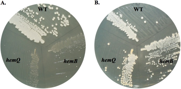
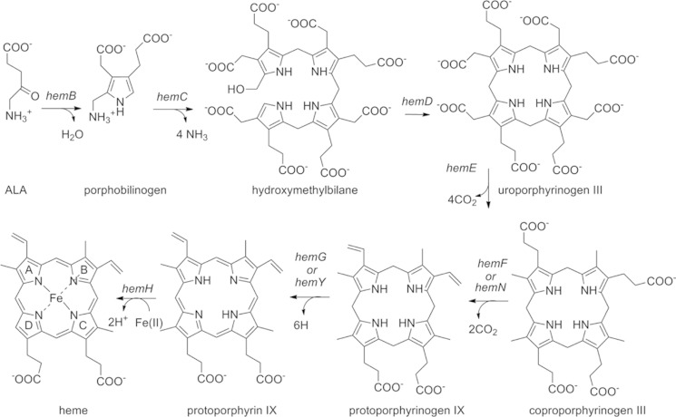
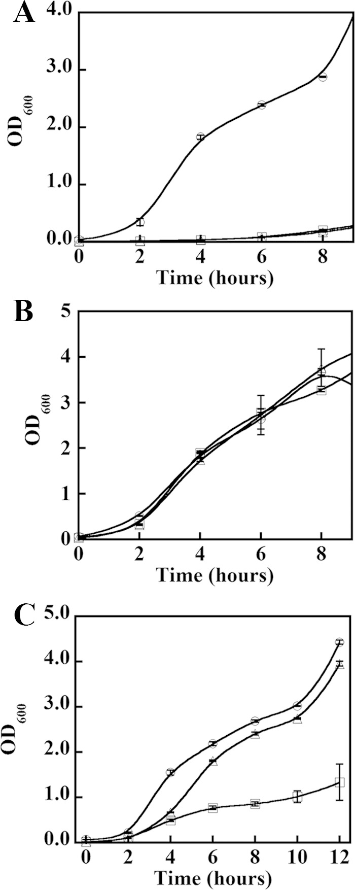
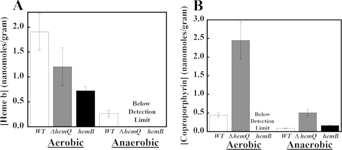
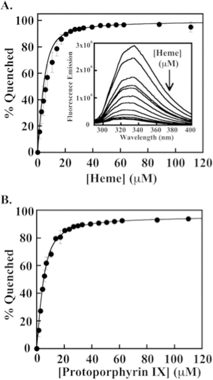
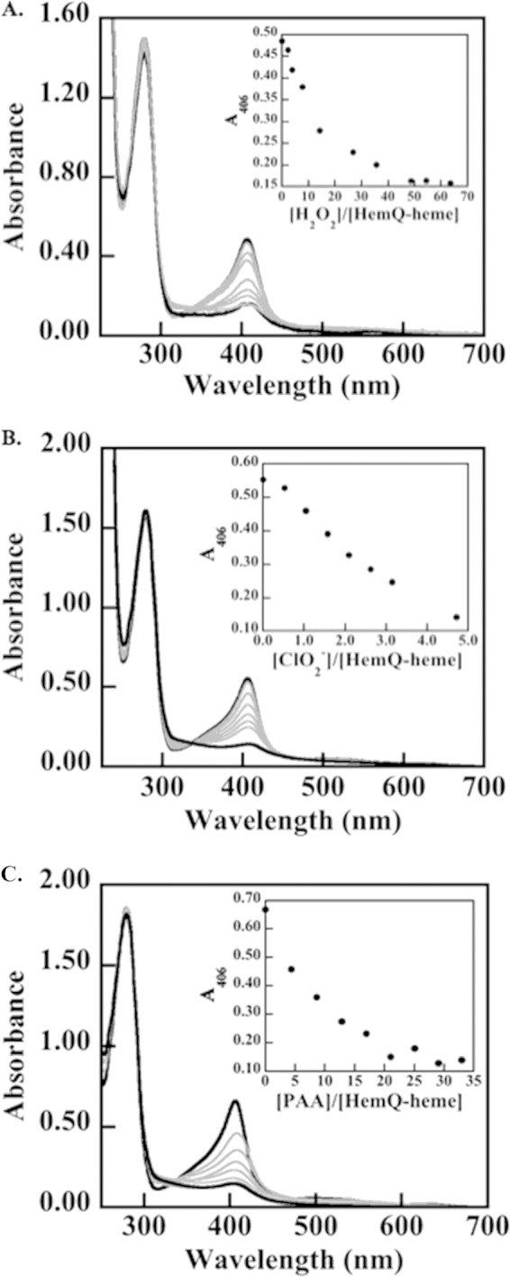
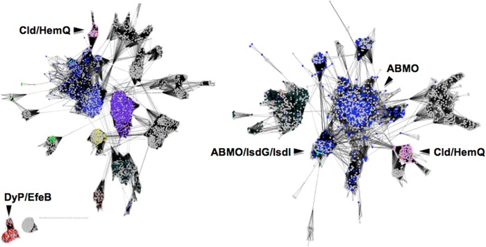
The chlorite dismutases (C-family proteins) are a widespread family of heme-binding proteins for which chemical and biological roles remain unclear. An association of the gene with heme biosynthesis in Gram-positive bacteria was previously demonstrated by experiments involving introduction of genes from two Gram-positive species into heme biosynthesis mutant strains of Escherichia coli, leading to the gene being renamed hemQ. To assess the gene product's biological role more directly, a Staphylococcus aureus strain with an inactivated hemQ gene was generated and shown to be a slow growing small colony variant under aerobic but not anaerobic conditions. The small colony variant phenotype is rescued by the addition of exogenous heme despite an otherwise wild type heme biosynthetic pathway. The ΔhemQ mutant accumulates coproporphyrin specifically under aerobic conditions. Although its sequence is highly similar to functional chlorite dismutases, the HemQ protein has no steady state reactivity with chlorite, very modest reactivity with H2O2 or peracetic acid, and no observable transient intermediates. HemQ's equilibrium affinity for heme is in the low micromolar range. Holo-HemQ reconstituted with heme exhibits heme lysis after <50 turnovers with peroxide and <10 turnovers with chlorite. The heme-free apoprotein aggregates or unfolds over time. IsdG-like proteins and antibiotic biosynthesis monooxygenases are close sequence and structural relatives of HemQ that use heme or porphyrin-like organic molecules as substrates. The genetic and biochemical data suggest a similar substrate role for heme or porphyrin, with possible sensor-regulator functions for the protein. HemQ heme could serve as the means by which S. aureus reversibly adopts an SCV phenotype in response to redox stress.

Keywords: Bacteria; Chlorite Dismutase; Heme; Peroxidase; Redox Regulation; Staphylococcus aureus; Superoxide Ion.


Figures

FIGURE 1.
FIGURE 1.
Heme-binding site of the heme-free Cld from G. stearothermophilus (carbon tan) a close sequence relative of HemQ, overlaid with the heme-bound Cld from D. aromatica (carbon blue). His-171 (G. stearothermophilus numbering) is a strictly conserved residue among all Cld/HemQ proteins that aligns with the heme-ligating His-170 from DaCld. Trp-156 is likewise strictly conserved among the entire family and is important for protein stability. Gln-184 aligns with DaCld-Arg-184. A glutamine at this position is strictly conserved among Clds/HemQ proteins from phylum Firmicutes. Figure was generated using PyMOL from Protein Data Bank codes 1T0T and 3Q08.
FIGURE 2.
FIGURE 2.
ΔhemQ mutant is a small colony variant that is rescued by addition of heme. WT S. aureus Newman, the hemB, mutant, and the ΔhemQ mutant were grown on TSA overnight at 37 °C in the absence (A) or the presence (B) of 5 μm hemin.
SCHEME 1.
SCHEME 1.
Heme biosynthesis pathway in bacteria.
FIGURE 3.
FIGURE 3.
Heme rescues slow growth of ΔhemQ under aerobic conditions, but iron does not. WT, hemB, and ΔhemQ strains of S. aureus Newman were grown in the absence of supplements (A), the presence of exogenous heme (B), and the presence of an equivalent concentration of exogenous Fe (C). Wild type (○), hemB (▵), and ΔhemQ (□) cultures were grown in TSB and monitored for 12 h in 50 ml of TSB in 150-ml Erlenmeyer flasks. Each point is an average of duplicate or triplicate measurements. Cultures in B were supplemented with 5 μm hemin (from 1 mm stock in 0.1 m NaOH). Cultures in C were supplemented with 5 μm Fe(III) citrate and 10 μm sodium citrate.
FIGURE 4.
FIGURE 4.
ΔhemQ cultures accumulate coproporphyrin under aerobic conditions. The bar graphs show measured amounts of heme (A) and coproporphyrin (B) in nanomoles of metabolite/g of wet cell pellet for wild type (white), ΔhemQ (dark gray), and hemB (black) strains. Error bars are the result of standard deviations taken from analysis of three separate cell cultures. Analyzed cell cultures (50 ml) were grown on a shaker incubator in TSB at 37 °C, 180 rpm, for ∼16 h before harvesting. Aerobic samples were grown in 150-ml Erlenmeyer flasks and anaerobic samples in filled, air-equilibrated 50-ml Falcon tubes that were subsequently sealed. Harvested cells were lysed and porphyrins extracted and quantitatively analyzed by exact mass LCMS as described in the text.
FIGURE 5.
FIGURE 5.
Equilibrium binding of apo-HemQ and heme/protoporphyrin IX measured by fluorescence quenching. A, binding isotherm for the quenching of apo-HemQ (∼5 μm) in 0.1 m potassium phosphate buffer, pH 6.8, by heme. The percent of quenching observed is plotted as a function of heme added and fit to a quadratic binding equation taking into account the concentration of protein (see “Experimental Procedures”). A Kd = 1.68 ± 0.16 μm was determined for heme. The inset shows quenching of apo-HemQ fluorescence over λ = 300–400 nm by addition of heme (excitation λ = 284 nm; 5 μm in 0.1 m potassium phosphate, pH 6.8, 25 °C). Heme was added from 1.4 to 111 μm, and spectra were collected after several minutes of incubation to allow equilibrium to be reached. B, binding isotherm for PPIX. The binding of PPIX was monitored in the same fashion as heme using a stock solution of PPIX (1 mm) dissolved in DMSO and diluted into 0.1 m potassium phosphate, pH 6.8. A Kd = 2.21 ± 0.1 μm was determined from the fit to the data.
FIGURE 6.
FIGURE 6.
Heme in HemQ degrades in the presence of relatively small numbers of equivalents of H2O2, chlorite, or PAA. The most dramatic effects are observed for the low pKa/anionic oxidants PAA and chlorite. A 30 μm solution of HemQ (heme-containing monomer) in 20 mm potassium phosphate buffer (pH 6.8, 25 °C) was titrated with aliquots of oxidant. UV-visible spectra were measured until the solution spectra stopped changing, and the final spectra recorded after equilibrium was reached. Spectra were adjusted for volume changes. A, titration with H2O2. B, titration with chlorite. C, titration with PAA. The protein absorbance at 280 nm is shown along with the heme Soret band at 406 nm. Notably, addition of oxidant leads to heme destruction without evidence for aggregation or precipitation of protein. Free heme has an absorbance maximum near 380 nm. Spectra at the beginning and end of the titrations are shown as dark black lines, and spectra measured following the addition of oxidant are shown in gray.
FIGURE 7.
FIGURE 7.
Network analysis of Cld/HemQ sequences and their homologs shows the highest levels of similarities to IsdG/IsdI and cofactor-independent monooxygenases. Important functional clusters of proteins are as labeled. Individual protein sequences are represented by dots colored according to their Uniprot-designated function as follows: chlorite dismutases (pink); antibiotic biosynthesis monooxygenases (dark blue); IsdG/IsdI proteins (cyan); DyP and EfeB proteins (red); uncharacterized proteins (gray). Left, network was generated at low stringency, with an E-value cutoff at 1e−5. DyP and EfeB proteins, the closest structural relatives of the Clds/HemQ proteins, remain unconnected to them on a sequence level even at the lowest stringency sampled. Right, network was generated at a higher stringency (E-value cutoff at 1e−3). The most closely related sequences to the Clds/HemQ proteins (pink cluster, lower right) are indicated by the many lines connecting this cluster to the antibiotic biosynthesis monooxygenase group. IsdG/IsdI protein sequences are interspersed with this group.
FIGURE 8.
FIGURE 8.
Cld homologs of S. aureus HemQ share strong sequence and structural similarities with IsdG and antibiotics associated monooxygenase proteins. A, DaCld like the Cld from G. stearothermophilus (GsCld, Protein Data Bank code 1T0T) forms a homopentamer. GsCld crystallizes as the apoprotein. B, structural overlay of GsCld (magenta) and DaCld (blue, Protein Data Bank code 3Q08) monomers shows the location of the DaCld heme (rendered as sticks) and illustrates the overall similarity of the two proteins. C, structural overlay of GsCld (magenta) and the S. aureus IsdG protein homodimer (cyan, Protein Data Bank code 2ZD0 (18)) illustrates the similarity of the two protein structures. IsdG-bound hemes (one per monomer) are rendered as sticks. D, overlay of the heme-binding domain of DaCld (blue) and the IsdG monomer (cyan) shows the location of the heme in each. E, overlay of the DaCld monomer (blue) and the biochemically characterized ABMO protein SnoaB (green, Protein Data Bank code 3KNG (19)). Figure was generated using Chimera.

References

    1. Goblirsch B., Kurker R. C., Streit B. R., Wilmot C. M., DuBois J. L. (2011) Chlorite dismutases, DyPs, and EfeB: 3 microbial heme enzyme families comprise the CDE structural superfamily. J. Mol. Biol. 408, 379–398 - PMC - PubMed
    1. Coates J. D., Achenbach L. A. (2004) Microbial perchlorate reduction: Rocket-fuelled metabolism. Nat. Rev. Microbiol. 2, 569–580 - PubMed
    1. Blanc B., Mayfield J. A., McDonald C. A., Lukat-Rodgers G. S., Rodgers K. R., DuBois J. L. (2012) Understanding how the distal environment directs reactivity in chlorite dismutase: Spectroscopy and reactivity of Arg183 mutants. Biochemistry 51, 1895–1910 - PMC - PubMed
    1. Goblirsch B. R., Streit B. R., DuBois J. L., Wilmot C. M. (2010) Structural features promoting dioxygen production by Dechloromonas aromatica chlorite dismutase. J. Biol. Inorg. Chem. 15, 879–888 - PMC - PubMed
    1. Lee A. Q., Streit B. R., Zdilla M. J., Abu-Omar M. M., DuBois J. L. (2008) Mechanism of and exquisite selectivity for O–O bond formation by the heme-dependent chlorite dismutase. Proc. Natl. Acad. Sci. U.S.A. 105, 15654–15659 - PMC - PubMed

Publication types

LinkOut - more resources