. 2013 Jul 12;288(28):20658-67.
doi: 10.1074/jbc.M113.450478. Epub 2013 Jun 4.

Transforming growth factor-β induces transcription factors MafK and Bach1 to suppress expression of the heme oxygenase-1 gene

Affiliations

Transforming growth factor-β induces transcription factors MafK and Bach1 to suppress expression of the heme oxygenase-1 gene

Yukari Okita et al. J Biol Chem. .

Abstract

Transforming growth factor-β (TGF-β) has multiple functions in embryogenesis, adult homeostasis, tissue repair, and development of cancer. Here, we report that TGF-β suppresses the transcriptional activation of the heme oxygenase-1 (HO-1) gene, which is implicated in protection against oxidative injury and lung carcinogenesis. HO-1 is a target of the oxidative stress-responsive transcription factor Nrf2. TGF-β did not affect the stabilization or nuclear accumulation of Nrf2 after stimulation with electrophiles. Instead, TGF-β induced expression of transcription factors MafK and Bach1. Enhanced expression of either MafK or Bach1 was enough to suppress the electrophile-inducible expression of HO-1 even in the presence of accumulated Nrf2 in the nucleus. Knockdown of MafK and Bach1 by siRNA abolished TGF-β-dependent suppression of HO-1. Furthermore, chromatin immunoprecipitation assays revealed that Nrf2 substitutes for Bach1 at the antioxidant response elements (E1 and E2), which are responsible for the induction of HO-1 in response to oxidative stress. On the other hand, pretreatment with TGF-β suppressed binding of Nrf2 to both E1 and E2 but marginally increased the binding of MafK to E2 together with Smads. As TGF-β is activated after tissue injury and in the process of cancer development, these findings suggest a novel mechanism by which damaged tissue becomes vulnerable to oxidative stress and xenobiotics.

Keywords: Cancer; Cancer Prevention; Heme Oxygenase; Nrf2; Oxidative Stress; Transcriptional Regulation; Transforming Growth Factor β (TGFβ).


Figures

FIGURE 1.
FIGURE 1.
TGF-β suppresses tBHQ-inducible expression of the HO-1. A, TGF-β suppresses tBHQ-inducible mRNA expression of HO-1. NMuMG cells were treated with TGF-β (5 ng/ml) for 1 h before stimulation with tBHQ (25 μm) and incubated for the indicated times. HO-1 and β-actin mRNAs were detected by semiquantitative RT-PCR (left panel). Representative mRNA expression was quantified using NIH ImageJ and normalized to β-actin (right panel). B, NMuMG cells were treated as in A. Immunoblot analysis was performed using anti-HO-1 antibody. β-Actin was examined as a loading control (left panel). Quantification of the protein levels was performed using NIH ImageJ and normalized to β-actin (right panel). All experiments were repeated more than three times to confirm their reproducibility.
FIGURE 2.
FIGURE 2.
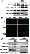
TGF-β does not affect the stabilization and nuclear accumulation of Nrf2. A, NMuMG cells were treated with TGF-β (5 ng/ml) for 1 h before stimulation with tBHQ (25 μm) and incubated for an additional 4 h. Immunoblot analysis was performed using anti-Nrf2, -phospho-Smad2 (P-Smad2), -Smad2 and -α-tubulin antibodies as indicated. Arrow, specific band for Nrf2; *, nonspecific bands. B, NMuMG cells were treated as in A. After fixation, cells were serially stained with anti-Nrf2 (green) and anti-Smad2 (red) antibodies. Nuclei were counterstained with DAPI. Scale bar, 50 μm. C, NMuMG cells were treated as in A. Nuclear and cytosolic fractions were isolated and analyzed by immunoblotting using antibodies for Nrf2 and Smad2. Lamin A/C was used as a nuclear protein marker, and α-tubulin was used as a cytosolic protein marker.
FIGURE 3.
FIGURE 3.
TGF-β induces MafK and Bach1. A, NMuMG cells were treated with TGF-β (5 ng/ml) for 1 h. mRNA for the small Maf family of transcription factors (MafF, MafG, and MafK), Bach1, Smad7, and β-actin was detected by semiquantitative RT-PCR. B, NMuMG cells were treated with TGF-β (5 ng/ml) for 4 h. Immunoblot analysis was performed using anti-MafK, -Bach1, -phospho-Smad2 (P-Smad2), -Smad2, and -β-actin antibodies as indicated. Bach1 was detected after immunoprecipitation (IP) with anti-Bach1 antibody to increase the sensitivity. C, NMuMG cells were treated with a TGF-β type I receptor kinase inhibitor, SD-208 (0, 0.03, 0.1, and 0.3 μm) for 30 min before treatment with TGF-β (5 ng/ml) for 1 h. MafK, Bach1, Smad7, and β-actin mRNAs were detected by semiquantitative RT-RCR.
FIGURE 4.
FIGURE 4.
MafK and MafG, but not MafF, suppress transcriptional activity of HO-1. A, pHO1-luc reporter activities were activated by overexpression of Nrf2, and the effects of MafF, MafG, and MafK were examined. Error bars represent S.D. B, interaction between Nrf2 and small Mafs was examined by coprecipitation assays in 293T cells. MafF, MafG, and MafK all coprecipitated Nrf2. C, binding of small Mafs to AREs from HO-1. HA-tagged MafF, MafG, and MafK were expressed in 293T cells, and the cell lysates were incubated with biotinylated double-stranded DNA fragments (Table 3) and precipitated with avidin beads. Coprecipitated proteins were detected with anti-HA antibody. D, chromatin immunoprecipitation analysis using anti-FLAG antibody detected binding of MafK and MafG to the HO-1 promoter region including ARE (E2) in NMuMG-MafK (K) and NMuMG-MafG (G) cells, respectively. DNAP, DNA affinity precipitation; IP, immunoprecipitation.
FIGURE 5.
FIGURE 5.
MafK and Bach1 regulate expression of HO-1. A and B, MafK suppresses HO-1 induction by tBHQ or DEM. A, establishment of NMuMG cells stably expressing FLAG-MafK (clones 4 and 10). Control represents NMuMG cells transfected with empty vector. B, impaired induction of HO-1 in NMuMG-MafK cells. NMuMG-MafK cells were treated with tBHQ (25 μm) or DEM (100 μm) for 4 h. HO-1 and β-actin mRNAs were detected by semiquantitative RT-PCR. C, pHO-1-luc activities are inactivated in NMuMG-MafK cells. Six hours after transfection with pHO-1-luc, cells were treated with tBHQ (25 μm) or DEM (100 μm) for 12 h. Error bars represent mean ± S.D. D, knockdown of MafK enhances induction of HO-1. NMuMG cells were transfected with MafK siRNA 1 or 2 as described under “Experimental Procedures.” Cells were then treated with TGF-β (5 ng/ml) for 1 h and stimulated with tBHQ (25 μm) for 4 h. HO-1, MafK, and β-actin mRNAs were detected by semiquantitative RT-PCR. E and F, Bach1 suppresses induction of HO-1 by tBHQ and DEM. E, establishment of NMuMG cells stably expressing FLAG-Bach1 (clones 2 and 7). F, impaired induction of HO-1 in NMuMG-Bach1 cells. NMuMG-Bach1 cells were treated with tBHQ (25 μm) or DEM (100 μm) for 4 h. HO-1 and β-actin mRNAs were examined by semiquantitative RT-PCR. G, pHO-1-luc activities are suppressed in NMuMG-Bach1 cells. Six hours after transfection with pHO-1-luc, cells were treated with tBHQ (25 μm) or DEM (100 μm) for 12 h. Error bars represent mean ± S.D. H, knockdown of Bach1 enhances induction of HO-1 and impairs the suppressive effects of TGF-β. NMuMG cells were transfected with Bach1 siRNA 1 or 2 as described under “Experimental Procedures.” Cells were treated with TGF-β (5 ng/ml) for 1 h before treatment with tBHQ (25 μm) for 4 h. HO-1, Bach1, and β-actin mRNAs were detected by semiquantitative RT-PCR. I, double knockdown of MafK and Bach1 enhances induction of HO-1 and almost completely impairs the suppressive effects of TGF-β. NMuMG cells were transfected with MafK siRNA 1 and Bach1 siRNA 2 as described under “Experimental Procedures.” Cells were then treated with TGF-β (5 ng/ml) for 1 h before treatment with tBHQ (25 μm) for 4 h. HO-1, MafK, Bach1, and β-actin mRNAs were detected by semiquantitative RT-PCR. Representative mRNA expression was quantified using NIH ImageJ and normalized to β-actin (right panel). J, knockdown of MafK or Bach1 enhances expression of HO-1 in breast cancer cells. JygMC(A) cells were transfected with MafK siRNA or Bach1 siRNA as indicated. HO-1, MafK, Bach1, and β-actin mRNAs were detected by semiquantitative RT-RCR.
FIGURE 6.
FIGURE 6.
Effects of TGF-β on the recruitment of Nrf2, MafK, and Bach1 to AREs (E1 and E2) in the HO-1 promoter. NMuMG cells were treated with TGF-β (5 ng/ml) for 1 h before stimulation with tBHQ (25 μm) for 4 h. After fixation, soluble chromatin was immunoprecipitated using anti-Nrf2 (A), anti-MafK (B), or anti-Bach1 (C) antibody as indicated. HO-1 promoter fragments containing AREs (E1 and E2) were amplified by PCR. Input, total chromatin solution analyzed as a control.
FIGURE 7.
FIGURE 7.
Cooperative functions of TGF-β/Smad signaling with MafK and Bach1. A and B, binding of Smad3 to MafK (A) and Bach1 (B). FLAG-MafK, FLAG-Bach1, Myc-Smad3, and a constitutively active TGF-β type I receptor (ALK5TD-V5) were transfected to 293T cells as indicated, and binding of MafK or Bach1 and Smad3 was examined by immunoprecipitation (IP) with anti-Myc antibody followed by immunoblotting with anti-FLAG antibody. Arrow, Myc-Smad3; *, IgG. C and D, chromatin immunoprecipitation analyses using anti-Smad2/3 antibody detected binding of Smad2/3 to the HO-1 promoter region including ARE (E2) in NMuMG cells (C). Binding of Smad2/3 to the Smad7 promoter region was used as a positive control (D). Error bars represent S.D.

Similar articles

Cited by

References

    1. Derynck R., Miyazono K. (eds) (2008) The TGF-β Family, Cold Spring Harbor Laboratory Press, Cold Spring Harbor, NY
    1. Massagué J. (2008) TGFβ in cancer. Cell 134, 215–230 - PMC - PubMed
    1. Massagué J., Seoane J., Wotton D. (2005) Smad transcription factors. Genes Dev. 19, 2783–2810 - PubMed
    1. Siegel P. M., Massagué J. (2003) Cytostatic and apoptotic actions of TGF-β in homeostasis and cancer. Nat. Rev. Cancer 3, 807–821 - PubMed
    1. Akhurst R. J., Derynck R. (2001) TGF-β signaling in cancer—a double-edged sword. Trends Cell Biol. 11, S44–S51 - PubMed

Publication types

MeSH terms