Review
. 2013 Apr 23:2013:973150.
doi: 10.1155/2013/973150. Print 2013.

Molecular imaging of experimental abdominal aortic aneurysms

Affiliations
Review

Molecular imaging of experimental abdominal aortic aneurysms

Aneesh K Ramaswamy et al. ScientificWorldJournal. .

Abstract

Current laboratory research in the field of abdominal aortic aneurysm (AAA) disease often utilizes small animal experimental models induced by genetic manipulation or chemical application. This has led to the use and development of multiple high-resolution molecular imaging modalities capable of tracking disease progression, quantifying the role of inflammation, and evaluating the effects of potential therapeutics. In vivo imaging reduces the number of research animals used, provides molecular and cellular information, and allows for longitudinal studies, a necessity when tracking vessel expansion in a single animal. This review outlines developments of both established and emerging molecular imaging techniques used to study AAA disease. Beyond the typical modalities used for anatomical imaging, which include ultrasound (US) and computed tomography (CT), previous molecular imaging efforts have used magnetic resonance (MR), near-infrared fluorescence (NIRF), bioluminescence, single-photon emission computed tomography (SPECT), and positron emission tomography (PET). Mouse and rat AAA models will hopefully provide insight into potential disease mechanisms, and the development of advanced molecular imaging techniques, if clinically useful, may have translational potential. These efforts could help improve the management of aneurysms and better evaluate the therapeutic potential of new treatments for human AAA disease.


Figures

Figure 1
Figure 1
Example of high-frequency anatomical ultrasound images of abdominal aortas obtained noninvasively. ((a)–(d)) Images of a suprarenal angiotensin II-induced abdominal aortic aneurysm (AAA). (a) Transverse ultrasound images of suprarenal and corresponding infrarenal aorta. (b) Longitudinal view of a suprarenal AAA. (c) Dissected abdominal aorta for anatomical comparison and (d) histological cross-section of the suprarenal aorta from the same animal in the dilated region. ((e)–(g)) Images of elastase-induced AAAs. (e) Longitudinal images of the vessel before surgery, after sham surgery, and after intraluminal elastase perfusion. (f) Corresponding transverse images and (g) histological cross-sections stained with H & E. Figure adapted from [45] for ((a)–(d)) and [46] for ((e)–(g)).
Figure 2
Figure 2
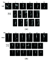
Coronal magnetic resonance maximum intensity projections showing lumen expansion in (a) angiotensin II-induced (AngII) and (b) elastase-induced abdominal aortic aneurysms (AAA). Angiotensin II-induced AAAs appear suddenly (arrowhead) and expand leftward directly above the right renal artery (arrow). Ten additional angiotensin II AAAs are shown at day 28. Elastase-induced AAAs expand slowly. Small region of signal hypointensity is seen at day 3 (triangle) due to a suture in the vessel. Twelve additional elastase AAAs are shown at day 28. The testicular artery is highlighted (arrow). Figure adapted from [77].
Figure 3
Figure 3
Transverse T2*-weighted magnetic resonance images, the spin-spin relaxation time measured in gradient echo sequences, of a single murine abdominal aortic aneurysm prior to iron oxide nanoparticle-labeled vascular smooth muscle cell delivery (a) and on postdelivery days 0 (b), 21 (c), and 28 (d). Arrows represent areas of hypointense signal in the aortic wall. The vessel lumen is highlighted with (★). Scale bar represents 1 mm. Figure adapted from [96].
Figure 4
Figure 4
Near-infrared fluorescence (NIRF) image of both angiotensin II-induced and elastase-induced aneurysms. (a) Control apolipoprotein-E deficient mouse aorta after injection of MMPSense 680. (b) Infrarenal aortic aneurysm induced via elastase infusion. (c) Suprarenal aortic aneurysm induced via angiotensin II infusion. (d) Dorsal and (e) ventral NIRF images of angiotensin II-induced abdominal aortic aneurysms showing asymmetric probe accumulation, suggestive of regional differences in protease activation and inflammation. Figure adapted from [77].
Figure 5
Figure 5
((a), (b)) In situ and ((c), (d)) ex vivo bioluminescent images of an infrarenal abdominal aortic aneurysm ((a), (c)) and control vessel ((b), (d)), with arbitrary units. Aneurysms were induced via elastase perfusion. Figure adapted from [97].
Figure 6
Figure 6
PET/CT imaging in mice with aortic aneurysms induced via angiotensin II infusion. Apolipoprotein E-deficient (apoE−/−) mice were compared to wild-type controls (apoE+/+). Dotted lines and yellow arrows outline the aneurysmal aorta. Liver signal highlighted with (∗). PET/CT images illustrate the ability of CT to add anatomical context for PET images. Figure adapted from [137].

References

    1. Belsley SJ, Tilson MD. Two decades of research on etiology and genetic factors in the abdominal aortic aneurysm (AAA)—with a glimpse into the 21st century. Acta Chirurgica Belgica. 2003;103(2):187–196. - PubMed
    1. Johnston WF, Salmon M, Su G, et al. Genetic and pharmacologic disruption of interleukin-1β signaling inhibits experimental aortic aneurysm formation. Arteriosclerosis, Thrombosis, and Vascular Biology. 2013;33(2):294–304. - PMC - PubMed
    1. Lederle FA, Johnson GR, Wilson SE, et al. Prevalence and associations of abdominal aortic aneurysm detected through screening. Annals of Internal Medicine. 1997;126(6):441–449. - PubMed
    1. von Allmen RS, Powell JT. The management of ruptured abdominal aortic aneurysms: screening for abdominal aortic aneurysm and incidence of rupture. The Journal of Cardiovascular Surgery. 2012;53(1):69–76. - PubMed
    1. Nanda S, Sharma SG, Longo S. Molecular targets and abdominal aortic aneurysms. Recent Patents on Cardiovascular Drug Discovery. 2009;4(2):150–159. - PubMed

Publication types

LinkOut - more resources