. 2013 May;9(5):e1003546.
doi: 10.1371/journal.pgen.1003546. Epub 2013 May 30.

CDK4 T172 phosphorylation is central in a CDK7-dependent bidirectional CDK4/CDK2 interplay mediated by p21 phosphorylation at the restriction point

Affiliations

CDK4 T172 phosphorylation is central in a CDK7-dependent bidirectional CDK4/CDK2 interplay mediated by p21 phosphorylation at the restriction point

Xavier Bisteau et al. PLoS Genet. 2013 May.

Abstract

Cell cycle progression, including genome duplication, is orchestrated by cyclin-dependent kinases (CDKs). CDK activation depends on phosphorylation of their T-loop by a CDK-activating kinase (CAK). In animals, the only known CAK for CDK2 and CDK1 is cyclin H-CDK7, which is constitutively active. Therefore, the critical activation step is dephosphorylation of inhibitory sites by Cdc25 phosphatases rather than unrestricted T-loop phosphorylation. Homologous CDK4 and CDK6 bound to cyclins D are master integrators of mitogenic/oncogenic signaling cascades by initiating the inactivation of the central oncosuppressor pRb and cell cycle commitment at the restriction point. Unlike the situation in CDK1 and CDK2 cyclin complexes, and in contrast to the weak but constitutive T177 phosphorylation of CDK6, we have identified the T-loop phosphorylation at T172 as the highly regulated step determining CDK4 activity. Whether both CDK4 and CDK6 phosphorylations are catalyzed by CDK7 remains unclear. To answer this question, we took a chemical-genetics approach by using analogue-sensitive CDK7(as/as) mutant HCT116 cells, in which CDK7 can be specifically inhibited by bulky adenine analogs. Intriguingly, CDK7 inhibition prevented activating phosphorylations of CDK4/6, but for CDK4 this was at least partly dependent on its binding to p21 (cip1) . In response to CDK7 inhibition, p21-binding to CDK4 increased concomitantly with disappearance of the most abundant phosphorylation of p21, which we localized at S130 and found to be catalyzed by both CDK4 and CDK2. The S130A mutation of p21 prevented the activating CDK4 phosphorylation, and inhibition of CDK4/6 and CDK2 impaired phosphorylations of both p21 and p21-bound CDK4. Therefore, specific CDK7 inhibition revealed the following: a crucial but partly indirect CDK7 involvement in phosphorylation/activation of CDK4 and CDK6; existence of CDK4-activating kinase(s) other than CDK7; and novel CDK7-dependent positive feedbacks mediated by p21 phosphorylation by CDK4 and CDK2 to sustain CDK4 activation, pRb inactivation, and restriction point passage.


Conflict of interest statement

The authors have declared that no competing interests exist.

Figures

Figure 1
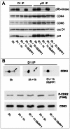
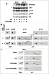
Figure 1. Specific inhibition of CDK7 by 1-NMPP1.
Specific inhibition of CDK7 by 1-NMPP1 prevents DNA synthesis (A), pRb phosphorylation (B), CDK4/6 pRb-kinase activity (C) and CDK4 activating phosphorylation (D). HCT116 WT or K7AS (K7as/as) cells were stimulated (+) or not stimulated (−) with FBS for the indicated times in the absence (−) or presence (+) of 1-NMPP1. (A) DNA synthesis was evaluated from duplicate dishes by counting the percentage of cells having incorporated BrdU during the last 30 min of stimulation. (B) Western blotting analysis with the indicated antibodies from whole-cell lysates. (C) Cell lysates were immunoprecipitated (IP) with anti-cyclin D1 (D1), anti-cyclin D3 (D3) or anti-p21 (p21) antibodies and were assayed for their pRb-kinase activity, separated by SDS-PAGE and immunoblotted with the indicated antibodies (C). (D) The same immunoprecipitates were separated by 2D gel electrophoresis followed by immunodetection using antibodies directed against CDK4 or T172-phosphorylated CDK4. Arrows, T172-phosphorylated form of CDK4.
Figure 2
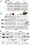
Figure 2. Effect of acute inhibition of CDK7 by 1-NMPP1.
Effect of acute inhibition of CDK7 by 1-NMPP1 on CDK4 activity (A) and phosphorylation (B). (A,B) HCT116 K7AS cells were stimulated with FBS for 5 h and 1-NMPP1 was then added for 1 or 3 h. Cell lysates were immunoprecipitated (IP) with anti-cyclin D1 (D1), or anti-p21 antibodies, assayed for their pRb-kinase activity, separated by SDS-PAGE and immunoblotted with the indicated antibodies (A), or separated by 2D gel electrophoresis followed by CDK4 immunodetection (B). In (B), T160 phosphorylation of CDK2 immunodetected from whole cell extracts is shown for comparison.
Figure 3
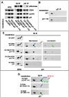
Figure 3. Specific inhibition of CDK7 by 1-NMPP1 prevents the main phosphorylation of p21, which is performed in vitro by both CDK4 and CDK2.
(A) HCT116 K7AS cells were stimulated (+) or not stimulated (−) with fetal bovine serum (FBS) for the indicated times in the absence (−) or presence (+) of 1-NMPP1. Cell lysates were immunoprecipitated (IP) with anti-p21, anti-cyclin D1 (D1) or anti-cyclin D3 (D3) antibodies and separated by 2D gel electrophoresis followed by p21 immunodetection. (B) CHO cells were transfected with a p21-expressing plasmid alone (left) or with plasmids encoding cyclin D3, CDK4-HA and p21 (right). Cell lysates were immunoprecipitated (IP) with anti-p21 (p21) or anti-cyclin D3 (D3) antibodies and incubated with the indicated recombinant kinases and ATP, before separation by 2D gel electrophoresis and p21 immunodetection. Arrows indicate the main phosphorylated forms of p21 as identified in Figure S4. (C) Schematic representation and comparison of p21 patterns from HCT116 K7AS cells that were restimulated for 3 and 20 h (in vivo) and from cyclin D3-CDK4-p21 complexes produced in CHO cells and phosphorylated by recombinant CDK2-cyclin E and CDK2-cyclin A (in vitro). The observed p21 forms are labeled according to their number of phosphorylations and the identified phosphorylated residues (e.g., 2P 98,130 means p21 phosphorylated at both S98 and S130; X, Y, unidentified phosphorylated sites).
Figure 4
Figure 4. Reciprocal impacts of p21 and cyclin D3-CDK4 on T172 phosphorylation of CDK4 and S130 phosphorylation of p21 in transfected CHO cells.
(A) p21 stabilizes cyclin D3-CDK4 complexes and inhibits their activity depending on its expression level. CHO cells were transfected with plasmids encoding cyclin D3 and CDK4-HA with or without p21 at high (p21) or low (p21 1/10) expression level. Cell lysates (WCE) were immunoprecipitated (IP) with anti-cyclin D3 (D3) or anti-p21 antibodies, assayed for their pRb-kinase activity, separated by SDS-PAGE and immunoblotted with the indicated antibodies. (B) p21 expressed alone is essentially unphosphorylated. CHO cells were transfected with a plasmid encoding p21 at high (p21) or low (p21 1/10) expression level. Cell lysates were immunoprecipitated (IP) with anti-p21 antibody and separated by 2D gel electrophoresis followed by p21 immunodetection. (C) Co-expression of cyclin D3-CDK4 induces the phosphorylation of p21, largely independently of CDK4 activity, whereas stronger expression of p21 inhibits phosphorylations of both p21 and CDK4. CHO cells were transfected with plasmids encoding cyclin D3 and CDK4-HA or its inactive mutants (T172A, K35R) with or without p21 at high (p21) or low (p21 1/10) expression level. Cell lysates were immunoprecipitated (IP) with anti-cyclin D3 antibody and separated by 2D gel electrophoresis followed by CDK4 and p21 immunodetection. (D) Identification of p21 phosphorylation sites. CHO cells were transfected with plasmids encoding cyclin D3, CDK4-HA and wild type (WT) or mutated (S130A, S98A) p21 at low (p21 1/10) expression level. Cell lysates were immunoprecipitated (IP) with anti-cyclin D3 antibody and separated by 2D gel electrophoresis followed by p21 immunodetection. Black arrows, T172-phosphorylated CDK4. Colored arrows indicate phosphorylated forms of p21.
Figure 5
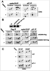
Figure 5. S130 phosphorylation of p21 is instrumental in CDK4 activation.
(A,B) Stably infected HCT116 K7AS cells for Tet-On inducible 3×HA-p21 wt or S130A mutant were treated with doxycycline (1 µg/ml) for 16 h prior cell restimulation with fetal bovine serum (FBS) for 16 h in the continuous presence of doxycycline. (A) Immunodetection of 3×HA-p21 and endogenous p21 from whole cell extracts (WCE) using a p21 antibody. (B) Cell lysates were immunoprecipitated (IP) with anti-HA antibody and separated by 2D gel electrophoresis followed by simultaneous immunodetection of ectopic 3×HA-p21 and 3×HA-p21-bound endogenous CDK4 using a mixture of anti-CDK4 and p21 antibodies. Black arrows, T172-phosphorylated CDK4. Colored arrows indicate phosphorylated forms of p21.
Figure 6
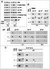
Figure 6. Inhibition of phosphorylations of p21 and CDK4 by CDK inhibitors.
HCT116 K7AS cells were stimulated (+) or not stimulated (−) with fetal bovine serum (FBS) for the indicated times in the absence (−) or presence (+) of roscovitine (rosco) (A,B) or CR8 (C). (A) Western blotting analysis with the indicated antibodies from whole-cell lysates. (B,C) Cell lysates were immunoprecipitated (IP) with anti-cyclin D1 (D1), anti-cyclin D3 (D3) or anti-p21 antibodies and separated by 2D gel electrophoresis followed by immunodetection of CDK4 and p21. Black arrows, T172-phosphorylated CDK4; green arrows, S130-phosphorylated p21.
Figure 7
Figure 7. Activities of both CDK4/6 and CDK2 are required for S130 phosphorylation of p21 and activation of p21-bound CDK4.
(A,B) HCT116 K7AS cells were stimulated (+) or not stimulated (−) with fetal bovine serum (FBS) for 5 h in the absence (−) or presence (+) of the CDK4/6 inhibitor PD0332991. Cell lysates were immunoprecipitated (IP) with anti-cyclin D1 (D1) or anti-p21 antibodies, assayed for their pRb-kinase activity, separated by SDS-PAGE and immunoblotted with the indicated antibodies (A), or separated by 2D gel electrophoresis followed by CDK4 and p21 immunodetection (B). (C) CHO cells were transfected with plasmids encoding cyclin D3, CDK4-HA and p21 at low (p21 1/10) expression level (see Figure 4), in the absence (DMSO as vehicle) or presence of CDK inhibitors: roscovitine (rosco), PD0332991 (1 µM) or their combination to inhibit both CDK2 and CDK4. Cell lysates were immunoprecipitated (IP) with anti-cyclin D3 (D3) antibody and separated by 2D gel electrophoresis followed by CDK4 and p21 immunodetection. Black arrows, T172-phosphorylated CDK4; colored arrows, phosphorylated forms of p21.
Figure 8
Figure 8. CDK7 inhibition does not prevent activating phosphorylations of p21-unbound CDK4 and S178P CDK6.
(A) HCT116 K7AS cells were transfected with plasmids encoding cyclin D3 and CDK4-HA in the absence or continuous presence of 1-NMPP1 for 48 h. Cell lysates were immunoprecipitated (IP) with anti-cyclin D3 or anti-p21 antibodies and separated by 2D gel electrophoresis followed by immunoblotting with anti-CDK4 antibody, which detected both ectopic and endogenous CDK4. (B) HCT116 K7AS cells were infected during 24 h by lentiviruses encoding Tet-On inducible wt or S178P CDK6-Flag with or without lentiviruses encoding Tet-On inducible cyclin D3. The expression of these proteins in the continuous presence (+) or absence (−) of 1-NMPP1 was induced by doxycycline 16 h prior to cell restimulation by fetal bovine serum (FBS) for 16 h. Western blotting detection of endogenous and ectopic (arrow) CDK6 from whole cell extracts is shown in the upper panel (NI, not infected). Cell lysates were immunoprecipitated (IP) with anti-Flag, anti-cyclin D3 or anti-p21 antibodies and separated by 2D gel electrophoresis followed by immunoblotting with anti-CDK6 antibody, which detected both ectopic and endogenous (e) CDK6. (C) T172 phosphorylation of endogenous CDK4 resists inhibition of CDK2 and CDK7 by roscovitine in p21-null (p21−/−) cells. Roscovitine (rosco; +) or vehicle (−) were administered for 16 h to asynchronously growing wild-type (WT) and p21−/− HCT116 cells cultured in parallel. Cell lysates were immunoprecipitated (IP) with anti-cyclin D1 (D1) or anti-cyclin D3 (D3) antibodies and separated by 2D gel electrophoresis followed by CDK4 immunodetection. Arrows, phosphorylated forms of CDK4 and CDK6-Flag; arrowheads in (B), unphosphorylated form of CDK6-Flag.
Figure 9
Figure 9. Model assembling the present observations with previous knowledge , , , , , .
CDK7 inhibition reveals novel positive feedbacks mediated by S130 phosphorylation of p21, which is catalyzed by active CDK4 and CDK2 to permit T172 phosphorylation and activation of CDK4 complexes stabilized by p21 binding. S130 phosphorylation of p21 thus amplifies CDK4 activation and subsequently leads to increased degradation of p21, which in turn facilitates CDK2 activation. Those retrocontrols of CDK4 activation should contribute to explaining the irreversibility of the cell cycle commitment at the R point. Green/red colors indicate a final positive/negative influence on R point passage. 1, 2, two different levels of CDK7 action demonstrated by the present observations. CDK4 AK?, hypothetical CDK4-activating kinase(s) phosphorylating p21-unbound CDK4 and S178P CDK6 during CDK7 inhibition.

References

    1. Sherr CJ (1995) D-type cyclins. Trends Biochem Sci 20: 187–190. - PubMed
    1. Bartek J, Bartkova J, Lukas J (1996) The retinoblastoma protein pathway and the restriction point. Curr Opin Cell Biol 8: 805–814. - PubMed
    1. Bockstaele L, Coulonval K, Kooken H, Paternot S, Roger PP (2006) Regulation of CDK4. Cell Division 1: 25. - PMC - PubMed
    1. Lundberg AS, Weinberg RA (1998) Functional inactivation of the retinoblastoma protein requires sequential modification by at least two distinct cyclin-cdk complexes. Mol Cell Biol 18: 753–761. - PMC - PubMed
    1. Yao G, Lee TJ, Mori S, Nevins JR, You L (2008) A bistable Rb-E2F switch underlies the restriction point. Nat Cell Biol 10: 476–482. - PubMed

Publication types

MeSH terms