. 2013:2013:575287.
doi: 10.1155/2013/575287. Epub 2013 May 8.

Antiteratogenic Effects of β-Carotene in Cultured Mouse Embryos Exposed to Nicotine

Affiliations

Antiteratogenic Effects of β-Carotene in Cultured Mouse Embryos Exposed to Nicotine

Chunmei Lin et al. Evid Based Complement Alternat Med. 2013.

Abstract

After maternal intake, nicotine crosses the placental barrier and causes severe embryonic disorders and fetal death. In this study, we investigated whether β -carotene has a beneficial effect against nicotine-induced teratogenesis in mouse embryos (embryonic day 8.5) cultured for 48 h in a whole embryo culture system. Embryos exposed to nicotine (1 mM) exhibited severe morphological anomalies and apoptotic cell death, as well as increased levels of TNF- α , IL-1 β , and caspase 3 mRNAs, and lipid peroxidation. The levels of cytoplasmic superoxide dismutase (SOD), mitochondrial manganese-dependent SOD, cytosolic glutathione peroxidase (GPx), phospholipid hydroperoxide GPx, hypoxia inducible factor 1 α , and Bcl-x L mRNAs decreased, and SOD activity was reduced compared to the control group. However, when β -carotene (1 × 10(-7) or 5 × 10(-7) μM) was present in cultures of embryos exposed to nicotine, these parameters improved significantly. These findings indicate that β -carotene effectively protects against nicotine-induced teratogenesis in mouse embryos through its antioxidative, antiapoptotic, and anti-inflammatory activities.


Figures

Figure 1
Figure 1
Representative images of mouse embryos exposed to nicotine and β-carotene. Normal control group (a). Embryos treated with 1 mM nicotine alone show typical abnormalities such as exposed brain, reduced forebrain, abnormal heart, deformed posterior trunk, and regressed forelimbs (b1−3). Embryos treated with nicotine plus β-carotene [1 × 10⁻⁷μM (c1 and c2) and 5 × 10⁻⁷μM (d1 and d2)] appear morphologically similar to the control group.
Figure 2
Figure 2
Protective effects of β-carotene against oxidative damage induced by nicotine in E8.5 mouse embryos treated in vitro for 2 days. Lipid peroxidation was evaluated by measuring the malondialdehyde (MDA) concentration in embryos treated with 1 mM nicotine in the absence or presence of 1 × 10⁻⁷ or 5 × 10⁻⁷μM β-carotene (β-car). Results are presented as mean ± SEM (n = 12). Significant differences (*control versus nicotine alone; #nicotine versus β-car + nicotine) were evaluated by one-way ANOVA at P < 0.05.
Figure 3
Figure 3
Superoxide dismutase (SOD) activity levels in E8.5 mouse embryos exposed to nicotine and β-carotene for 2 days in vitro. SOD activity in embryos treated with 1 mM nicotine in the absence or presence of 1 × 10⁻⁷ or 5 × 10⁻⁷μM β-carotene (β-car) was measured. Results are presented as mean ± SEM (n = 6). Significant differences (*control versus nicotine alone; #nicotine versus β-car + nicotine) were evaluated by one-way ANOVA at P < 0.05.
Figure 4
Figure 4
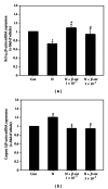
Gene expression levels of antioxidant enzymes in E8.5 mouse embryos exposed to nicotine and β-carotene for 2 days in vitro. Levels of mRNA for cytoplasmic superoxide dismutase (SOD1, (a)), manganese SOD (SOD2, (b)), cytoplasmic glutathione peroxidase (GPx1, (c)), and phospholipid hydroperoxide GPx (GPx4, (d)) in embryos exposed to 1 mM nicotine in the absence or presence of 1 × 10⁻⁷ or 5 × 10⁻⁷μM β-carotene (β-car) were measured by quantitative RT-PCR. Results are mean ± SEM (n = 8). β-actin was used as an internal standard to normalize target transcript expression. Significant differences (*control versus nicotine alone; #nicotine versus β-car + nicotine) were evaluated by one-way ANOVA at P < 0.05.
Figure 5
Figure 5
Hypoxia inducible factor-1 α expression levels in E8.5 mouse embryos exposed to nicotine and β-carotene for 2 days in vitro. HIF-1α mRNA in embryos exposed to 1 mM nicotine in the absence or presence of 1 × 10⁻⁷ or 5 × 10⁻⁷μM β-carotene (β-car) was measured by quantitative RT-PCR. Results are mean ± SEM (n = 8). β-actin was used as an internal standard to normalize target transcript expression. Significant differences (*control versus nicotine alone; #nicotine versus β-car + nicotine) were evaluated by one-way ANOVA at P < 0.05.
Figure 6
Figure 6
Gene expression levels of proinflammatory cytokines in E8.5 mouse embryos exposed to nicotine and β-carotene for 2 days in vitro. Levels of TNF-α (a) and IL-1β (b) mRNA in embryos exposed to 1 mM nicotine in the absence or presence of 1 × 10⁻⁷ or 5 × 10⁻⁷μM β-carotene (β-car) were measured by quantitative RT-PCR. Results are mean ± SEM (n = 8). β-actin was used as an internal standard to normalize target transcript expression. Significant differences (*control versus nicotine alone; #nicotine versus β-car + nicotine) were evaluated by one-way ANOVA at P < 0.05.
Figure 7
Figure 7
Gene expression levels of apoptosis related factors in E8.5 mouse embryos exposed to nicotine and β-carotene for 2 days in vitro. Levels of Bcl-x L (a) and caspase 3 (b) mRNA in embryos exposed to 1 mM nicotine in the absence or presence of 1 × 10⁻⁷ or 5 × 10⁻⁷μM β-carotene (β-car) were measured by quantitative RT-PCR. Results are mean ± SEM (n = 8). β-actin was used as an internal standard to normalize target transcript expression. Significant differences (*control versus nicotine alone; #nicotine versus β-car + nicotine) were evaluated by one-way ANOVA at P < 0.05.
Figure 8
Figure 8
Representative images of apoptotic embryos exposed to nicotine and β-carotene by Nile blue staining. Nile blue staining was performed to observe apoptotic nuclei and dead cells which stained dark blue. Normal control embryos (a). Embryos treated with 1 mM nicotine exhibit increased levels of apoptosis (b). Embryos treated with 1 mM nicotine plus β-carotene [1 × 10⁻⁷μM (c) and 5 × 10⁻⁷μM (d)] appear similar to the control group.

References

    1. Lambers DS, Clark KE. The maternal and fetal physiologic effects of nicotine. Seminars in Perinatology. 1996;20(2):115–126. - PubMed
    1. Comhair SAA, Erzurum SC. Antioxidant responses to oxidant-mediated lung diseases. American Journal of Physiology. 2002;283(2):L246–L255. - PubMed
    1. Ornoy A. Embryonic oxidative stress as a mechanism of teratogenesis with special emphasis on diabetic embryopathy. Reproductive Toxicology. 2007;24(1):31–41. - PubMed
    1. Cogswell ME, Weisberg P, Spong C. Cigarette smoking, alcohol use and adverse pregnancy outcomes: implications for micronutrient supplementation. Journal of Nutrition. 2003;133:1722S–1731S. - PubMed
    1. Lin C, Yon JM, Jung AY, et al. Resveratrol prevents nicotine-induced teratogenesis in cultured mouse embryos. Reproductive Toxicology. 2012;34(3):340–360. - PubMed

LinkOut - more resources