. 2013 May 30;8(5):e64480.
doi: 10.1371/journal.pone.0064480. Print 2013.

Effect of propofol in the immature rat brain on short- and long-term neurodevelopmental outcome

Affiliations

Effect of propofol in the immature rat brain on short- and long-term neurodevelopmental outcome

Tanja Karen et al. PLoS One. .

Abstract

Background: Propofol is commonly used as sedative in newborns and children. Recent experimental studies led to contradictory results, revealing neurodegenerative or neuroprotective properties of propofol on the developing brain. We investigated neurodevelopmental short- and long-term effects of neonatal propofol treatment.

Methods: 6-day-old Wistar rats (P6), randomised in two groups, received repeated intraperitoneal injections (0, 90, 180 min) of 30 mg/kg propofol or normal saline and sacrificed 6, 12 and 24 hrs following the first injection. Cortical and thalamic areas were analysed by Western blot and quantitative real-time PCR (qRT-PCR) for expression of apoptotic and neurotrophin-dependent signalling pathways. Long-term effects were assessed by Open-field and Novel-Object-Recognition at P30 and P120.

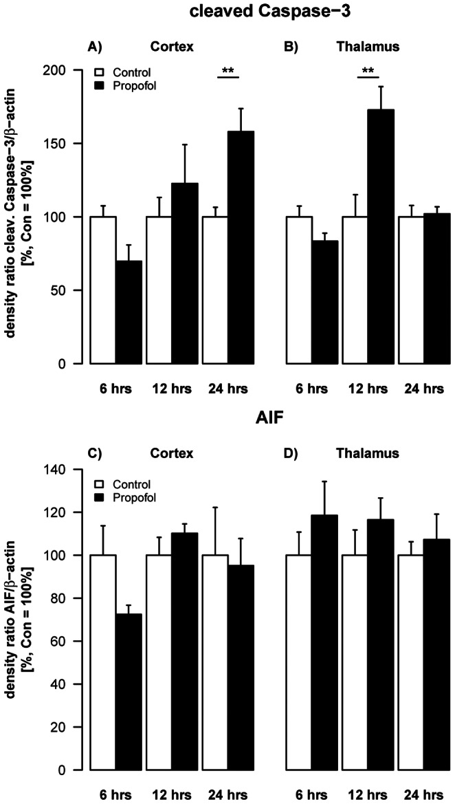
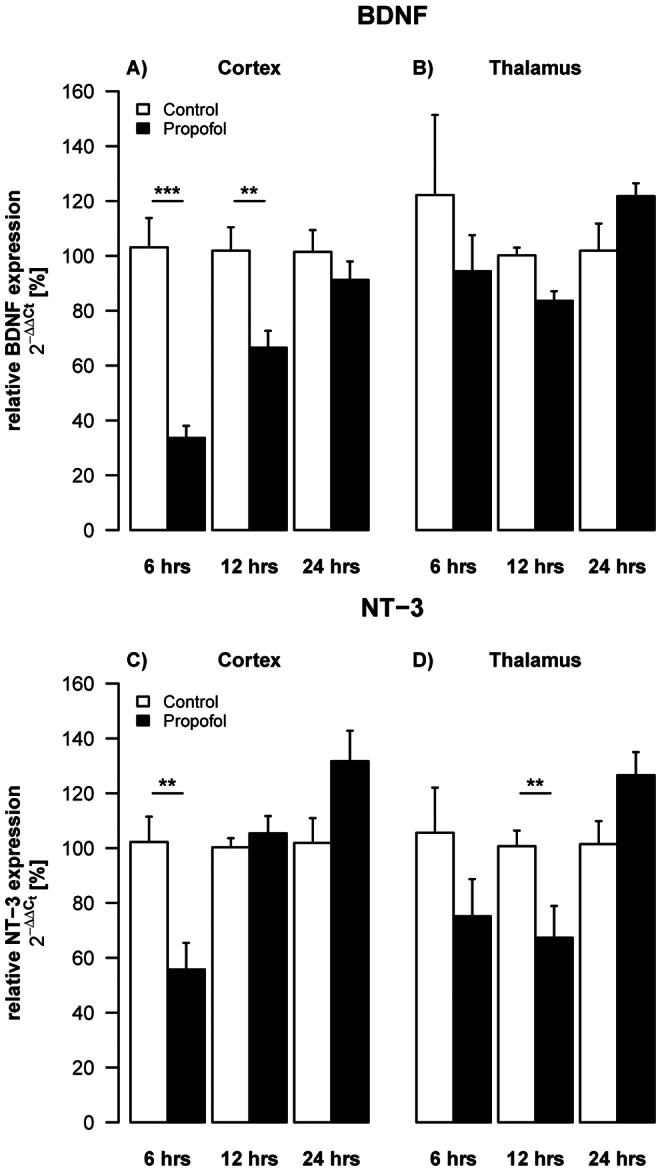
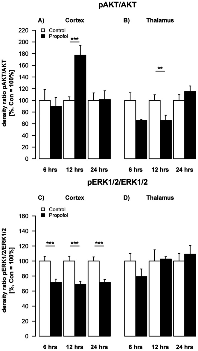
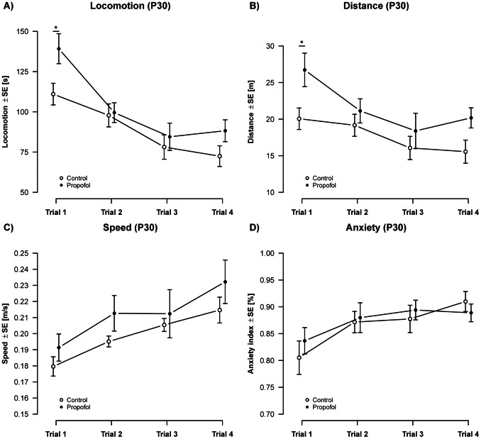
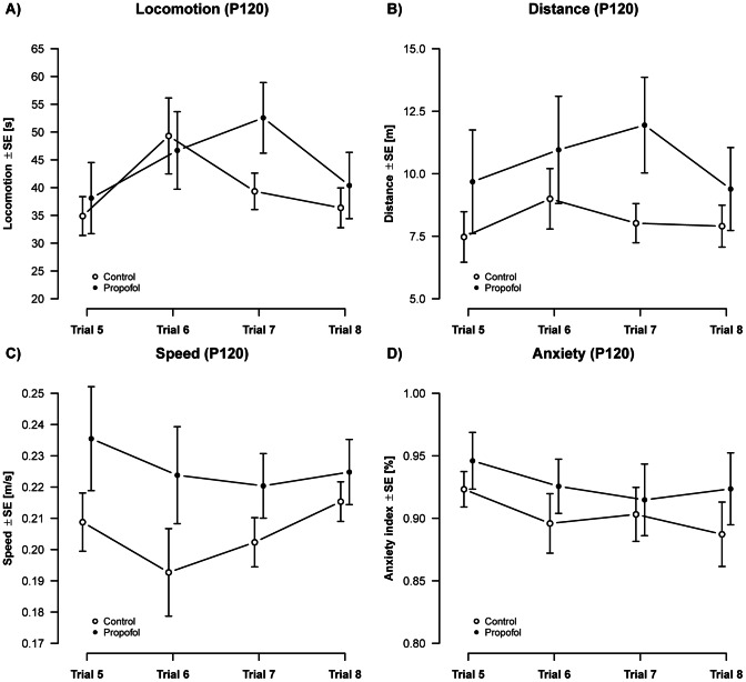
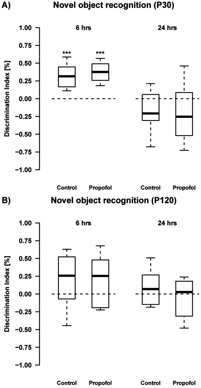
Results: Western blot analyses revealed a transient increase of activated caspase-3 in cortical, and a reduction of active mitogen-activated protein kinases (ERK1/2, AKT) in cortical and thalamic areas. qRT-PCR analyses showed a down-regulation of neurotrophic factors (BDNF, NGF, NT-3) in cortical and thalamic regions. Minor impairment in locomotive activity was observed in propofol treated adolescent animals at P30. Memory or anxiety were not impaired at any time point.

Conclusion: Exposing the neonatal rat brain to propofol induces acute neurotrophic imbalance and neuroapoptosis in a region- and time-specific manner and minor behavioural changes in adolescent animals.


Conflict of interest statement

Competing Interests: The authors have declared that no competing interests exist.

Figures

Figure 1
Figure 1. Impact of propofol on key proteins involved in apoptotic signalling.
Densitometric quantifications of caspase-3 and AIF in cortex and thalamus of P6 rats as analysed by Western blotting. Values represent mean normalised ratios of the densities of caspase-3 and AIF bands compared to densities of the control group (n = 5–6/point+SE). There was an effect of propofol treatment on caspase-3 levels over time, which was significant after 24 hrs in the cortex [F(1,29) = 3.63, p = 0.06] and after 12 hrs in the thalamus [F(1,28) = 3.1, p = 0.09).
Figure 2
Figure 2. Impact of propofol on neurotrophins.
Densitometric quantifications of mRNA levels of BDNF and NT-3 in cortex and thalamus of P6 rats, analysed by qRT-PCR. Values represent mean normalised ratios of the densities of BDNF and NT-3 bands compared to the density of the control group (n = 6–7/point+SE). There was an effect of propofol treatment with a decrease of BDNF levels over time, which was significant after 6 hrs in the cortex [F(1,30) = 66.5, p<0.001]. There was also a decrease in NT-3 levels, which was significant in the cortex after 6 hrs [F(1,28) = 12.7, p = 0.004] and after 12 hrs in the thalamus [F(1,24) = 3.5, p = 0.06].
Figure 3
Figure 3. Impact of propofol on survival promoting proteins.
Densitometric quantifications of pAKT and pERK1/2 in the cortex and thalamus of P6 rats, analysed by Western blotting. Values represent mean normalised ratios of the densities of pAKT and pERK1/2 bands compared to the density of the control group (n = 6/point+SE). There was an effect of propofol treatment in decrease of pAKT levels over time in the thalamus, which was significant after 12 hrs [F(1,28) = 5.6, p = 0.06]. Post-hoc analysis showed most pronounced decrease after 12 hrs (2-sample t-test). In the cortex there was a significant decrease of pERK1/2 levels over the time, which was significant after 6, 12 and 24 hrs [F(1,29) = 12.7, p = 0.013].
Figure 4
Figure 4. Activity (P30): Analysis of activity over 4 repeated measurements showed an overall increase in A) locomotion [F(1,71) = 7.12, p = 9×10⁻³] and B) distance [F(1,71) = 7.36, p = 8.37×10³], but no change on C) speed [F(1,74) = 1.92, p = 1.69] in propofol treated animals.
Propofol treatment did not alter D) anxiety related behavior in adolescent animals [F(1,74) = 0.02, p = 0.89]. An overall change in activity was observed over individual measurements, resulting in a significant decrease in locomotion [F(3,71) = 13.6, p = 4.08×10⁻⁷] and distance [F(3,71) = 5.35, p = 2.23×10⁻³] and a significant increase in speed [F(3,74) = 15.7, p = 5.53×10⁻⁸] and the index of anxiety [F(3,74) = 7.25, p = 3×10⁻⁴]. (ncontrols = 12 animals, npropofol = 8 animals).
Figure 5
Figure 5. Activity (P120): Analysis of activity over 4 repeated measurements showed no treatment effects on A) locomotion [F(1,73) = 0.94, p = 0.33], B) distance [F(1,74) = 1.86, p = 0.18], C) speed [F(1,74) = 0.44, p = 0.51] or D) anxiety related behavior [F(1,62) = 0.57, p = 0.45] in propofol treated animals.
Apart from a transient effect on locomotion [F(3,71) = 5.92, p = 1.13×10⁻³], no significant changes over repeated measurements were observed in adult aged animals. (ncontrols = 12 animals, npropofol = 8 animals).
Figure 6
Figure 6. Novel object recognition on P30 and P120: At the age of 30 days, both propofol treated animals (t(7) = 7.45, ***q = 4.3×10⁻⁴) and controls (t(10) = 6.30, ***q = 3.6×10⁻⁴) spent significantly more time with the novel object indicating their ability to discriminate the novel from the old object.
Propofol (t(7) = −1.44, q = 0.192) as well as control animals (t(10) = −1.92, q = 0.168) failed to do so after a 24 hrs inter-trial interval. At P120 both groups spent a random amount of time with either of the objects after 6 hrs and also after a 24 hrs interval, indicating that they were unable to remember the old object. (ncontrols = 12 animals, npropofol = 8 animals).

References

    1. Motsch J, Roggenbach J (2004) [Propofol infusion syndrome]. Anaesthesist 53: 1009–1022; quiz 1023–1004. - PubMed
    1. Shah PS, Shah VS (2011) Propofol for procedural sedation/anaesthesia in neonates. Cochrane Database Syst Rev: CD007248. - PubMed
    1. Henschel O, Gipson KE, Bordey A (2008) GABAA receptors, anesthetics and anticonvulsants in brain development. CNS Neurol Disord Drug Targets 7: 211–224. - PMC - PubMed
    1. Young Y, Menon DK, Tisavipat N, Matta BF, Jones JG (1997) Propofol neuroprotection in a rat model of ischaemia reperfusion injury. Eur J Anaesthesiol 14: 320–326. - PubMed
    1. Gelb AW, Bayona NA, Wilson JX, Cechetto DF (2002) Propofol anesthesia compared to awake reduces infarct sise in rats. Anesthesiology 96: 1183–1190. - PubMed

Publication types

MeSH terms