Review
. 2013:2013:285825.
doi: 10.1155/2013/285825. Epub 2013 Apr 23.

Plasma lipoproteins as mediators of the oxidative stress induced by UV light in human skin: a review of biochemical and biophysical studies on mechanisms of apolipoprotein alteration, lipid peroxidation, and associated skin cell responses

Affiliations
Review

Plasma lipoproteins as mediators of the oxidative stress induced by UV light in human skin: a review of biochemical and biophysical studies on mechanisms of apolipoprotein alteration, lipid peroxidation, and associated skin cell responses

Paulo Filipe et al. Oxid Med Cell Longev. 2013.

Abstract

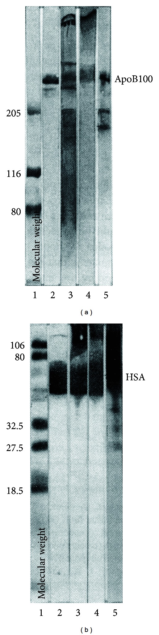
There are numerous studies concerning the effect of UVB light on skin cells but fewer on other skin components such as the interstitial fluid. This review highlights high-density lipoprotein (HDL) and low-density lipoprotein (LDL) as important targets of UVB in interstitial fluid. Tryptophan residues are the sole apolipoprotein residues absorbing solar UVB. The UVB-induced one-electron oxidation of Trp produces (•)Trp and (•)O2 (-) radicals which trigger lipid peroxidation. Immunoblots from buffered solutions or suction blister fluid reveal that propagation of photooxidative damage to other residues such as Tyr or disulfide bonds produces intra- and intermolecular bonds in apolipoproteins A-I, A-II, and B100. Partial repair of phenoxyl tyrosyl radicals (TyrO(•)) by α -tocopherol is observed with LDL and HDL on millisecond or second time scales, whereas limited repair of α -tocopherol by carotenoids occurs in only HDL. More effective repair of Tyr and α -tocopherol is observed with the flavonoid, quercetin, bound to serum albumin, but quercetin is less potent than new synthetic polyphenols in inhibiting LDL lipid peroxidation or restoring α -tocopherol. The systemic consequences of HDL and LDL oxidation and the activation and/or inhibition of signalling pathways by oxidized LDL and their ability to enhance transcription factor DNA binding activity are also reviewed.


Figures

Figure 1
Figure 1
TBARS production as a function of the initial rate of Trp photolysis. TBARS expressed in nmol/mg of protein and the Trp photolysis initial rate expressed in nM/s have been determined with air-saturated pH 7 buffered solutions of HDL at concentrations up to 1 μM. The incident UVB light dose in these experiments was 6.7 J/min. Drawn from data in [14].
Figure 2
Figure 2
Immunoblots of air-saturated solutions of HDL with antibodies specific for apoA-I or apoA-II. Lanes A: unirradiated samples bubbled with air; lanes B: as in A but irradiated with 6.7 J/minof UVB during the indicated times; lanes C: same as B but the solutions contained 50 μM desferrioxamine, a strong Fe(III) complexing agent. Adapted from [15].
Figure 3
Figure 3
Immunoblots of apoB100 and albumin from air-saturated suction blister fluid before and after irradiation with UVB (absorbed light dose: 12 J/mL). (a) Unmodified apoB100 migration is indicated as apoB100. Lane 1: molecular weight standards; lane 2: unirradiated suction blister fluid (130 μg); lane 3: irradiated suction blister fluid; lane 4: reconstituted blister fluid; lane 5: isolated LDL as reference (prepared from human serum and irradiated). (b) same as (a) but with 20 μg of proteins. See [25] for full experimental details.
Figure 4
Figure 4
Time courses of Car and αTocOH consumption. Lower time scale: carotenoid consumption during Cu2+-catalyzed oxidation of 240 nM of LDL in the absence () or presence (■) of 0.75 μM quercetin. Upper time scale: Car (□, ∆) or αTocOH (,  ) consumption under irradiation of 400 nM of LDL with UVB (□,  ) or UVA (∆,  ). Drawn from data in [14, 28].
Figure 5
Figure 5
(a) Absorbance of apolipoprotein and quercetin radicals in HDL3. () Transient absorbance spectra of 12.5 μM HDL3 in N2O saturated 10 mM pH 7 phosphate buffer containing 0.1 M KBr recorded 200 ms after oxidation with 3.2 μM of Br2 radical-anions. (□, ■) The same but solutions contained 18.75 μM HDL3, 5 μM HSA, and 5 μM QH. Spectra were recorded at 30 ms (□) and 700 ms (■) after oxidation with 2.9 μM of Br2 radical-anions. (b) Absorbance of apoB100 and quercetin radicals in LDL. () Transient absorbance spectra of 1.6 μM LDL in N2O saturated 10 mM, pH 7, recorded 16 ms after oxidation with 4.0 μM of Br2 radical-anions. (□, ■) The same but the solutions contained 2.4 μM LDL, 5 μM HSA, and 5 μM QH. Spectra were recorded at 30 ms (□) and 1.2 s (■) after oxidation with 3.2 μM of Br2 radical-anions. Redrawn from data in [32].
Figure 6
Figure 6
(a) Decay of transient absorbance of αTocO radicals at 430 nm (, ) and bleaching of the carotenoid absorbance at 470 nm (□,  ∆) after oxidation of 20 μM HDL by Br2 radical-anions in 10 mM pH 7 phosphate buffer. Solutions were saturated with N2O (□,  ) and O2 (,  ∆). (b) Transient absorbance changes measured at 430 nm and 470 nm for solutions containing 1.6 μM LDL. In (a) and (b), [Br2 ] = 3.0 μM for N2O-saturated solutions and [Br2 ] = 5.0 μM for O2-saturated solutions (see [31] for full experimental details).
Figure 7
Figure 7
Immunoblots using specific antibodies for ERK, phospho-ERK, Akt, phospho-Akt, and electrophoretic mobility shift assays showing the concentration-dependent effect of oxidized LDL (oxLDL) in presence (Ins) or absence (−Ins) of insulin on signalling kinases ERK and Akt (A) and on transcription factors AP1 and NFκB (see full experimental details in [33]).

References

    1. Epstein JH. Photocarcinogenesis, skin cancer, and aging. Journal of the American Academy of Dermatology. 1983;9(4):487–502. - PubMed
    1. Nishi J, Ogura R, Sugiyama M, Hidaka T, Kohno M. Involvement of active oxygen in lipid peroxide radical reaction of epidermal homogenate following ultraviolet light exposure. The Journal of Investigative Dermatology. 1991;97(1):115–119. - PubMed
    1. Tyrrell RM. UVA (320-380 nm) radiation as an oxidative stress. In: Sies H, editor. Oxidative Stress. Oxidants and Antioxidants. London, UK: Academic Press; 1991. pp. 57–83.
    1. Morlière P, Moysan A, Tirache I. Action spectrum for UV-induced lipid peroxidation in cultured human skin fibroblasts. Free Radical Biology and Medicine. 1995;19(3):365–371. - PubMed
    1. Matsumoto K, Sugiyama M, Ogura R. Non-dimer DNA damage in Chinese hamster V-79 cells exposed to ultraviolet-B light. Photochemistry and Photobiology. 1991;54(3):389–392. - PubMed

Substances

LinkOut - more resources