

Peroneus longus tear and its relation to the peroneal tubercle: A review of the literature
- PMID: 23738264
- PMCID: PMC3666483
Peroneus longus tear and its relation to the peroneal tubercle: A review of the literature
Abstract
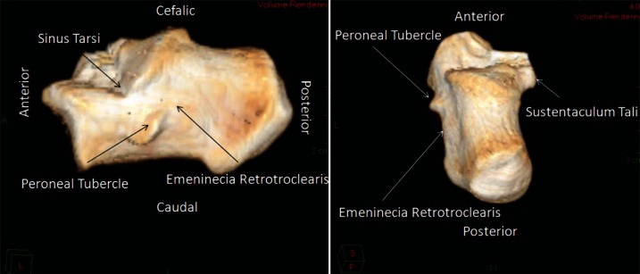
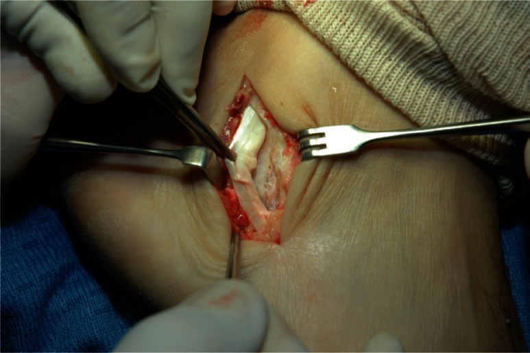
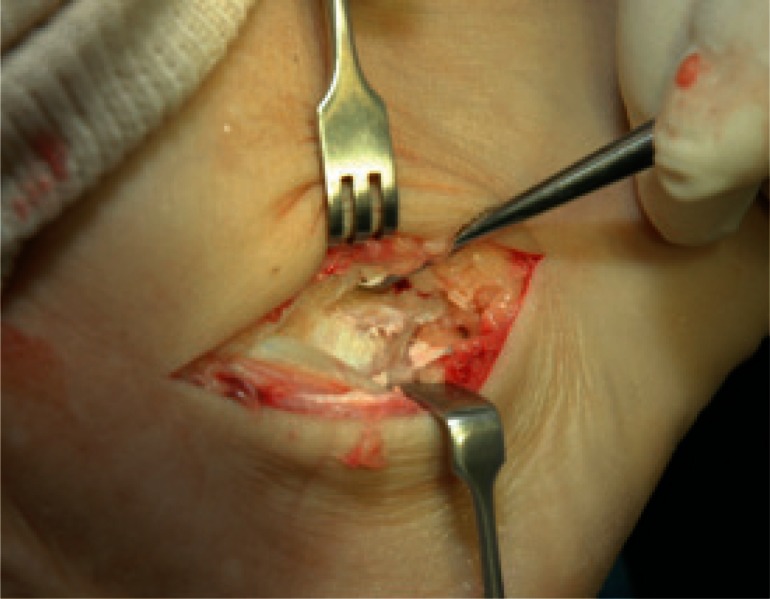
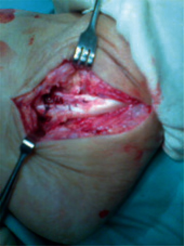
Tear of the peroneal tendon may occur in different anatomical sites. The most prevalent site is around the lateral malleolus. Tear of the peroneus longus at the level of the peroneal tubercle is unusual. Anatomically, the lateral surface of the calcaneous can be divided into thirds. The middle third includes the peroneal tubercle, which separates the peroneus longus tendon from the peroneus brevis. An anatomic variation of the peroneal tubercle may lead to chronic irritation of the peroneus longus tendon that could ultimately cause a longitudinal tear. We conducted this review aiming to clarify the anatomy, biomechanics of the tendon, and the clinical features of tear of the peroneus longus tendon on the lateral surface of the calcaneous due to an enlarged peroneal tubercle. In addition, we reviewed the diagnostic and treatment options of peroneal tendon tears at this site.
Keywords: calcaneous; peroneal tubecle; peroneus longus tear.
Figures
References
-
- Sarrafian SK. Osteology/Myology. Anatomy of the Foot and Ankle
-
- Agarwal AK, Jeyasingh P, Gupta SC, Gupta CD, Sahai A. Peroneal tubercle and its variations in the Indian calcanei. Anat Anz. 1984;156:241–244. - PubMed
-
- Pierson JL, Inglis AE. Stenosing tenosynovitis of the peroneous longus tendon associated with hypertrophy of peroneal tubercle and os perineum. A case report. J Bone Joint Surg Am. 1992;74:440–442. - PubMed
-
- Bruce WD, Christofersen MR, Phillips DL. Stenosing tenosynovitis and impingement of the peroneal tendons associated with hypertrophy of the peroneal tubercle. Foot Ankle Int. 1999;20:464–467. - PubMed
-
- Chen YJ, Hsu RW, Huang TJ. Hypertrophic peroneal tubercle with stenosing tenosynovitis: the results of surgical treatment. Changgeng Yi Xue Za Zhi. 1998;21:442–446. - PubMed
LinkOut - more resources
Full Text Sources