. 2012 Oct 16;2(3):169-80.
Print 2012 Jul.

Mesenchymal stem cell responses to mechanical stimuli

Affiliations

Mesenchymal stem cell responses to mechanical stimuli

Robin M Delaine-Smith et al. Muscles Ligaments Tendons J. .

Abstract

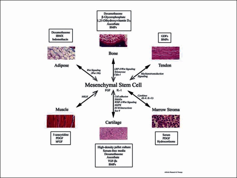
Mesenchymal stem cells (MSCs) have the potential to replace or restore the function of damaged tissues and offer much promise in the successful application of tissue engineering and regenerative medicine strategies. Optimising culture conditions for the pre-differentiation of MSCs is a key goal for the research community, and this has included a number of different approaches, one of which is the use of mechanical stimuli. Mesenchymal tissues are subjected to mechanical stimuli in vivo and terminally differentiated cells from the mesenchymal lineage respond to mechanical stimulation in vivo and in vitro. MSCs have also been shown to be highly mechanosensitive and this may present an ideal method for controlling MSC differentiation. Here we present an overview of the response of MSCs to various mechanical stimuli, focusing on their differentiation towards the mesenchymal tissue lineages including bone, cartilage, tendon/ligament, muscle and adipose tissue. More research is needed to elucidate the complex interactions between biochemically and mechanically stimulated differentiation pathways.

Keywords: mechanical stimuli; mesenchymal stem cell; osteogenesis; tenogenesis.


Figures

Figure 1
Figure 1
Diagram summarising the lineage potential of adult human MSC. The figure depicts the in vitro culture conditions (boxed) used to promote the differentiation into the lineage indicated and some of the signalling pathways and transcription factors involved in the process (italics). Reprinted from Arthritis Research and Therapy (4), BioMed Central, with kind permission of Professor Tuan.
Figure 2
Figure 2
Compression loading of human MSCs in polyurethane foam scaffolds. A: Fluorescent micrograph of a pore of the scaffold with MSCs attached (blue = cell nucleus stained with DAPI, red = cell cytoskeleton stained with TRITC-phaloidin). B: PCR analysis of mRNA for RUNX2, OPN and ALP showed that these genes were only slightly upregulated by the short (2 hour) loading period and not as much as by continuous dex treatment. However Col 1 was upregulated by loading and inhibited by dex which was also reflected in collagen analysis by Sirius red at a later time-point (data not shown). ALP activity was stimulated by loading to levels seen in dex treated cells as was calcium secretion which was highest with a combination of dex and loading. Adapted from (20) reproduced with kind permission from eCM journal (www.ecmjournal.org).
Figure 3
Figure 3
Osteogenic progenitor cells of the hES-MP line were subjected to oscillatory fluid flow induced shear stress (FFSS) using a simple rocking platform. ALP activity (A) was significantly increased at day 14 with FSS for cells cultured in osteogenic media containing dex (OM). Matrix deposition at day 21 (B) was highest in both FFSS groups for Sirius Red (collagen) and in FSS + OM group for Alizarin Red (calcium). Adapted from (62) reproduced with kind permission from eCM journal (www.ecmjournal.org).
Figure 4
Figure 4
MSCs of the cell line hES-MP (embryonic derived mesenchymal progenitors) stained for acetylated alpha tubulin on day 7 of culture. The structures with a high aspect ratio that stain brightly are primary cilia (yellow arrows), the dispersed, background green staining is the cell microtubules. Nuclei are stained blue with DAPI.

References

    1. Sioud M, Mobergslien A, Boudabous A, Floisand Y. Mesenchymal stem cell-mediated T cell suppression occurs through secreted galectins. Int J Oncol. 2011;38(2):385–390. - PubMed
    1. Zuk PA, Zhu M, Mizuno H, Huang J, Futrell JW, Katz AJ, et al. Multilineage cells from human adipose tissue: Implications for cell-based therapies. Tissue Eng. 2001;7(2):211–228. - PubMed
    1. Vater C, Kasten P, Stiehler M. Culture media for the differentiation of mesenchymal stromal cells. Acta Biomater. 2011;7(2):463–477. - PubMed
    1. Tuan RS, Boland G, Tuli R. Adult mesenchymal stem cells and cell-based tissue engineering. Arthrit Res Ther. 2003;5(1):32–45. - PMC - PubMed
    1. Nombela-Arrieta C, Ritz J, Silberstein LE. The elusive nature and function of mesenchymal stem cells. Nat Rev Mol Cell Biol. 2011;12:126–131. - PMC - PubMed

LinkOut - more resources