. 2013 Aug 16;8(8):1841-51.
doi: 10.1021/cb400289x. Epub 2013 Jun 17.

A high-throughput functional screen identifies small molecule regulators of temperature- and mechano-sensitive K2P channels

Affiliations

A high-throughput functional screen identifies small molecule regulators of temperature- and mechano-sensitive K2P channels

Sviatoslav N Bagriantsev et al. ACS Chem Biol. .

Abstract

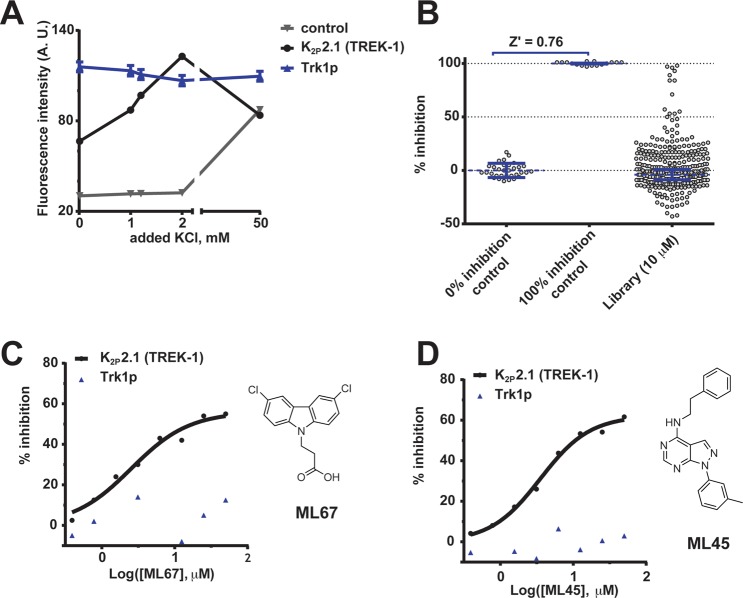
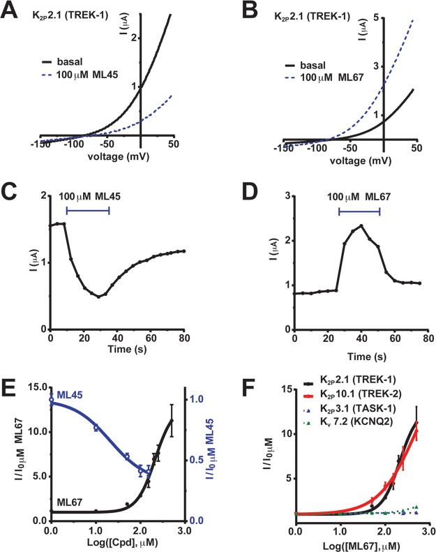
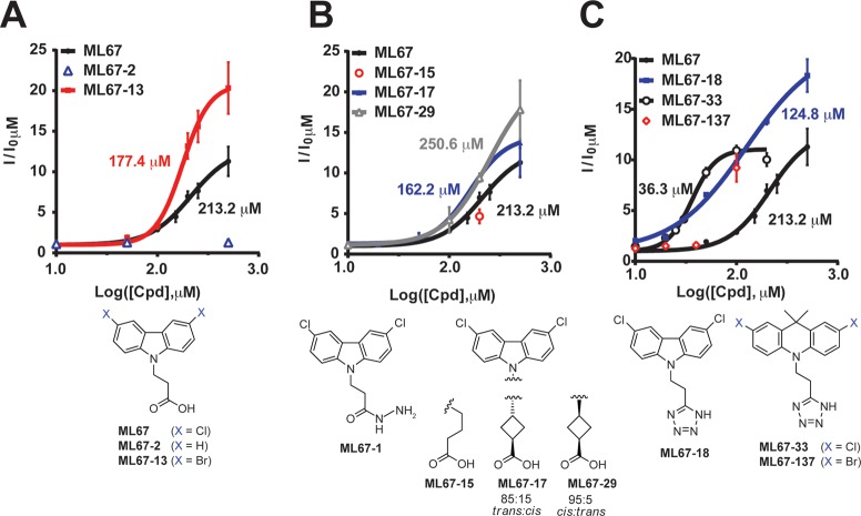
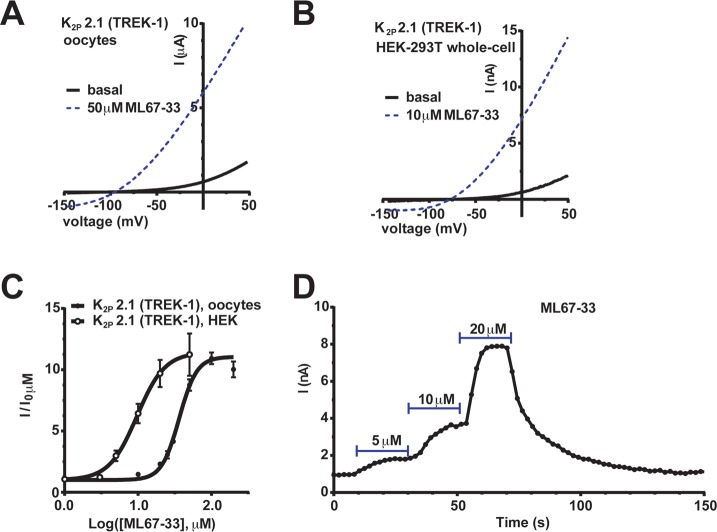
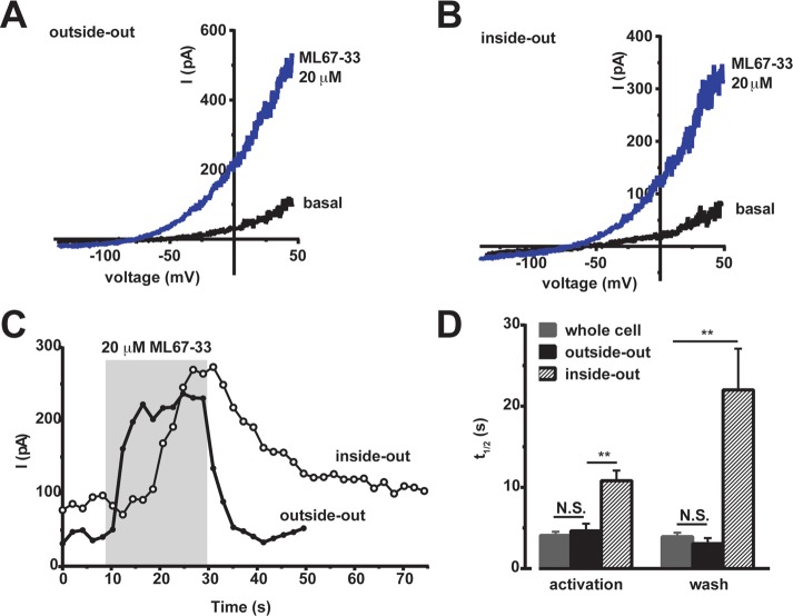
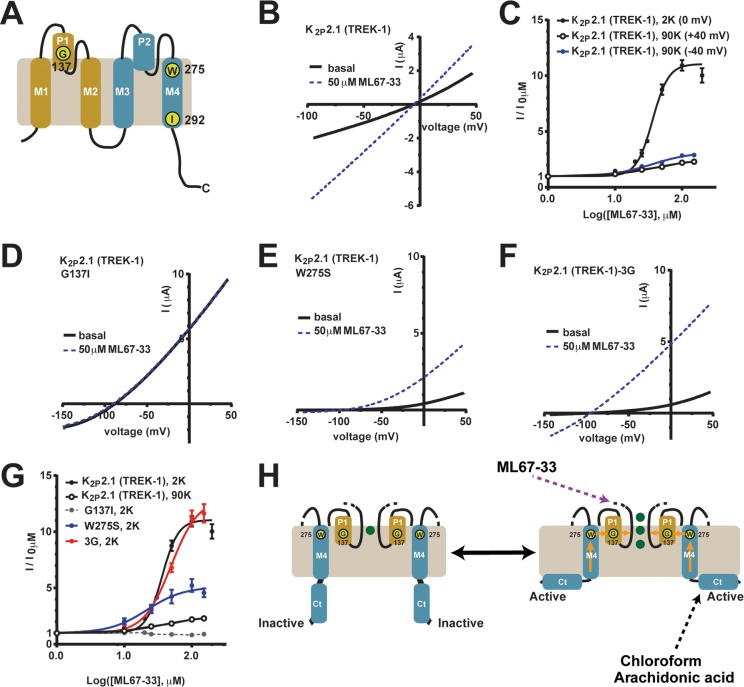
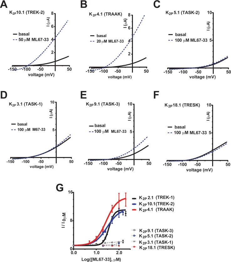
K2P (KCNK) potassium channels generate "leak" potassium currents that strongly influence cellular excitability and contribute to pain, somatosensation, anesthesia, and mood. Despite their physiological importance, K2Ps lack specific pharmacology. Addressing this issue has been complicated by the challenges that the leak nature of K2P currents poses for electrophysiology-based high-throughput screening strategies. Here, we present a yeast-based high-throughput screening assay that avoids this problem. Using a simple growth-based functional readout, we screened a library of 106,281 small molecules and identified two new inhibitors and three new activators of the mammalian K2P channel K2P2.1 (KCNK2, TREK-1). By combining biophysical, structure-activity, and mechanistic analysis, we developed a dihydroacridine analogue, ML67-33, that acts as a low micromolar, selective activator of temperature- and mechano-sensitive K2P channels. Biophysical studies show that ML67-33 reversibly increases channel currents by activating the extracellular selectivity filter-based C-type gate that forms the core gating apparatus on which a variety of diverse modulatory inputs converge. The new K2P modulators presented here, together with the yeast-based assay, should enable both mechanistic and physiological studies of K2P activity and facilitate the discovery and development of other K2P small molecule modulators.


Figures

Figure 1
Figure 1
Yeast screen identifies K2P2.1 (TREK-1) small molecule modulators. (A) Resazurin (Alamar blue) measurement of potassium concentration growth effects on SGY1528 yeast expressing the indicated constructs. Error bars show ± SE, n = 16. For some points, error bars are smaller than symbols. (B) Exemplar scatter plot showing growth inhibition score distribution from a 384-well screening plate. Each point represents end-point normalized resazurin fluorescence. Error bars show ± SD. (C, D) Dose–response for (C) ML67 and (D) ML45 on growth inhibition of yeast expressing K2P2.1 (TREK-1) (black circles) or Trk1p (blue triangles). Compound structures are shown.
Figure 2
Figure 2
ML45 and ML67 reversibly modulate K2P activity in Xenopus oocytes. (A, B) Exemplar two-electrode voltage clamp I–V curves for application of 100 μM (A) ML45 or (B) ML67 measured using a −150 to 50 mV ramp from a −80 mV holding potential in 2 mM [K+]o. (C, D) Exemplar K2P2.1 (TREK-1) responses to 100 μM (C) ML45 or (D) ML67 measured at 20 mV and 0 mV for ML45 and ML67, respectively. (E) ML45 and ML67 dose–response for K2P2.1 (TREK-1). “Cpd” denotes tested compound. Data were normalized to basal activity and fit with the Hill equation. (F) Dose–responses measured by two-electrode voltage clamp for ML67 against K2P2.1 (TREK-1), black; K2P10.1 (TREK-2), red; K2P3.1 (TASK-1), green; and Kv7.2 (KCNQ2), blue. Error bars show SE, n ≥ 6, N ≥ 2, where n and N is the number of oocytes or independent oocyte batches, respectively.
Figure 3
Figure 3
Studies to improve ML67 potency. Effects of changes to ML67 (A) halogen positions, (B) linker region, and (C) acidic group measured against K2P2.1 (TREK-1) by a −150 to 50 mV ramp from a −80 mV holding potential using two-electrode voltage clamp in Xenopus oocytes in 2 mM [K+]o. “Cpd” denotes tested compound. Data (mean ± SE, n ≥ 6, N ≥ 2) from 0 mV were normalized to basal activity and fitted to the Hill equation. EC50 values are ML67-13, 177.4 ± 1.1 μM; ML67-17, 162.2 ± 1.2 μM; ML67-29, 250.6 ± 2.0 μM μM; ML67-18, 124.8 ± 1.2 μM; ML67-33, 36.3 ± 1.0 μM. Error bars show SE, n ≥ 6 and N ≥ 2 except for ML67-2 and ML67-15 where n = 4 and N = 2. Compound structures are shown.
Figure 4
Figure 4
ML67-33 reversibly activates K2P2.1 (TREK-1) independent of expression system. (A, B) Exemplar I–V curves showing the effect of ML67-33 on K2P2.1 (TREK-1) activity in (A) Xenopus oocytes (two-electrode voltage clamp) or (B) HEK-293T cells (whole cell patch clamp). In both, the external solution contained 2 mM [K+]o, pH 7.4. Currents were elicited by a −150 to 50 mV voltage ramp from a −80 mV (oocytes) or −40 mV (HEK-293T) holding potential. (C) Quantification of the effect of ML67-33 on the indicated channels. Data (mean ± SE, n ≥ 6, N ≥ 2) were normalized to basal channel activity and fit with the Hill equation. EC50 36.3 ± 1.0 μM, 9.7 ± 1.2 μM and Emax at 100 μM 11.1 ± 0.4, 11.4 ± 1.1 for oocytes and HEK cells, respectively. (D) Exemplar reversible activation of K2P2.1 (TREK-1) by ML67-33 measured at 0 mV in HEK-293T cells.
Figure 5
Figure 5
ML67-33 activates K2P2.1 (TREK-1) in excised membrane patches. (A, B) Exemplar I–V curves showing ML67-33 effects on K2P2.1 (TREK-1) in (A) outside-out and (B) inside-out excised patches from HEK-293T cells. Currents were elicited by a −100 to 50 mV ramp from a −40 mV holding potential. (C) Exemplar responses to ML67-33 measured at 0 mV in the indicated configurations. Gray indicates presence of 20 μM ML67-33. (D) Time to half-maximal activation following ML67-33 application and recovery from activation (wash) following ML67-33 removal, measured in HEK-293T cells at 0 mV. Error bars: mean ± SE n ≥ 6, N ≥ 2. ** p ≤ 0.01; N.S. indicates not significant (p ≥ 0.05) as determined by t test.
Figure 6
Figure 6
ML67-33 activates the K2P2.1 (TREK-1) extracellular C-type gate. (A) K2P2.1 (TREK-1) subunit cartoon diagram. Key residue positions, transmembrane segments (M1–M4), and pore helices (P1 and P2) are indicated. First and second pore-forming regions are tan and blue, respectively. (B–G) Exemplar two-electrode voltage clamp I–V curves in Xenopus oocytes and dose response curves showing ML67-33 responses in channels having perturbed gating elements. (B) C-type gate stabilization by 90 mM [K+]o. (C) ML67-33 dose response at +40 and −40 mV in 90 mM [K+]o (90K) and 0 mV in 2 mM [K+]o (2K). (D, E) C-type gate stabilization by (D) G137I and (E) W275S. (F) Uncoupling Ct from the pore by the K2P2.1 (TREK-1)-3G mutant. (G) ML67-33 dose responses for the indicated channels at +40 mV (90K) or 0 mV (2K), normalized to basal channel activity and fit with the Hill equation. Error bars indicate SE, n ≥ 6, N ≥ 2. (H) Model of K2P2.1 (TREK-1) activation after ref (21). Green spheres indicate potassium ions. Positions of Gly137 and Trp275 are indicated. Orange arrows indicate pathway for coupling Ct activation to the C-type gate. Elements involved in activation by ML67-33, chloroform, and arachidonic acid are indicated. I–V curves were measured in (B) 90 mM [K+]o or (D–F) 2 mM [K+]o. Currents were elicited by a −100 to 50 mV ramp from a 0 mV holding potential (90K) or by a −150 to 50 mV ramp, from a −80 mV holding potential (2K).
Figure 7
Figure 7
ML67-33 is a selective activator of temperature- and mechanosensitive K2P channels. (A–F) Exemplar I–V curves showing ML67-33 effects on (A) K2P10.1 (TREK-2), (B) K2P4.1 (TRAAK), (C) K2P5.1 (TASK-2), (D) K2P3.1 (TASK-1), (E) K2P9.1 (TASK-3), and (F) K2P18.1 (TRESK) measured in Xenopus oocytes using a −150 to 50 mV ramp from a −80 mV holding potential in 2 mM [K+]o. (G) ML67-33 dose responses for the indicated channels. Data (mean ± SE, n ≥ 6, N ≥ 2) were normalized to basal activity and fit with the Hill equation. EC50 values: K2P2.1 (TREK-1) 36.3 ± 1.0 μM, K2P10.1 (TREK-2) 30.2 ± 1.4 μM, and K2P4.1 (TRAAK) 27.3 ± 1.2 μM. Emax values at 100 μM are K2P2.1 (TREK-1) 11.1 ± 0.4, K2P10.1 (TREK-2) 11.4 ± 1.8, K2P4.1 (TRAAK) 14.7 ± 1.1, K2P5.1 (TASK-2) 2.0 ± 0.1, K2P9.1 (TASK-3) 1.7 ± 0.3, K2P3.1 (TASK-1) 1.1 ± 0.0, K2P18.1 (TRESK) 0.9 ± 0.1. Error bars indicate SE, n ≥ 6, N ≥ 2.

References

    1. Enyedi P.; Czirjak G. (2010) Molecular background of leak K+ currents: two-pore domain potassium channels. Physiol. Rev. 90, 559–605. - PubMed
    1. Lesage F.; Barhanin J. (2011) Molecular physiology of pH-sensitive background K(2P) channels. Physiology 26, 424–437. - PubMed
    1. Es-Salah-Lamoureux Z.; Steele D. F.; Fedida D. (2010) Research into the therapeutic roles of two-pore-domain potassium channels. Trends Pharmacol. Sci. 31, 587–595. - PubMed
    1. Lotshaw D. P. (2007) Biophysical, pharmacological, and functional characteristics of cloned and native mammalian two-pore domain K+ channels. Cell Biochem. Biophys. 47, 209–256. - PubMed
    1. Fink M.; Duprat F.; Lesage F.; Reyes R.; Romey G.; Heurteaux C.; Lazdunski M. (1996) Cloning, functional expression and brain localization of a novel unconventional outward rectifier K+ channel. EMBO J. 15, 6854–6862. - PMC - PubMed

Publication types

MeSH terms

Substances