Review
. 2013 Jun;19(3 Epilepsy):571-97.
doi: 10.1212/01.CON.0000431377.44312.9e.

The 2010 revised classification of seizures and epilepsy

Affiliations
Review

The 2010 revised classification of seizures and epilepsy

Anne T Berg et al. Continuum (Minneap Minn). 2013 Jun.

Abstract

Purpose of review: Classifications of epilepsies (1989) and seizures (1981) took a central role in epilepsy care and research. Based on nearly century-old concepts, they were abandoned in 2010, and recommendations for new concepts and terminology were made in accordance with a vision of what a future classification would entail. This review outlines the major changes, the ways these changes relate to the earlier systems, the implications for the practicing health care provider, and some of the recommendations for future classification systems.

Recent findings: New terminology for underlying causes (genetic, structural-metabolic, and unknown) was introduced to replace the old (idiopathic, symptomatic, and cryptogenic) in 2010. The use of generalized and focal to refer to the underlying epilepsy was largely abandoned, but the terms were retained in reference to mode of seizure initiation and presentation. The terms "complex" and "simple partial" for focal seizures were abandoned in favor of more descriptive terms. Electroclinical syndromes were highlighted as specific epilepsy diagnoses and distinguished from nonsyndromic-nonspecific diagnoses. The importance of diagnosis (a clinical goal focused on the individual patient) over classification (an intellectual system for organizing information) was emphasized.

Summary: Accurate description and diagnosis of the seizures, causes, and specific type of epilepsy remain the goal in clinical epilepsy care. While terminology and concepts are being revised, the implications for patient care currently are minimal; however, the gains in the future of clear, accurate terminology and a multidomain classification system could potentially be considerable.


Figures

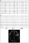
Figure 1-1.
Figure 1-1.
Epoch of wake EEG of a patient at 3 years old showing brief bursts of generalized, bifrontal predominant, irregular 2- to 3-Hz polyspike/spike and slow-wave discharges. At times, the epileptiform discharges had a focal right hemisphere predominance or lead-in (arrows).
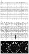
Figure 1-2.
Figure 1-2.
EEG and neuroimaging studies for a patient with infantile spasms. A, EEG epoch from sleep showing hypsarhythmia, characterized by symmetric discontinuous high-voltage (>300 μV) polymorphic delta slow-wave activity with admixed multifocal spike-wave discharges. B, EEG epoch showing three representative infantile spasms from a cluster that lasted for 11 minutes with more than 20 spasms. Each clinical spasm was accompanied by a concurrent high-voltage, broad, diffuse slow wave with attenuation of voltage (arrows). The cluster was associated with a period of relative decrease in interictal epileptiform activity. C, T2-weighted, axial brain MRI showing hyperintensity in the left middle frontal gyrus extending toward the left lateral ventricle (arrows).
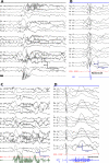
Figure 1-3.
Figure 1-3.
Typical examples of seizure types. A, EEG of epileptic spasm; B, EEG of myoclonic seizure; C, EEG of tonic seizure; D, EEG of myoclonic-tonic seizure.
Figure 1-4.
Figure 1-4.
Epilepsies secondary to a structural cause. A, Serial T2-weighted brain MRI showing changes over time in untreated Rasmussen syndrome. Earliest abnormal MRI at 3 years 2 months of age during the acute phase shows increased cortical signal (arrows). B, C, MRIs at 13 years 9 months in the residual phase show severe unilateral atrophy of the entire right hemisphere with normal signal intensity. D, Brain MRI of a 10-year-old girl with pharmacoresistant epilepsy with gelastic seizures secondary to hypothalamic hamartoma. This is a T2 fast-spin echo, coronal view, and E, postcontrast T1 inversion-recovery fast-spin gradient echo, sagittal view, showing a lesion centered in the left aspect of the hypothalamus and extending into the third ventricle (arrows). F, Brain MRI performed on a 3-Tesla scanner for presurgical evaluation of an 11-year-old boy with pharmacoresistant epilepsy with focal seizures arising from the left temporal lobe. This is a T2 fast-spin echo, coronal view, showing asymmetrical volume loss of the left medial temporal lobe with hyperintensity and obscuration of the hippocampal internal architecture (arrow). Panels A, B, and C with permission from Millichap J J, Goldstein J L, Neurology. © 2010, American Academy of Neurology. www.neurology.org/content/75/20/e85.full.
Figure 1-5.
Figure 1-5.
Distribution of epilepsy diagnoses overall and by age (<3, 3–9, and 10–15 years). Blue shaded groups are nonsyndromic and nonspecific diagnoses. Pink shaded groups are specific syndromic diagnoses that are common and in which the seizures are generally easily controlled and, in many cases, the epilepsy fully resolves in time and neurodisability is not a feature associated with the epilepsy. The solid colors represent other syndromes, many of which are pharmacoresistant and associated with moderate to severe developmental disability. The frequency of these different types of epilepsy diagnoses varies considerably by age at onset. In adult-onset epilepsy, 90% to 95% of epilepsies would fall in the blue-shaded regions. GGE = genetic generalized epilepsy (contains other specific electroclinical syndromes but also less-specific presentations that are considered likely part of the GGE spectrum); JME = juvenile myoclonic epilepsy; JAE = juvenile absence epilepsy; CAE = childhood absence epilepsy; BECTS = benign epilepsy with centrotemporal spikes; LKS = Landau-Kleffner syndrome; EMA = epilepsy with myoclonic absence seizures; MAE = myoclonic-atonic epilepsy; LGS = Lennox-Gastaut syndrome; Dravet = Dravet syndrome; BFN-IS = benign familial neonatal-infantile seizures; West = West syndrome; NSE-GEN = nonsyndromic epilepsy with generalized features; NSE-focal-unknown = nonsyndromic epilepsy with focal features and unknown cause; NSE-foc/plus = nonsyndromic epilepsy with focal features with underlying structural metabolic condition or neuroimpairment. Data from Berg AT, et al, Brain, brain.oxfordjournals.org/content/132/10/2785.full and Berg AT et al, Epilepsia. onlinelibrary.wiley.com/doi/10.1111/j.1528-1157.2000.tb04604.x/abstract.
Figure 1-6.
Figure 1-6.
EEG and neuroimaging studies in a case of N-methyl-D-aspartate (NMDA) receptor antibody encephalitis with seizure-associated cardiac asystole (sensitivity: 10 μV/mm; bars = 1 s). A, Epoch of EEG recorded just after the onset of seizure in left anterior-midtemporal region with rhythmic-theta-alpha pattern that spread to the left frontal region. B, An EEG of the same seizure 80 seconds after onset showing generalized epileptiform discharges with asystole (arrow) that lasted 28 seconds. C, Sequential axial fluid-attenuated inversion recovery (FLAIR) MRIs of the brain 2 weeks after initial presentation. Subtle increased FLAIR signal in the left posterior temporal lobe cortex (arrows), with associated subtle gyral thickening is shown. Also note the mild increased FLAIR signal in the medial temporal lobes. Reprinted with permission from Millichap JJ, et al, Pediatrics. © 2011, American Academy of Pediatrics. pediatrics.aappublications.org/content/127/3/e781.full?sid=84eceb04-945a-46dc-b7a2-708bd2e8122e.

References

    1. Merlis JK. Proposal for an international classification of the epilepsies. Epilepsia 1970; 11 (1): 114–119. - PubMed
    1. Commission on Classification and Terminology of the International League Against Epilepsy. Proposal for the international classification of the epilepsies. Epilepsia 1970; 11: 114–119.
    1. Commission on Classification and Terminology of the International League Against Epilepsy. Proposal for revised clinical and electrographic classification of epileptic seizures. Epilepsia 1981; 22 (4): 489–501. - PubMed
    1. Commission on Classification and Terminology of the International League Against Epilepsy. Proposal for revised classification of epilepsies and epileptic syndromes. Epilepsia 1989; 30 (4): 389–399. - PubMed
    1. Berg AT,, Berkovic SF,, Brodie MJ, et al. Revised terminology and concepts for organization of seizures and epilepsies: Report of the ILAE Commission on Classification and Terminology, 2005–2009. Epilepsia 2010; 51 (4): 676–685. - PubMed