. 2013 Jul;140(14):2923-32.
doi: 10.1242/dev.093534. Epub 2013 Jun 5.

An exclusively mesodermal origin of fin mesenchyme demonstrates that zebrafish trunk neural crest does not generate ectomesenchyme

Affiliations

An exclusively mesodermal origin of fin mesenchyme demonstrates that zebrafish trunk neural crest does not generate ectomesenchyme

Raymond Teck Ho Lee et al. Development. 2013 Jul.

Abstract

The neural crest is a multipotent stem cell population that arises from the dorsal aspect of the neural tube and generates both non-ectomesenchymal (melanocytes, peripheral neurons and glia) and ectomesenchymal (skeletogenic, odontogenic, cartilaginous and connective tissue) derivatives. In amniotes, only cranial neural crest generates both classes, with trunk neural crest restricted to non-ectomesenchyme. By contrast, it has been suggested that anamniotes might generate derivatives of both classes at all axial levels, with trunk neural crest generating fin osteoblasts, scale mineral-forming cells and connective tissue cells; however, this has not been fully tested. The cause and evolutionary significance of this cranial/trunk dichotomy, and its absence in anamniotes, are debated. Recent experiments have disputed the contribution of fish trunk neural crest to fin osteoblasts and scale mineral-forming cells. This prompted us to test the contribution of anamniote trunk neural crest to fin connective tissue cells. Using genetics-based lineage tracing in zebrafish, we find that these fin mesenchyme cells derive entirely from the mesoderm and that neural crest makes no contribution. Furthermore, contrary to previous suggestions, larval fin mesenchyme cells do not generate the skeletogenic cells of the adult fin, but persist to form fibroblasts associated with adult fin rays. Our data demonstrate that zebrafish trunk neural crest does not generate ectomesenchymal derivatives and challenge long-held ideas about trunk neural crest fate. These findings have important implications for the ontogeny and evolution of the neural crest.

Keywords: Dermomyotome; Ectomesenchyme; Fibroblast; Fin mesenchyme; Neural crest.


Figures
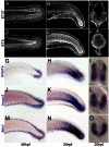
Fig. 1.
Fig. 1.
Paraxial mesoderm expression often precedes fin mesenchyme expression. (A-F) Confocal images of the trunk and tail regions of the ET37 (A-C) and ET5 (D-F) zebrafish lines immunofluorescently stained for eGFP. Lateral views at 48 hpf (A,D) and 24 hpf (B,E) show that expression in fin mesenchyme follows earlier expression in the mesoderm. Transverse confocal images of the trunk of ET37 (C) and ET5 (F) at 20 hpf show expression in paraxial mesoderm as well as at other sites. (G-O) Micrographs of embryos stained by in situ hybridisation with probes against bmp1a (G-I), hmcn2 (J-L) and fbln1 (M-O). Lateral views show the fin mesenchyme expression at 48 hpf (G,J,M) and the preceding mesodermal expression at 24 hpf (H,K,N). Mesodermal expression is paraxial as shown in images of transverse sections of 20-hpf embryos viewed with Nomarski optics (I,L,O).
Fig. 2.
Fig. 2.
Fin mesenchyme cells emerge from trunk mesoderm. Stills of time-lapse Movies 1-3 (see supplementary material Movies 1-3) of the tail of the ET37 line alone (A,B) or crossed to the ntla:lyn-tdtomato transgenic line (C). eGFP is shown in green and membrane-tdTomato is in magenta. Left panels are taken at ∼26 hpf (A), 29 hpf (B) and 22 hpf (C), with subsequent time points (indicated in minutes) shown in panels to the right. Examples of fin mesenchyme cells are indicated (arrows) emerging from the ventral (A,C) and dorsal (B) mesoderm into the adjacent fin. First panel of C is shown without eGFP signal to highlight the epithelial nature of the cells within the mesoderm prior to fin immigration.
Fig. 3.
Fig. 3.
Extensive contribution of mesoderm to fin mesenchyme. (A-F′) Tail of tbx6:gal4; uas:kaede embryos at 24 hpf (A-B′,D-E′) and 48 hpf (C,C′,F,F′) both prior to (A,A′,D,D′) and after (B-C′,E-F′) Kaede photoconversion. Unconverted Kaede protein is in green, overlaid with UV-photoconverted Kaede in red (A-F); the red channel is additionally displayed alone for clarity (A′-F′). Ventral (A-C′) and dorsal (D-F′) regions converted by UV laser are outlined in A and D. At 48 hpf, converted cells can be seen in the adjacent fins (magnified in insets in each channel and merged, C,F) and muscle blocks (C,C′,F,F′).
Fig. 4.
Fig. 4.
Neural crest cells do not contribute to fin mesenchyme. (A-B′) Lateral trunk (A-A′) and tail region (B,B′) of 48-hpf sox10:gal4; uas:kaede transgenic embryos. Kaede protein fluorescence (green) is observed in melanophores (A-A′), nascent dorsal root ganglia and spinal nerves (A), as well as in the fin (B,B′). (C-C′) Immunofluorescent labelling of Kaede (red, C), eGFP (green, C′) and merged image (C′) of the fin of a sox10:gal4; uas:kaede; ET37 transgenic embryo. (D-G) Overviews (D,E) and Nomarski images (F,G) of 48-hpf wild-type (WT) (D,F) and mos-/-; mob-/- (E,G) embryos showing loss of pigment but presence of a fully formed medial fin (E) with fin mesenchyme cells (G) in the double mutant. Red brackets indicate the region used for quantifying fin mesenchyme cells in H. (H) Quantification of fin mesenchyme cells in 12 WT and 12 mos-/-; mob-/- embryos at 48 hpf. No significant differences (n.s.) were observed (two-tailed Student’s t-test). Bars indicate the mean.
Fig. 5.
Fig. 5.
All fin mesenchyme cells derive from paraxial mesoderm. (A) Confocal image of the tail region of a 48-hpf ntla:gal4; uas:kaede embryo. (B-B′) Immunofluorescent staining of 48-hpf ntla:gal4; uas:kaede; ET37 triple transgenic embryo showing total overlap (B′) of Kaede signal (red, B,B′) and eGFP (green, B′,B′) in the fin. (C-E′) Fluorescent images alone (C,D,E) and superimposed on Nomarski images (C′,D′,E′) of the trunk/tail of 48-hpf embryos. The myotome (C,C′), sclerotome (D,D′) and dermomyotome (E,E′) are labelled by actc1b:Gal4i269; uas:kaede (C,C′), Ola-Twist:Gal4; uas:kaede (D,D′) and TgBAC(pax3a:EGFP)i150 (E,E′) transgenics, respectively. (F) Stills taken from time-lapse Movie 4 (see supplementary material Movie 4) of the tail region of a TgBAC(pax3a:EGFP)i150 embryo at 24 hpf (left panel) with subsequent time points at given intervals (in minutes) in the panels to the right. Two fin mesenchyme cells can be tracked (arrows) from the dermomyotome into the fins. Note that eGFP expression is higher in neural crest and dorsal neural tube than in dermomyotome.
Fig. 6.
Fig. 6.
Fin mesenchyme contributes to the adult fin fibroblasts. (A,B) tbx6:CreERt2; ubi:switch transgenics treated with 4-hydroxytamoxifen and imaged at 5 dpf (A) and 21 dpf (B). Arrowheads indicate fin mesenchyme cells in the larval fin (A) and retained in juvenile fin (B). Chains of cells are seen invading at 21 dpf (arrow in B). (C-E) Images of ubi:switch transgenics injected with hmcn2:CreERt2 BAC transgene and treated with 4-hydroxytamoxifen from 3-4 dpf. (C) Tail region of 7-dpf larva shows mCherry in fin mesenchyme cells. The extent of the fin is outlined. (D,E) Adult fins immunostained for mCherry (red) and with zns-5 antibody (green) imaged in lateral whole-mount (D) or in transverse view following cryosectioning and counterstaining with DAPI (E). mCherry cells are in locations consistent with fibroblasts and are zns-5 negative. They reside within the fin rays (open arrowheads in D,E) or can be seen in the inter-ray region (white arrowheads in D,E).

References

    1. Abzhanov A., Tzahor E., Lassar A. B., Tabin C. J. (2003). Dissimilar regulation of cell differentiation in mesencephalic (cranial) and sacral (trunk) neural crest cells in vitro. Development 130, 4567–4579 - PubMed
    1. Asharani P. V., Keupp K., Semler O., Wang W., Li Y., Thiele H., Yigit G., Pohl E., Becker J., Frommolt P., et al. (2012). Attenuated BMP1 function compromises osteogenesis, leading to bone fragility in humans and zebrafish. Am. J. Hum. Genet. 90, 661–674 - PMC - PubMed
    1. Baroffio A., Dupin E., Le Douarin N. M. (1991). Common precursors for neural and mesectodermal derivatives in the cephalic neural crest. Development 112, 301–305 - PubMed
    1. Blentic A., Tandon P., Payton S., Walshe J., Carney T., Kelsh R. N., Mason I., Graham A. (2008). The emergence of ectomesenchyme. Dev. Dyn. 237, 592–601 - PMC - PubMed
    1. Breau M. A., Pietri T., Stemmler M. P., Thiery J. P., Weston J. A. (2008). A nonneural epithelial domain of embryonic cranial neural folds gives rise to ectomesenchyme. Proc. Natl. Acad. Sci. USA 105, 7750–7755 - PMC - PubMed

Publication types

LinkOut - more resources