. 2014 Aug;73(8):1566-74.
doi: 10.1136/annrheumdis-2012-202925. Epub 2013 Jun 5.

Evidence that autophagy, but not the unfolded protein response, regulates the expression of IL-23 in the gut of patients with ankylosing spondylitis and subclinical gut inflammation

Affiliations

Evidence that autophagy, but not the unfolded protein response, regulates the expression of IL-23 in the gut of patients with ankylosing spondylitis and subclinical gut inflammation

Francesco Ciccia et al. Ann Rheum Dis. 2014 Aug.

Abstract

Objectives: Interleukin (IL)-23 has been implicated in the pathogenesis of ankylosing spondylitis (AS). The aim of the study was to clarify the mechanisms underlying the increased IL-23 expression in the gut of AS patients.

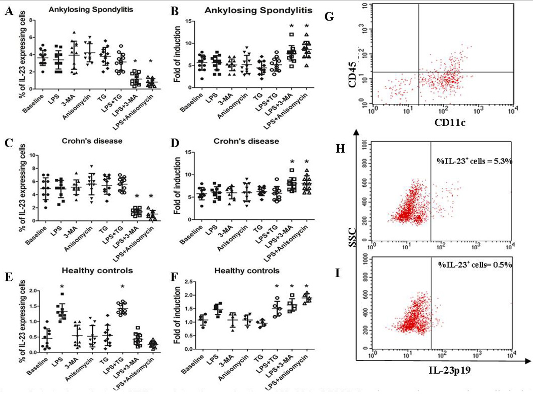
Methods: Consecutive gut biopsies from 30 HLA-B27(+) AS patients, 15 Crohn's disease (CD) patients and 10 normal subjects were obtained. Evidence for HLA-B27 misfolding was studied. Unfolded protein response (UPR) and autophagy were assessed by RT-PCR and immunohistochemistry. The contribution of UPR and autophagy in the regulation of IL-23 expression was evaluated in in vitro experiments on isolated lamina propria mononuclear cells (LPMCs).

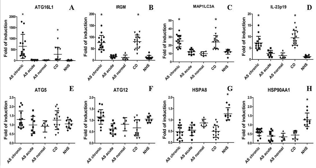
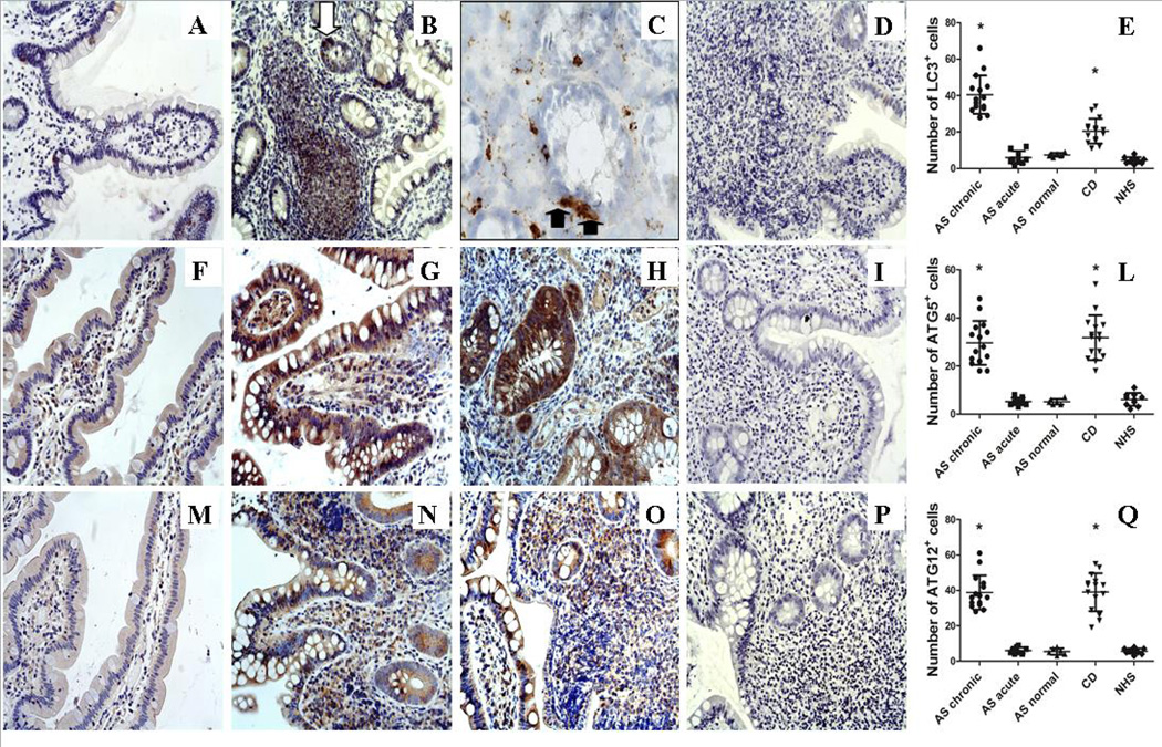
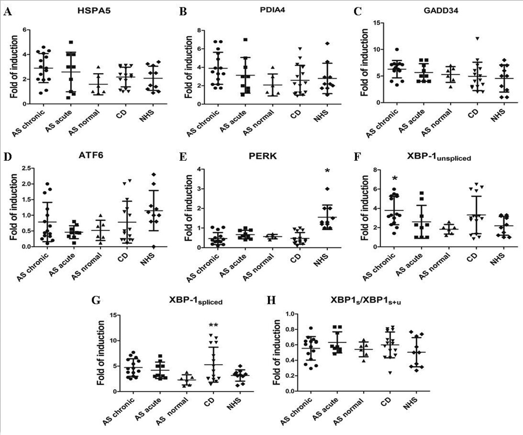
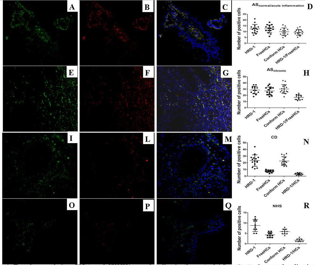
Results: Intracellular colocalisation of SYVN1 and FHCs but not a significant overexpression of UPR genes was observed in the gut of AS patients. Conversely, upregulation of the genes involved in the autophagy pathway was observed in the gut of AS and CD patients. Immunohistochemistry showed an increased expression of LC3II, ATG5 and ATG12 but not of SQSTM1 in the ileum of AS and CD patients. LC3II was expressed among infiltrating mononuclear cells and epithelial cells resembling Paneth cells (PC) and colocalised with ATG5 in AS and CD. Autophagy but not UPR was required to modulate the expression of IL-23 in isolated LPMCs of AS patients with chronic gut inflammation, CD patients and controls.

Conclusions: Our data suggest that HLA-B27 misfolding occurs in the gut of AS patients and is accompanied by activation of autophagy rather than a UPR. Autophagy appears to be associated with intestinal modulation of IL-23 in AS.

Keywords: Cytokines; Inflammation; Spondyloarthritis.


Figures

Figure 1
Figure 1. Macroautophagy and chaperone-mediated autophagy in the gut in the gut of AS patients
A–H: Relative m-RNA quantification of ATG16L1 (A), IRGM (B), MAP1LC3A (C), IL-23p19 (D), ATG5 (E), ATG12 (F), HSPA8 (G) and HSP90AA1 (H) was assessed by quantitative rt-PCR in ileal biopsy specimens obtained from 30 AS, 15 CD patients and 10 controls. Patients with AS were further divided into 3 groups: those with normal histologic findings, those with acute inflammation and those with chronic inflammation. Data are shown as mean (SD). *p<0.0001
Figure 2
Figure 2. Autophagy is up-regulated in the gut in the gut of AS patients
Five-µm–thick paraffin embedded sections of ileal biopsies obtained from AS, CD patients and controls were stained with anti-LC3II, anti-ATG5 and anti-ATG12 antibodies. A–C: Representative microphotographs showing LC3II immunostainings in HC (A), and AS patients with chronic inflammation (B–C). C: representative immunostaining of LC3II staining on frozen samples obtained from AS patients. E–G: Representative microphotographs showing ATG5 immunostainings in HC (E), AS patients with chronic inflammation (F) and CD patients (G). I–M: Representative microphotographs showing ATG12 immunostainings in HC (I), AS patients with chronic inflammation (L) and CD patients (M). Diffuse expression of LC3II, ATG5 and ATG12 was observed in epithelial cells and infiltrating mononuclear cells of AS patients with chronic gut inflammation and CD patients compared to controls. Intense LC3 expression was observed in some epithelial cells of AS patients highly resembling Paneth cells for their pyramidal shape (black arrow) (C). The classic punctate staining was highly evident among infiltrating mononuclear cells and Paneth cells (arrows). A–B, E–G, I–M original magnification ×250; C original magnification × 630. D, H, N: Number of LC3+, ATG5+, ATG12+ cells in the ileal mucosa; *p<0.0001
Figure 3
Figure 3. Unfolded protein response in the gut of AS and CD patients and healthy subjects
A–H: Relative m-RNA quantification of HSPA5 (A), PDIA4 (B), GADD34 (C), PERK (D), ATF6 (E), XBP1-unspliced (F), XBP1-spliced (G) and XBP1 ratio (H) was assessed by quantitative rt-PCR in ileal biopsy specimens from AS patients, CD patients and controls. Data are shown as mean (SD). *p<0.0001; **p<0.05
Figure 4
Figure 4. Free heavy chains (HCs) and SYVN1 co-colocalize in the gut of AS patients
Immunolocalization by confocal microscopy of SYVN1 (green staining) and HCs (red staining) in AS, CD and control ileal biopsies. Paraffin-embedded section from patients and controls were stained with rabbit anti-human SYVN-1 and mouse anti-human free HCs (HC10) antibodies and treated with FITC-conjugated anti-rabbit antibody and Rhodamine Red–conjugated anti-mouse antibody. SYVN1 and free HCs expression was significantly increased in the gut of AS patients independently by the degree of intestinal inflammation (A–B, D and E–F, H) compared to controls (O–P, R). Significant co-localization of SYVN1 and free HCs was detected in the gut of AS patients independently by the degree of intestinal inflammation (C–D, G–H) compared to CD (M–N) and controls (Q–R). (A–C, E–G, I–M, O–Q: original magnification × 250).
Figure 5
Figure 5. Autophagy but not UPR regulates the production of IL-23 in LPMC
Lamina propria mononuclear cells isolated from patients with AS, CD and HCs were cultured in the presence of LPS, 3-MA/anisomycin (to block autophagy) and TG (to induce UPR) alone and with LPS+TG and LPS+3-MA. The percentage of IL-23-producing cells and the m-RNA levels of IL-23p19 were evaluated by flow cytometry and RT-PCR respectively. The percentage of IL-23 producing cells and the m-RNA levels were modified in AS (A) and CD (C) only by the combination of 3MA/anisomycin and LPS. In normal controls LPS alone increased the percentage of IL-23-producing cells and the levels of IL-23 that were not further increased by the co-incubation with TG. Block of autophagy and chaperone mediated autophagy significantly reduced the number of IL-23-producing cells in AS and CD and restrained the LPS-dependent IL-23 expression in LPMC from controls, also increasing the IL-23p19 m-RNA levels (B, D, F) *p<0.05. G: representative dot plot of CD45+ versus CD11c+ cells among LPMC from AS patients. H–I: representative dot plot showing the percentage of IL-23 expressing cells before (H) and after (I) 3-MA/LPS incubation.

References

    1. Mielants H, Veys EM, Cuvelier C, de Vos M. Ileocolonoscopic findings in seronegative spondylarthropathies. Br J Rheumatol. 1988;27:95–105. - PubMed
    1. Mielants H, Veys EM, De Vos M, et al. The evolution of spondyloarthropathies in relation to gut histology. I. Clinical aspects. J Rheumatol. 1995;22:2266–2272. - PubMed
    1. Mielants H, Veys EM, Cuvelier C, et al. The evolution of spondyloarthropathies in relation to gut histology. II. Histological aspects. J Rheumatol. 1995;22:2273–2278. - PubMed
    1. Becker C, Wirtz S, Blessing M, et al. Constitutive p40 promoter activation and IL-23 production in the terminal ileum mediated by dendritic cells. J Clin Invest. 2003;112:693–706. - PMC - PubMed
    1. Ciccia F, Bombardieri M, Principato A, et al. Overexpression of interleukin-23, but not interleukin-17, as an immunologic signature of subclinical intestinal inflammation in ankylosing spondylitis. Arthritis Rheum. 2009;60:955–965. - PubMed

Publication types

MeSH terms