. 2013 Jul 19;288(29):21197-21207.
doi: 10.1074/jbc.M113.484576. Epub 2013 Jun 5.

An undesired effect of chemotherapy: gemcitabine promotes pancreatic cancer cell invasiveness through reactive oxygen species-dependent, nuclear factor κB- and hypoxia-inducible factor 1α-mediated up-regulation of CXCR4

Affiliations

An undesired effect of chemotherapy: gemcitabine promotes pancreatic cancer cell invasiveness through reactive oxygen species-dependent, nuclear factor κB- and hypoxia-inducible factor 1α-mediated up-regulation of CXCR4

Sumit Arora et al. J Biol Chem. .

Abstract

Recently, we have shown that CXCL12/CXCR4 signaling plays an important role in gemcitabine resistance of pancreatic cancer (PC) cells. Here, we explored the effect of gemcitabine on this resistance mechanism. Our data demonstrate that gemcitabine induces CXCR4 expression in two PC cell lines (MiaPaCa and Colo357) in a dose- and time-dependent manner. Gemcitabine-induced CXCR4 expression is dependent on reactive oxygen species (ROS) generation because it is abrogated by pretreatment of PC cells with the free radical scavenger N-acetyl-L-cysteine. CXCR4 up-regulation by gemcitabine correlates with time-dependent accumulation of NF-κB and HIF-1α in the nucleus. Enhanced binding of NF-κB and HIF-1α to the CXCR4 promoter is observed in gemcitabine-treated PC cells, whereas their silencing by RNA interference causes suppression of gemcitabine-induced CXCR4 expression. ROS induction upon gemcitabine treatment precedes the nuclear accumulation of NF-κB and HIF-1α, and suppression of ROS diminishes these effects. The effect of ROS on NF-κB and HIF-1α is mediated through activation of ERK1/2 and Akt, and their pharmacological inhibition also suppresses gemcitabine-induced CXCR4 up-regulation. Interestingly, our data demonstrate that nuclear accumulation of NF-κB results from phosphorylation-induced degradation of IκBα, whereas HIF-1α up-regulation is NF-κB-dependent. Lastly, our data demonstrate that gemcitabine-treated PC cells are more motile and exhibit significantly greater invasiveness against a CXCL12 gradient. Together, these findings reinforce the role of CXCL12/CXCR4 signaling in gemcitabine resistance and point toward an unintended and undesired effect of chemotherapy.

Keywords: CXCR4; Chromatography; Hypoxia-inducible Factor (HIF); NF-κB; Pancreatic Cancer; Reactive Oxygen Species (ROS).


Figures

FIGURE 1.
FIGURE 1.
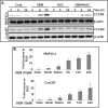
Gemcitabine induces CXCR4 expression in human pancreatic cancer cells. Pancreatic cancer cells were grown in 6-well plates and treated with different doses of gemcitabine (GEM, 0–20 μm) for 24 h (A) or with a constant dose (10 μm) of gemcitabine for different time intervals as indicated (B). Total RNA and protein were extracted and analyzed for CXCR4 expression by quantitative RT-PCR and immunoblot analysis, respectively. GAPDH (quantitative RT-PCR) and β-actin (immunoblot analysis) were used as internal controls. The data show that gemcitabine induces a dose- and time-dependent expression of CXCR4 at both transcript and protein levels. Bars represent the mean of triplicates ± S.D. *, p < 0.05; **, p < 0.01.
FIGURE 2.
FIGURE 2.
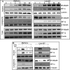
Gemcitabine-induced expression of CXCR4 is mediated through generation of ROS in pancreatic cancer cells. A, pancreatic cancer cells (MiaPaCa and Colo357) were pretreated with 10 mm of NAC (a ROS scavenger) for 1 h followed by treatment with gemcitabine (GEM, 10 μm) for 6 and 24 h. Total protein was isolated and subjected to immunoblot analysis to assess the expression of CXCR4. β-Actin was used as an internal control (Cont.). Data indicate that gemcitabine-induced CXCR4 expression is mediated through generation of ROS in pancreatic cancer cells. B, MiaPaCa and Colo357 cells were treated with gemcitabine (10 μm) at the indicated time intervals (10 min-48 h). Post-treatment, 2′,7′-dichloro-fluorescin diacetate (20 μm) was added to the wells and allowed to be incorporated into the cells for 30 min at 37 °C. Thereafter, cells were washed twice with PBS, resuspended in PBS, and analyzed by flow cytometry. Bars represent the mean of triplicates ± S.D. *, p < 0.05; **, p < 0.01. The data show that gemcitabine treatment generates ROS in a time-dependent manner in pancreatic cancer cells.
FIGURE 3.
FIGURE 3.
Gemcitabine-induced ROS enhances nuclear localization of NF-κB/p65 and HIF-1α. A, MiaPaCa and Colo357 cells were treated with gemcitabine at different time intervals (0–48 h). Thereafter, nuclear, cytoplasmic and total extracts were prepared, and expression and localization of NF-κB/p65 and HIF-1α were examined by immunoblot analysis. B, cells were pretreated with NAC as described earlier, followed by treatment with gemcitabine (GEM, 10 μm) for 12 h. Nuclear and cytoplasmic extracts were prepared, and effects on the localization of NF-κB/p65 and HIF-1α were examined by immunoblot analysis. Laminin (for nuclear fraction), α-tubulin (for cytoplasmic fraction), and β-actin (for total fraction) were used as loading controls. The data indicate an increased level of NF-κB/p65 and HIF-1α in the nuclei of pancreatic cancer cells treated with gemcitabine, which is mediated through gemcitabine-induced ROS generation.
FIGURE 4.
FIGURE 4.
NF-κB/p65 and HIF-1α are both involved in gemcitabine-induced CXCR4 expression in pancreatic cancer cells. Subconfluent pancreatic cancer cells were transiently transfected with non-target (NT) or NF-κB/p65-targeted (A) or HIF-1α-targeted (B) siRNAs. Following 48 h of transfection, cells were treated with gemcitabine (GEM, 10 μm) for the next 12 h (for NF-κB/p65 and HIF-1α) and 24 h (for CXCR4). Total and nuclear proteins were isolated and subjected to immunoblot analysis to assess the expression of NF-κB/p65, HIF-1α, and CXCR4. Laminin and β-actin were used as loading controls for nuclear and total protein, respectively. The data indicate specific silencing of NF-κB/p65 (A) andHIF-1α (B) by target-specific siRNAs and clearly demonstrate that gemcitabine-induced CXCR4 expression is mediated through the additive action of NF-κB/p65 and HIF-1α in pancreatic cancer cells. C, pancreatic cancer cells were treated with gemcitabine (10 μm), and proteins and DNA were cross-linked with formaldehyde. Cross-linked chromatin was sheared and immunoprecipitated with an anti-NF-κB/p65 or anti-HIF-1α or nonspecific IgG. Immunoprecipitated chromatin was subjected to PCR amplification using CXCR4 promoter-specific primers, and amplified products were resolved by electrophoresis. Data show an increased binding of both NF-κB/p65 and HIF-1α on the CXCR4 promoter upon gemcitabine treatment.
FIGURE 5.
FIGURE 5.
ROS-induced nuclear localization of NF-κB/p65 and HIF-1α is mediated through the Akt and ERK pathways. A, pancreatic cancer cells were pretreated with Akt inhibitor (LY294002, 20 μm) or ERK inhibitor (PD98059, 25 μm) for 1 h, followed by treatment with gemcitabine (GEM, 10 μm) for either 15 min, 12 h, or 24 h. Total or nuclear protein was isolated, and expression of Akt, p-Akt, ERK, p-ERK (after a 15-min exposure in total lysate), NF-κB/p65 and HIF-1α (after a 12-h exposure in nuclear lysate), and CXCR4 (after a 24-h exposure in total lysate) was examined by immunoblot analysis. β-Actin and laminin were used as loading controls for total and nuclear lysates, respectively. B, cells were pretreated with NAC as described earlier, followed by treatment with gemcitabine (10 μm) for 15 min. Total protein was isolated, and the effects on the expression of Akt, p-Akt, ERK, and p-ERK were examined by immunoblot analysis. β-Actin was used as a loading control. Altogether, the data demonstrate the selective efficacy of inhibitors and indicate that the ROS-induced nuclear localization of both NF-κB/p65 and HIF-1α occurs through the Akt and ERK pathways after gemcitabine treatment.
FIGURE 6.
FIGURE 6.
NF-κB/p65 enhances expression of HIF-1α in pancreatic cancer cells. A, MiaPaCa and Colo357 cells were pretreated with gemcitabine (GEM, 10 μm) for 6 h, followed by cycloheximide (CHX, 50 μm) treatment for 30 min. Thereafter, NAC (10 mm) was added to the wells for different time intervals (15–90 min). As a control, one of the wells treated with gemcitabine and cycloheximide lacked NAC for each time point. Total protein was isolated, and expression of HIF-1α was examined by immunoblot assay. β-Actin was used as an internal control. The data show that there is no effect of the gemcitabine treatment on the stability of HIF-1α. B, cells were treated with gemcitabine (10 μm) for the indicated time intervals, and total RNA was extracted. c-DNA was prepared, and the expression of HIF-1α was analyzed by quantitative RT-PCR. GAPDH was used as an internal control. The data show that gemcitabine induces expression of HIF-1α in a time-dependent manner. Bars represent the mean of triplicates ± S.D. *, p < 0.05; **, p < 0.01. C, cells were transiently transfected with non-target (NT) or NF-κB/p65-targeted siRNAs. Following 48 h of transfection, cells were treated with gemcitabine (10 μm) for the next 12 h. Nuclear protein was isolated and subjected to immunoblot analysis to assess the expression of NF-κB/p65 and HIF-1α. Laminin was used as a loading control. D, pancreatic cancer cells were treated with gemcitabine (10 μm), and proteins and DNA were cross-linked with formaldehyde. Cross-linked chromatin was sheared and immunoprecipitated with an anti-NF-κB/p65 or nonspecific IgG. Immunoprecipitated chromatin was subjected to PCR to amplify the HIF-1α promoter region harboring a putative NF-κB site. The data show an increased binding of NF-κB/p65 to the HIF-1α promoter upon gemcitabine treatment, suggesting its direct involvement in gemcitabine-induced HIF-1α expression.
FIGURE 7.
FIGURE 7.
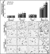
Gemcitabine promotes CXCL12-induced migration and invasion of pancreatic cancer cells. MiaPaCa and Colo357 cells grown in 6-wells plate were treated with gemcitabine (GEM, 10 μm) for 24 h. After treatment, cells were trypsinized, counted, and an equal number of cells was seeded on the non-coated (for migration) and Matrigel-coated (for invasion) membranes. Medium containing 5% FBS alone or supplemented with CXCL12 (100 ng/ml) was added in the lower chamber as a chemoattractant, cells were allowed to migrate/invade overnight through the membrane/Matrigel. Cells that migrated/invaded to the bottom of the insert were fixed, stained, imaged, and counted in ten random view fields. Bars represent the mean ± S.D., n = 3 of migrated (M)/invaded (I) cells per field. *, p < 0.05; **, p < 0.01. The data indicate that the response to CXCL12 in gemcitabine-treated cells was higher compared with control cells.

References

    1. Long J., Zhang Y., Yu X., Yang J., LeBrun D. G., Chen C., Yao Q., Li M. (2011) Overcoming drug resistance in pancreatic cancer. Expert. Opin. Ther. Targets 15, 817–828 - PMC - PubMed
    1. Siegel R., Naishadham D., Jemal A. (2013) Cancer statistics, 2013. CA-Cancer J. Clin. 63, 11–30 - PubMed
    1. Bardeesy N., DePinho R. A. (2002) Pancreatic cancer biology and genetics. Nat. Rev. Cancer 2, 897–909 - PubMed
    1. Olive K. P., Jacobetz M. A., Davidson C. J., Gopinathan A., McIntyre D., Honess D., Madhu B., Goldgraben M. A., Caldwell M. E., Allard D., Frese K. K., Denicola G., Feig C., Combs C., Winter S. P., Ireland-Zecchini H., Reichelt S., Howat W. J., Chang A., Dhara M., Wang L., Rückert F., Grützmann R., Pilarsky C., Izeradjene K., Hingorani S. R., Huang P., Davies S. E., Plunkett W., Egorin M., Hruban R. H., Whitebread N., McGovern K., Adams J., Iacobuzio-Donahue C., Griffiths J., Tuveson D. A. (2009) Inhibition of Hedgehog signaling enhances delivery of chemotherapy in a mouse model of pancreatic cancer. Science 324, 1457–1461 - PMC - PubMed
    1. Wang Z., Li Y., Ahmad A., Banerjee S., Azmi A. S., Kong D., Sarkar F. H. (2011) Pancreatic cancer. Understanding and overcoming chemoresistance. Nat. Rev. Gastroenterol. Hepatol. 8, 27–33 - PubMed

Publication types

MeSH terms