. 2013 Jul 19;288(29):21341-21350.
doi: 10.1074/jbc.M113.481093. Epub 2013 Jun 5.

The MID1 E3 ligase catalyzes the polyubiquitination of Alpha4 (α4), a regulatory subunit of protein phosphatase 2A (PP2A): novel insights into MID1-mediated regulation of PP2A

Affiliations

The MID1 E3 ligase catalyzes the polyubiquitination of Alpha4 (α4), a regulatory subunit of protein phosphatase 2A (PP2A): novel insights into MID1-mediated regulation of PP2A

Haijuan Du et al. J Biol Chem. .

Abstract

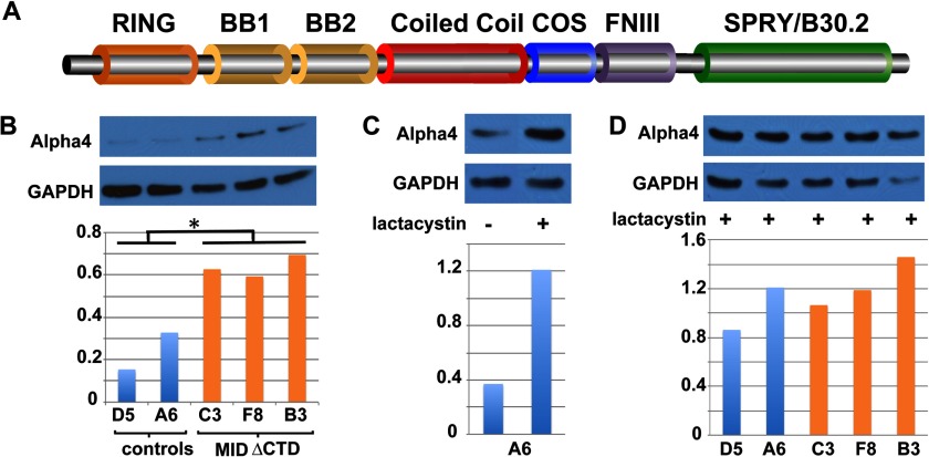
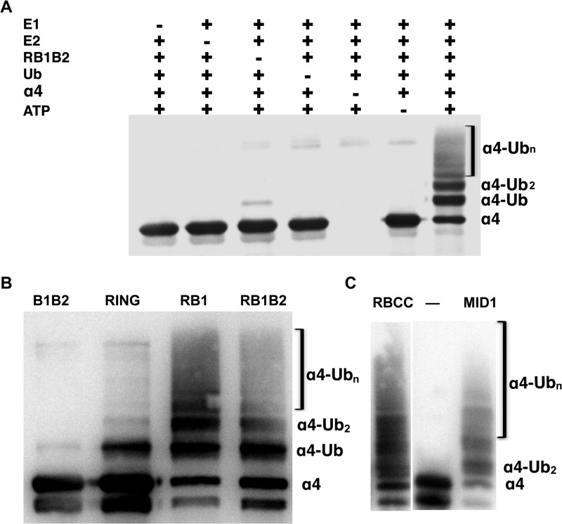
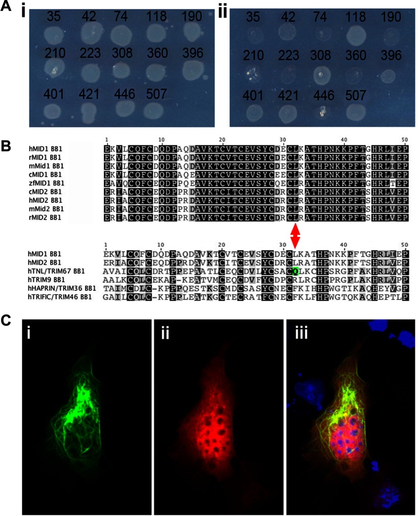
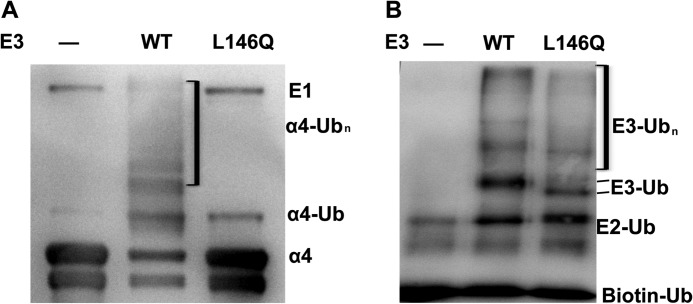
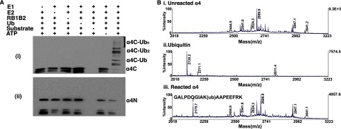
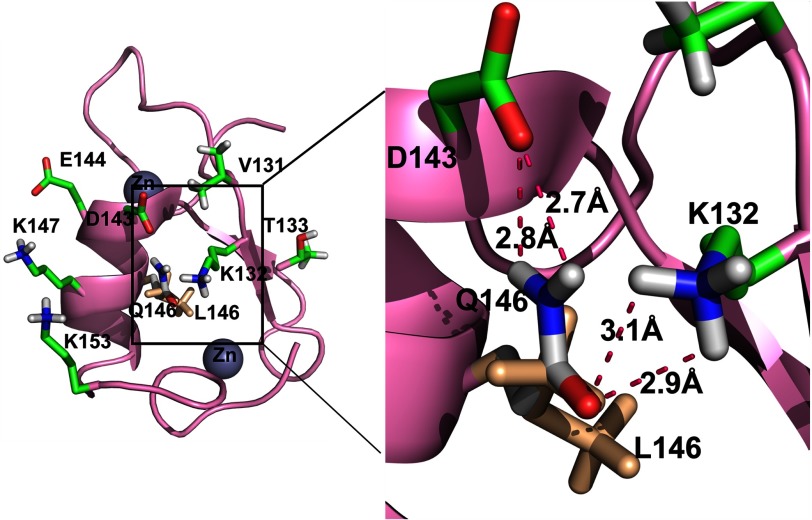
Alpha4 (α4) is a key regulator of protein phosphatase 2A (PP2A) and mTOR in steps essential for cell-cycle progression. α4 forms a complex with PP2A and MID1, a microtubule-associated ubiquitin E3 ligase that facilitates MID1-dependent regulation of PP2A and the dephosphorylation of MID1 by PP2A. Ectopic overexpression of α4 is associated with hepatocellular carcinomas, breast cancer, and invasive adenocarcinomas. Here, we provide data suggesting that α4 is regulated by ubiquitin-dependent degradation mediated by MID1. In cells stably expressing a dominant-negative form of MID1, significantly elevated levels of α4 were observed. Treatment of cells with the specific proteasome inhibitor, lactacystin, resulted in a 3-fold increase in α4 in control cells and a similar level in mutant cells. Using in vitro assays, individual MID1 E3 domains facilitated monoubiquitination of α4, whereas full-length MID1 as well as RING-Bbox1 and RING-Bbox1-Bbox2 constructs catalyzed its polyubiquitination. In a novel non-biased functional screen, we identified a leucine to glutamine substitution at position 146 within Bbox1 that abolished MID1-α4 interaction and the subsequent polyubiquitination of α4, indicating that direct binding to Bbox1 was necessary for the polyubiquitination of α4. The mutant had little impact on the RING E3 ligase functionality of MID1. Mass spectrometry data confirmed Western blot analysis that ubiquitination of α4 occurs only within the last 105 amino acids. These novel findings identify a new role for MID1 and a mechanism of regulation of α4 that is likely to impact the stability and activity level of PP2Ac.

Keywords: Cancer; Cell Signaling; Cellular Immune Response; E3 Ubiquitin Ligase; PP2A; Protein Degradation; Ubiquitination.


Figures

FIGURE 1.
FIGURE 1.
MID1 facilitates the in vivo proteasome degradation of α4. A, shown is a schematic of MID1 depicting the different domains. The RING and Bbox domains are cysteine- and histidine-rich regions that coordinate two zinc ions. The coiled-coil domain is important for MID1 dimerization. The COS box is an extended region of the coiled-coil region that is required for microtubule association. The MID1 protein also contains a fibronectin type III (FNIII) motif and a C-terminal SPRY/B30.2 domain. The latter domain received its name based on homology to a protein domain encoded by exon B30–2 in human MHC-I and to regions found within the spla and the ryanodine receptor. B, levels of α4 are significantly elevated (p = 0.135) in permanent MDCK cells expressing EGFP-tagged MID1delCTD, a dominant negative MID1, when compared with control cells expressing EGFP only. Results from multiple independent cell lines (mutant: C3, F8, B3; control: D5, A6) are shown. The level of α4 was determined by Western blot and quantified as described under “Materials and Methods.” C, control MDCK cells (A6 line) were cultured in the presence (+) or absence (−) of lactacystin for 3 h, and the levels of α4 protein were compared. D, after the addition of lactacystin to control (EGFP) and EGFP-MID1delCTD-expressing MDCK cells, there was no significant difference in α4 levels (p = 0.3846). All experiments were performed at least twice with multiple cell lines.
FIGURE 2.
FIGURE 2.
MID1 catalyzes the polyubiquitination of α4 in a RING- and B-box1-dependent manner. A, a Western blot shows in vitro ubiquitination of full-length α4. Lane 1 consists of all components of the ubiquitination assay except the E1-activating enzyme (E1). The second through fifth lanes represent a control experiment in which specific components of the assay were omitted. The high molecular weight-shifted bands or smear pattern represented polyubiquitinated α4. Antibody was specific for the C terminus. Ub, ubiquitin. B, a Western blot shows ubiquitination assays of α4 with the RING and Bbox domains of MID1. From left to right, the assay was performed separately with His6-Bbox1-Bbox2 (B1B2, residues 71–214), His6-RING (RING, residues1–92), His6-RING-Bbox1 (RB1, residues1–164), and His6-RING-Bbox1-Bbox2 (RB1B2, residues 1–214). Antibody for the Western blot was specific for the C-terminal domain of α4. The band slightly lower than the band labeled α4 indicates partial degraded product of α4. C, a Western blot shows the polyubiquitination of α4 by a RING-Bbox1-Bbox2-Coiled-Coil (RBCC) construct and full-length MID1. The first lane shows the control assay in which E3 ligase is omitted.
FIGURE 3.
FIGURE 3.
Bbox1 domain is essential for MID1-α4 interaction. A, a random mutagenesis based bacterial two-hybrid screen was conducted to identify point mutations that abolished interaction between the MID1 Bbox1 and α4. 14 of 850 colonies from the original screen were re-spotted onto a master (i) and selection (ii) plates for validation. Those showing reduced or no growth on selection plates were sequenced to identify the underlying mutation. B, shown is sequence alignment of MID1 and MID2 Bbox1 domain sequences from human (h), rat (r), mouse (m), chicken (c), and zebrafish (z). Fully conserved residues are highlighted in black. Dark and light gray shading highlight slightly less conservation. In the bottom half of the alignment, the human MID1 and MID2 Bbox1 domains are compared with the Bbox1 domains of the other C-I TRIM subfamily proteins. Note the presence of a glutamine residue (Q, highlighted with green shading) in human TNL/TRIM67 in the equivalent place to Leu-146 in MID1 and MID2. C, shown is ectopic co-expression of EGFP-MID1-L146Q (i) and myc-tagged α4 in Cos1 cells (ii) and the merged images (iii). EGFP-MID1-L146Q expression was detected by GFP fluorescence (green). The α4 expression was visualized by immunocytochemistry using an anti-myc antibody (red). The L146Q mutant shows a microtubule distribution indistinguishable from wild-type MID1 (data not shown). However, in contrast to when α4 was co-expressed with wild-type MID1, α4 was not recruited to the microtubules when co-expressed with the L146Q mutant.
FIGURE 4.
FIGURE 4.
Leucine 146 of Bbox1 domain is essential for MID1-α4 interaction and α4 polyubiquitination. A, a Western blot assay shows the ubiquitination of α4 in the presence of wt RB1B2 (WT) and mutant RB1B2_L146Q in which Leu-146 was mutated to glutamine. The first lane represents a control assay in which MID1 was omitted. Antibody was specific for α4 C-terminal region. B, a Western blot shows the autoubiquitination of RB1B2_L146Q and wt RB1B2 (WT). Antibody was specific for the N-terminal domain of MID1.
FIGURE 5.
FIGURE 5.
Identification of α4 ubiquitination site. Ai, a Western blot assay shows the ubiquitination of the C-terminal domain (α4C, residues 236–339) of α4. Antibody was C-terminal-specific. ii, a Western blot assay shows the ubiquitination of the N-terminal domain (α4N, residues 1–235) of α4. Antibody was specific for the N-terminal region of α4. Ub, ubiquitin. B, a portion of the tandem mass spectra shows fragmentations of unmodified α4 (i), ubiquitinated α4 (ii), and ubiquitin (iii). The peptide (GALPDQGIAKAAPEEFRK) was identified showing Lys-287 as the site of ubiquitination. Tandem mass spectrometry was performed in the linear ion trap, and y-ion and b-ion series were assigned by a database search assuming potential Gly-Gly residues covalently attached to any of the Lys residues on the protein.
FIGURE 6.
FIGURE 6.
Model of L146Q mutation of Bbox1. A ribbon representation of MID1 Bbox1 shows a proposed model of the effect of the L146Q mutation. Labeled are the residues on one surface of Bbox1 that are shown by NMR studies to be involved with binding the C terminus of α4 (21). The position of Leu-146 overlay with Gln-146 is shown in greater magnification to depict potential hydrogen bonds that can result between Gln-146 and Asp-143 and Lys-132.

References

    1. Chuang E., Fisher T. S., Morgan R. W., Robbins M. D., Duerr J. M., Vander Heiden M. G., Gardner J. P., Hambor J. E., Neveu M. J., Thompson C. B. (2000) The CD28 and CTLA-4 receptors associate with the serine/threonine phosphatase PP2A. Immunity 13, 313–322 - PubMed
    1. Inui S., Kuwahara K., Mizutani J., Maeda K., Kawai T., Nakayasu H., Sakaguchi N. (1995) Molecular cloning of a cDNA clone encoding a phosphoprotein component related to the Ig receptor-mediated signal transduction. J. Immunol. 154, 2714–2723 - PubMed
    1. Murata K., Wu J., Brautigan D. L. (1997) B cell receptor-associated protein α4 displays rapamycin-sensitive binding directly to the catalytic subunit of protein phosphatase 2A. Proc. Natl. Acad. Sci. U.S.A. 94, 10624–10629 - PMC - PubMed
    1. Inui S., Sanjo H., Maeda K., Yamamoto H., Miyamoto E., Sakaguchi N. (1998) Ig receptor binding protein 1 (α4) is associated with a rapamycin-sensitive signal transduction in lymphocytes through direct binding to the catalytic subunit of protein phosphatase 2A. Blood 92, 539–546 - PubMed
    1. Onda M., Inui S., Maeda K., Suzuki M., Takahashi E., Sakaguchi N. (1997) Expression and chromosomal localization of the human α4/IGBP1 gene, the structure of which is closely related to the yeast TAP42 protein of the rapamycin-sensitive signal transduction pathway. Genomics 46, 373–378 - PubMed

Publication types

MeSH terms

LinkOut - more resources