. 2013 Sep;65(9):2290-300.
doi: 10.1002/art.38026.

Doxycycline-induced expression of transgenic human tumor necrosis factor α in adult mice results in psoriasis-like arthritis

Affiliations

Doxycycline-induced expression of transgenic human tumor necrosis factor α in adult mice results in psoriasis-like arthritis

Eugen Retser et al. Arthritis Rheum. 2013 Sep.

Abstract

Objective: To generate doxycycline-inducible human tumor necrosis factor α (TNFα)-transgenic mice to overcome a major disadvantage of existing transgenic mice with constitutive expression of TNFα, which is the limitation in crossing them with various knockout or transgenic mice.

Methods: A transgenic mouse line that expresses the human TNFα cytokine exclusively after doxycycline administration was generated and analyzed for the onset of diseases.

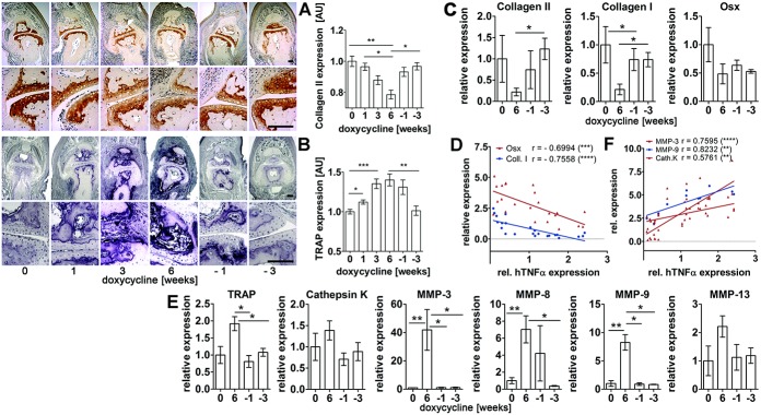
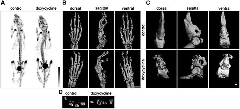
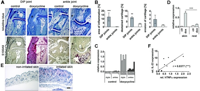
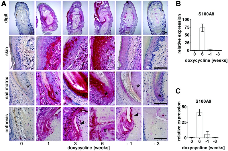
Results: Doxycycline-inducible human TNFα-transgenic mice developed an inflammatory arthritis- and psoriasis-like phenotype, with fore and hind paws being prominently affected. The formation of "sausage digits" with characteristic involvement of the distal interphalangeal joints and nail malformation was observed. Synovial hyperplasia, enthesitis, cartilage and bone alterations, formation of pannus tissue, and inflammation of the skin epidermis and nail matrix appeared as early as 1 week after the treatment of mice with doxycycline and became aggravated over time. The abrogation of human TNFα expression by the removal of doxycycline 6 weeks after beginning stimulation resulted in fast resolution of the most advanced macroscopic and histologic disorders, and 3-6 weeks later, only minimal signs of disease were visible.

Conclusion: Upon doxycycline administration, the doxycycline-inducible human TNFα-transgenic mouse displays the major features of inflammatory arthritis. It represents a unique animal model for studying the molecular mechanisms of arthritis, especially the early phases of disease genesis and tissue remodeling steps upon abrogation of TNFα expression. Furthermore, unlimited crossing of doxycycline-inducible human TNFα-transgenic mice with various knockout or transgenic mice opens new possibilities for unraveling the role of various signaling molecules acting in concert with TNFα.


Figures

Figure 1
Figure 1
Expression of human tumor necrosis factor α (hTNFα) in doxycycline (Dox)–inducible human TNFα–transgenic mice after doxycycline administration. A and B, Systemic doxycycline-induced human TNFα (n = 8, 4, 9, and 6 mice for increasing concentrations shown) (A) and S100A8/A9 proteins (n = 6, 4, 5, and 6 mice for increasing concentrations shown) (B) in mouse serum, as determined by enzyme-linked immunosorbent assay. C, Transcriptional expression of human TNFα in tissues of untreated control mice and mice treated with doxycycline (0.5 mg/ml for 6 weeks), as analyzed by TaqMan quantitative reverse transcription–polymerase chain reaction. The amount of mRNA in fore paws of control mice was assigned a value of 1. Numbers of animals analyzed are shown above columns. D and E, Changes in body weight (D) and swelling of fore paws (E) during stimulation with various concentrations of doxycycline (n = 4 mice per time point for each group of mice). Values in A–E are the mean ± SEM. ∗∗ = P < 0.01; ∗∗∗ = P < 0.001 for doxycycline-treated mice versus untreated mice. F, Magnetic resonance imaging of fore paws before and after injection of contrast agent (CA; 0.5 mmoles/kg) (n = 5 mice per group). Arrowhead indicates prominent swelling of digits after doxycycline treatment. Bar = 1 mm. G, 18F-fluorodeoxyglucose (18F-FDG) positron emission tomography imaging showing locally increased 18F-FDG uptake as a surrogate marker for inflammatory activity in distal interphalangeal joints of doxycycline-treated (arrowheads) (n = 5 mice per experiment) but not untreated (control; n = 3 mice per experiment) doxycycline-inducible human TNFα–transgenic mice. In addition, a typical high uptake of the tracer by the heart and its excretion-based accumulation in the urinary bladder was always observed in both control and doxycycline-treated mice. Color figure can be viewed in the online issue, which is available at http://onlinelibrary.wiley.com/doi/10.1002/art.38026/abstract.
Figure 2
Figure 2
Inflammation in paws of doxycycline-inducible human tumor necrosis factor α (hTNFα)–transgenic mice is reversible. Six-week-old doxycycline-inducible human TNFα–transgenic mice received water with 0.5 mg/ml doxycycline for 6 weeks, after which they received water without the antibiotic (weeks labeled −1 to −5). A, Changes in grip strength of hind paws during doxycycline treatment (n = 4 mice for each time point). B, Alteration in transcription of the human TNFα transgene in paws (n = 5, 5, 4, and 4 mice for the respective time points shown). C, Levels of soluble human TNFα cytokine in serum (n = 4–9 mice at each time point). D, Fore paws of doxycycline-inducible human TNFα–transgenic mice before (0 weeks), during (6 weeks), and after (–5 weeks) doxycycline treatment. E, Histologic changes in toes of hind paws. Images of proximal interphalangeal (PIP) joints are shown (toluidine blue staining). Note the progression of synovitis (asterisks), cartilage destaining (arrowheads), and attachment and invasion of pannus-like tissue into bone (hatchmark). Bars = 200 μm. F and G, Histomorphometric quantification of inflamed synovium (F) and cartilage loss of proteoglycan (G) in PIP joints shown in E (n = 4 mice for each time point). Values are the mean ± SEM. ∗ = P < 0.05; ∗∗ = P < 0.01; ∗∗∗ = P < 0.001.
Figure 3
Figure 3
Distal interphalangeal (DIP) joints but not ankle joints are severely inflamed in doxycycline-inducible human TNFα–transgenic mice after stimulation with doxycycline. Doxycycline-inducible human TNFα–transgenic mice were treated with 0.5 mg/ml doxycycline for 6 weeks or left untreated (control). A, Staining of serial sections of hind paws with toluidine blue (top) or antibodies to S100A9 protein (red) (bottom). Asterisks indicate a strong synovitis in DIP joints but not ankle joints of doxycycline-treated mice. B, Quantification of inflammation (left), cartilage destaining (middle), and cartilage erosion (right) by morphometry (n = 4 mice per group). ∗∗ = P < 0.01; ∗∗∗ = P < 0.001 versus ankle joints. C, Expression of human TNFα transcripts in hind paw digits and ankles, quantified by TaqMan quantitative reverse transcription–polymerase chain reaction (RT-PCR). Values for separate numbered animals are presented. D, Quantitative RT-PCR analysis of human TNFα expression in the skin and the rest of the tissues of hind paws (n = 5 mice per group). ∗∗∗ = P < 0.001. E, S100A9 staining (red) of a healthy skin section and an irritated (see http://zmbe.uni-muenster.de/vwixler/retser.zip) skin section from a doxycycline-treated mouse. Nuclei (blue) were counterstained with hematoxylin. F, Relationship between human TNFα and interleukin-23 (IL-23) mRNA transcripts in skin samples from doxycycline-treated mice (n = 12 mice). ∗∗∗ = P < 0.001. Bars = 100 μm. Values in B and D are the mean ± SEM. See Figure 1 for other definitions.
Figure 4
Figure 4
Inflammation in the toes of doxycycline-inducible human tumor necrosis factor α–transgenic mice during (0–6 weeks) and after (weeks labeled −1 to −3) doxycycline treatment, analyzed by S100A9 (red) expression. A, Immunohistochemistry of S100A9. Representative images of S100A9-stained sections of hind paw toes are depicted. Note the inflammation of nail matrix (asterisks), chondrocytes (arrowheads), and bone tissue (hatchmark). Bars = 100 μm. Samples were counterstained with hematoxylin to reveal nuclei (blue). Three to 5 animals per each time point were analyzed. B and C, Changes in S100A8 (B) and S100A9 (C) transcripts in paws during doxycycline treatment, determined by TaqMan quantitative reverse transcription–polymerase chain reaction (n = 5 mice for each time point). Mean mRNA levels in untreated mice were assigned a value of 1. Values are the mean ± SEM.
Figure 5
Figure 5
Alterations in gene expression are reversible. A and B, Immunostaining (left) and morphometric quantification (right) of type II collagen (top) and tartrate-resistant acid phosphatase (TRAP) (bottom) in distal interphalangeal joints during (0–6 weeks) and after (weeks labeled −1 to −3) doxycycline stimulation (n = 4 mice per group). Bars = 100 μm. C and E, Transcriptional alterations of the indicated genes in paws of doxycycline-inducible human tumor necrosis factor α (hTNFα)–transgenic mice (n = 9 paws per group). Values are the mean ± SEM. D and F, Relationship between human TNFα and indicated mRNAs in paws of doxycycline-inducible human TNFα–transgenic mice treated for 6 weeks with doxycycline (n = 23 paws). Mean mRNA levels in untreated mice were assigned a value of 1. Osx = osterix; Coll. I = type I collagen; MMP-3 = matrix metalloproteinase 3; Cath. K = cathepsin K. ∗ = P < 0.05; ∗∗ = P < 0.01; ∗∗∗ = P < 0.001; ∗∗∗∗ = P < 0.0001.
Figure 6
Figure 6
Bone phenotype of control (unstimulated) doxycycline-inducible human tumor necrosis factor α (TNFα)–transgenic mice and doxycycline-inducible human TNFα–transgenic mice treated with doxycycline for 6 weeks. A, In vivo methylene diphosphonate (MDP) single-photon–emission computed tomography images showing a similar distribution pattern of MDP in doxycycline-treated (n = 3) and control (n = 2) doxycycline-inducible human TNFα–transgenic mice. B, Representative micro–computed tomography (micro-CT) images of fore paws of control (n = 2) and doxycycline-treated (n = 4) doxycycline-inducible human TNFα–transgenic mice. Bar = 1 mm. C, Representative micro-CT images of the first 2 distal phalanges of the middle digits. Bar = 100 μm. D, Cross-section of metacarpals.

References

    1. Locksley RM, Killeen N, Lenardo MJ. The TNF and TNF receptor superfamilies: integrating mammalian biology. Cell. 2001;104:487–501. - PubMed
    1. Bradley JR. TNF-mediated inflammatory disease. J Pathol. 2008;214:149–60. - PubMed
    1. Efimov GA, Kruglov AA, Tillib SV, Kuprash DV, Nedospasov SA. Tumor necrosis factor and the consequences of its ablation in vivo. Mol Immunol. 2009;47:19–27. - PubMed
    1. Li P, Schwarz EM. The TNF-α transgenic mouse model of inflammatory arthritis. Springer Semin Immunopathol. 2003;25:19–33. - PubMed
    1. Pei Y, Harvey A, Yu XP, Chandrasekhar S, Thirunavukkarasu K. Differential regulation of cytokine-induced MMP-1 and MMP-13 expression by p38 kinase inhibitors in human chondrosarcoma cells: potential role of Runx2 in mediating p38 effects. Osteoarthritis Cartilage. 2006;14:749–58. - PubMed

Publication types

Substances