

Establishment and serial passage of cell cultures derived from LuCaP xenografts
- PMID: 23740600
- PMCID: PMC3720815
- DOI: 10.1002/pros.22610
Establishment and serial passage of cell cultures derived from LuCaP xenografts
Abstract
Background: LuCaP serially transplantable xenografts derived from primary and metastatic human prostate cancer encompass the molecular and cellular heterogeneity of the disease and are an invaluable resource for in vivo preclinical studies. A limitation of this model, however, has been the inability to establish and passage cell cultures derived from the xenografts. Here, we describe a novel spheroid culture system that supports long-term growth of LuCaP cells in vitro.
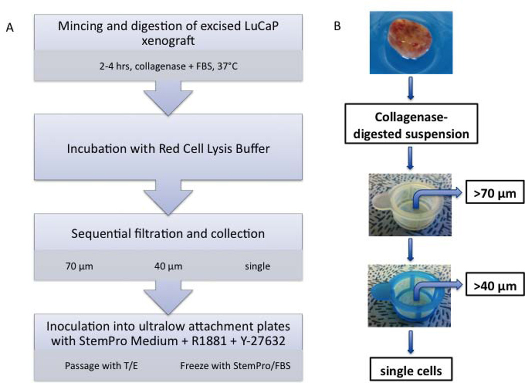
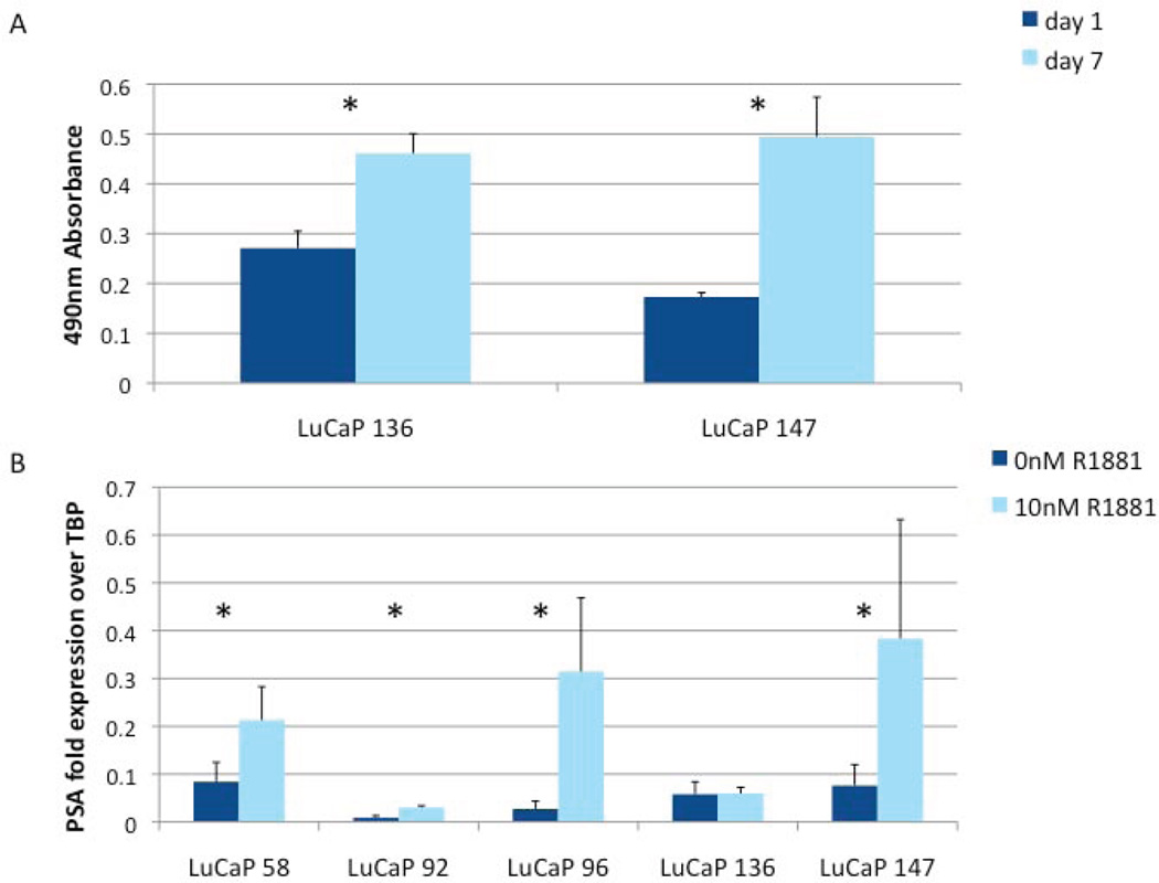
Methods: Xenografts were minced and digested with collagenase. Tissue dissociation was terminated while the majority of cells remained as clusters rather than single cells. The cell clusters were suspended in StemPro medium supplemented with R1881 and Y-27632, a Rho kinase inhibitor, and placed in ultralow attachment dishes for spheroid culture. Serial passage was achieved by partial digestion to small clusters with trypsin/EDTA in the presence of Y-27632. Cell viability, growth and phenotype were monitored with LIVE/DEAD®, MTS, qRT-PCR, and immunocytochemical assays.
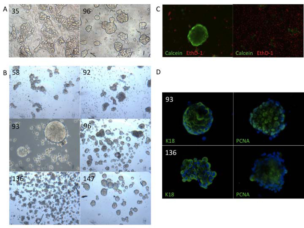
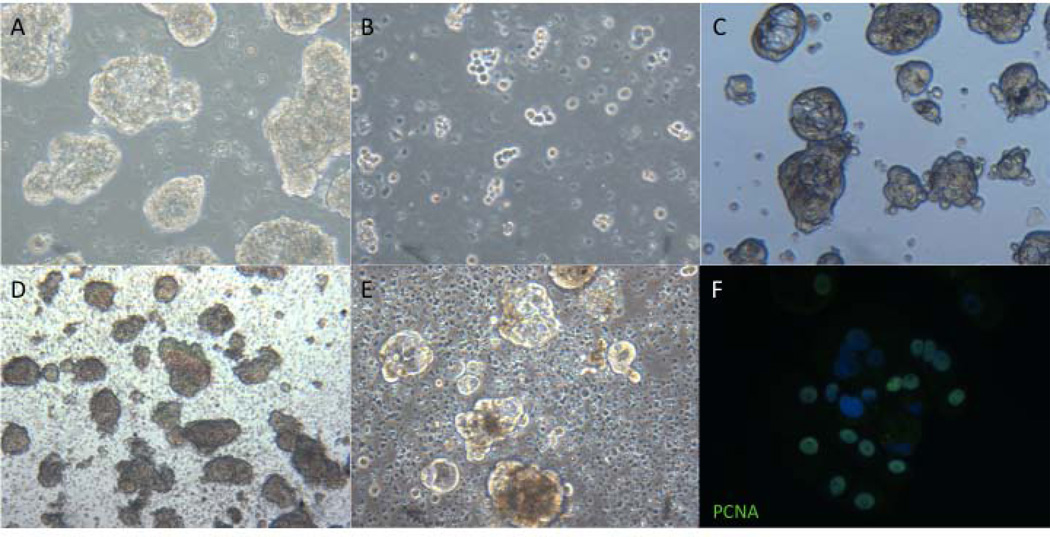
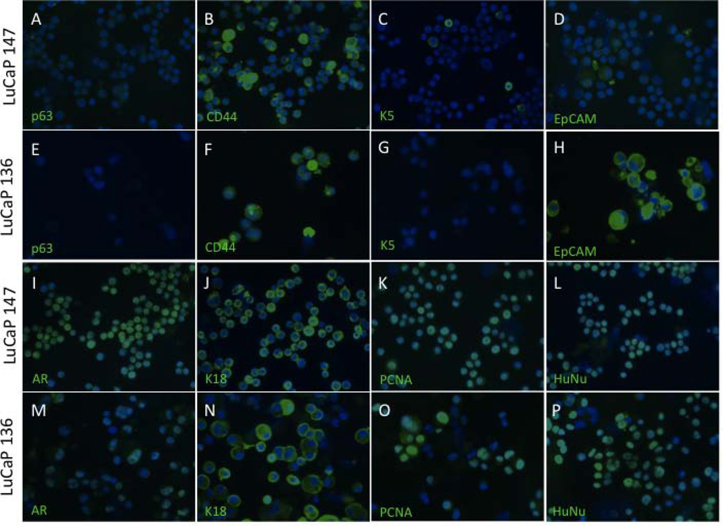
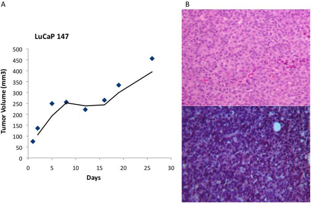
Results: Cells from six LuCaP xenografts formed proliferating spheroids that were serially passaged a minimum of three times and cryopreserved. Two of the cell lines, LuCaP 136 and LuCaP 147, were further passaged and characterized. Both expressed biomarkers characteristic of the xenografts of origin, were determined to be of independent origin by STR fingerprinting, and were free of mycoplasma. LuCaP 147 formed tumors similar to the original xenograft when injected into mice.
Conclusions: The ability to culture LuCaP cells affords new opportunities for fast, cheap, and efficient preclinical studies and extends the value of the LuCaP xenograft models.
Keywords: preclinical model; prostate cancer; spheroids.
Copyright © 2013 Wiley Periodicals, Inc.
Conflict of interest statement
Disclosure statement: The authors declare that they have no affiliations with any organization that may have a direct interest in the research described, or a real or perceived conflict of interest. The funders had no role in study design, data collection and analysis, decision to publish, or preparation on the manuscript.
Figures
References
-
- Sobel RE, Sadar MD. Cell lines used in prostate cancer research: a compendium of old and new lines--part 1. J Urol. 2005;173(2):342–359. - PubMed
-
- Brenner JC, Ateeq B, Li Y, Yocum AK, Cao Q, Asangani IA, Patel S, Wang X, Liang H, Yu J, Palanisamy N, Siddiqui J, Yan W, Cao X, Mehra R, Sabolch A, Basrur V, Lonigro RJ, Yang J, Tomlins SA, Maher CA, Elenitoba-Johnson KS, Hussain M, Navone NM, Pienta KJ, Varambally S, Feng FY, Chinnaiyan AM. Mechanistic rationale for inhibition of poly(ADP-ribose) polymerase in ETS gene fusion-positive prostate cancer. Cancer Cell. 2011;19(5):664–678. - PMC - PubMed
Publication types
MeSH terms
Grants and funding
LinkOut - more resources
Full Text Sources
Other Literature Sources
Medical
