. 2013 Aug;51(8):2599-607.
doi: 10.1128/JCM.00719-13. Epub 2013 Jun 5.

Cell extract-containing medium for culture of intracellular fastidious bacteria

Affiliations

Cell extract-containing medium for culture of intracellular fastidious bacteria

Sudhir Singh et al. J Clin Microbiol. 2013 Aug.

Abstract

The culture of fastidious microorganisms is a critical step in infectious disease studies. As a proof-of-concept experiment, we evaluated an empirical medium containing eukaryotic cell extracts for its ability to support the growth of Coxiella burnetii. Here, we demonstrate the exponential growth of several bacterial strains, including the C. burnetii Nine Mile phase I and phase II strains, and C. burnetii isolates from humans and animals. Low-oxygen-tension conditions and the presence of small hydrophilic molecules and short peptides were critical for facilitating growth. Moreover, bacterial antigenicity was conserved, revealing the potential for this culture medium to be used in diagnostic tests and in the elaboration of vaccines against C. burnetii. We were also able to grow the majority of previously tested intracellular and fastidious bacterial species, including Tropheryma whipplei, Mycobacterium bovis, Leptospira spp., Borrelia spp., and most putative bioterrorism agents. However, we were unable to culture Rickettsia africae and Legionella spp. in this medium. The versatility of this medium should encourage its use as a replacement for the cell-based culture systems currently used for growing several facultative and putative intracellular bacterial species.


Figures

Fig 1
Fig 1
Growth of C. burnetii in VCEM. Growth of the Nine Mile phase I strain and the Nine Mile phase II strain in liquid VCEM with or without a replacement with fresh medium (RM) every 48 h. The growth of C. burnetii was monitored by microscopy using acridine orange staining.
Fig 2
Fig 2
Identification of C. burnetii by mass spectrometry. MALDI-TOF spectra of the Nine Mile phase II strain obtained from liquid VCEM culture (a) and ACCM culture (b) and purified from Vero cells (c).
Fig 3
Fig 3
Isolation of C. burnetii from a human cardiac valve specimen. C. burnetii was successfully cultured from a human cardiac valve and isolated at 17 days postinoculation in VCEM. C. burnetii appears regrouped in clusters of 10 to 20 viable orange bacilli with acridine orange staining (×100).
Fig 4
Fig 4
C. burnetii serological assays. (a) ELISA with positive and negative sera from patients using the C. burnetii antigen purified from C. burnetii grown in cell culture systems and antigens from C. burnetii grown in VCEM. The results are expressed as a normalized ratio (NR) (see Materials and Methods for explanation). An NR of <1 was assigned for negative sera (green), and an NR of >1 was assigned for positive sera (red). (b) IFA assay performed with polyclonal rabbit antiserum on Nine Mile phase I (right) and phase II (left) strain antigens grown in liquid VCEM (bottom) and purified from Vero cells (top).
Fig 5
Fig 5
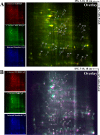
Representative 2D-DIGE analysis of C. burnetii proteins. (A) Each sample of C. burnetii grown in Vero cells and in ACCM was labeled with Cy5 and Cy3, respectively, and (B) each sample of C. burnetii grown in Vero cells, VCEM, and a pooled reference sample was labeled with Cy5, Cy3, and Cy2, respectively, and the samples were then separated on the same 2D-DIGE system. Three images were obtained from each gel, and an overlay of the dye-scanned images was obtained. Selected protein spots exhibiting an ANOVA score of ≤0.05 and a change in intensity of ≥1.5-fold are indicated by arrows and numbers and are listed in Table S1 in the supplemental material.

References

    1. Singh S, Eldin C, Kowalczewska M, Raoult D. 2013. Axenic culture of fastidious and intracellular bacteria. Trends Microbiol. 21:92–99 - PubMed
    1. Renesto P, Crapoulet N, Ogata H, La Scola B, Vestris G, Claverie JM, Raoult D. 2003. Genome-based design of a cell-free culture medium for Tropheryma whipplei. Lancet 362:447–449 - PubMed
    1. Omsland A, Cockrell DC, Howe D, Fischer ER, Virtaneva K, Sturdevant DE, Porcella SF, Heinzen RA. 2009. Host cell-free growth of the Q fever bacterium Coxiella burnetii. Proc. Natl. Acad. Sci. U. S. A. 106:4430–4434 - PMC - PubMed
    1. Omsland A, Beare PA, Hill J, Cockrell DC, Howe D, Hansen B, Samuel JE, Heinzen RA. 2011. Isolation from animal tissue and genetic transformation of Coxiella burnetii are facilitated by an improved axenic growth medium. Appl. Environ. Microbiol. 77:3720–3725 - PMC - PubMed
    1. Rappé MS, Connon SA, Vergin KL, Giovannoni SJ. 2002. Cultivation of the ubiquitous SAR11 marine bacterioplankton clade. Nature 418:630–633 - PubMed

Publication types

LinkOut - more resources