. 2013 Aug;22(8):1037-48.
doi: 10.1002/pro.2290. Epub 2013 Jun 24.

Structural transitions in tau k18 on micelle binding suggest a hierarchy in the efficacy of individual microtubule-binding repeats in filament nucleation

Affiliations

Structural transitions in tau k18 on micelle binding suggest a hierarchy in the efficacy of individual microtubule-binding repeats in filament nucleation

Patrick Barré et al. Protein Sci. 2013 Aug.

Abstract

The protein tau is found in an aggregated filamentous state in the intraneuronal paired helical filament deposits characteristic of Alzheimer's disease and other related dementias and mutations in tau protein and mRNA cause frontotemproal dementia. Tau isoforms include a microtubule-binding domain containing either three or four imperfect tandem microtubule binding repeats that also form the core of tau filaments and contain hexapaptide motifs that are critical for tau aggregation. The tau microtubule-binding domain can also engage in direct interactions with detergents, fatty acids, or membranes, which can greatly facilitate tau aggregation and may also mediate some tau functions. Here, we show that the alternatively spliced second microtubule-binding repeat exhibits significantly different structural characteristics compared with the other three repeats in the context of the intact repeat domain. Most notably, the PHF6* hexapeptide motif located at the N-terminus of repeat 2 has a lower propensity to form strand-like structure than the corresponding PHF6 motif in repeat 3, and unlike PHF6 converts to partially helical structure in the micelle-bound state. Interestingly, the behavior of the Module-B motif, located at the beginning of repeat 4, resembles that of PHF6* rather than PHF6. Our observations, combined with previous results showing that PHF6* and Module-B are both less effective than PHF6 in nucleating tau aggregation, suggest a hierarchy in the efficacy of these motifs in nucleating tau aggregation that originates in differences in their intrinsic propensities for extended strand-like structure and the resistance of these propensities to changes in tau's environment.

Keywords: Alzheimer's; PHF6; PHF6*; amyloid; microtubule-binding domain; microtubule-binding repeat; paired helical filaments; protein aggregation; tau.


Figures

Figure 1
Figure 1
Schematic representation of the primary sequence of tau protein, indicating the alternatively spliced exons (2, 3, and 10), the two proline-rich domains (P1 and P2) the microtubule-binding repeats (R1-R4) and the pseudorepeat R'. The amino acid sequence of the tau K18 construct used in these studies is shown, with each repeat on a separate line for ease of comparison. The K19 construct is identical to K18 except that R2 is excised. The PHF6* (in R2) and PHF6 (in R3) hexapeptide motifs are shown in boldface and underlined.
Figure 2
Figure 2
Tau K18 gains helical structures on binding to SDS micelles and PAPC lipid vesicles. The far UV CD spectrum of the free state has a lineshape characteristic of disordered peptides with a minimum at 198 nm. When bound to SDS, the lineshape corresponds to a mixture of disordered and helical secondary structure with a shoulder at 222 nm and a deeper minimum at around 205 nm. In the presence of PAPC lipid vesicles, the spectrum shifts slightly from that of the micelle-bound state toward the free state spectrum, likely reflecting the presence of a small amount of unbound protein.
Figure 3
Figure 3
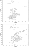
Tau K18 adopts a more ordered, but likely nonglobular structure in the micelle-bound state. NMR proton-nitrogen correlation (HSQC) spectra of tau K18 in the free state (a) and bound to SDS micelles (b) reveal increased dispersion in the micelle-bound state. The increase indicates the formation of some degree of ordered structure on micelle-binding, but does not approach that typically seen for a well-structured globular protein. Backbone resonance assignments are indicated for each state of the protein.
Figure 4
Figure 4
Residual secondary structure in the free state of tau K18 is altered on micelle-binding. NMR secondary chemical shifts are shown for tau K18 Cα (a) and CO (b) nuclei in the free state and Cα (c) and CO (d) nuclei in the micelle-bound state. In (a) small but contiguous positive Cα deviations for residues 284-297 in the free state of R2 are indicated by a horizontal black bar. The first 9 residues of R2 (275-283), exhibiting smaller but mostly positive deviations, are indicated by a horizontal red bar. In (b) the first 8 residues of R2, exhibiting negative CO deviations, are also indicated by a horizontal red bar. In (c) and (d), residues 284-291 of R2 become highly helical, exhibiting large positive Cα and CO deviations, indicated by horizontal black bars. Residues 275-283 also exhibit increased (positive) Cα deviations. Microtubule binding repeats are delineated by dashed orange lines. Corresponding data from our previous studies of K19 are shown in black lines. In (c) the free state data of panel (a) is also shown as a green line for purposes of comparison.
Figure 5
Figure 5
Extended structure propensity is strongest in PHF6. NMR residual dipolar couplings (RDCs) for bicelle-aligned K18 show strong negative RDCs at the N-terminal region of each repeat, including a proline-rich region at the beginning of R1, the PHF6* containing region of R2, the PHF6 region of R3 and the Module-B region of R4. The highest amplitude RDCs are in the PHF6 region. Subsequent to each region of negative RDCs is a region of smaller amplitude mixed positive and negative signals. Microtubule binding repeats are delineated by dashed orange lines and the Cα secondary shifts from Figure 4, scaled arbitrarily for ease of comparison, are shown for reference.
Figure 6
Figure 6
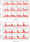
Regions that become helical on micelle-binding also exhibit decreased fast timescale backbone motions. NMR relaxation parameters R1, R2 and the heteronuclear NOE for the free (a) and micelle-bound (b) states of K18 show that regions that become highly helical on micelle-binding, such as residues 284-291, also experience an increase in the R2 and heteronuclear NOE values. Microtubule binding repeats are delineated by dashed orange lines and the Cα secondary shifts from Figure 4, scaled arbitrarily for ease of comparison, are shown for reference.
Figure 7
Figure 7
PHF6* becomes compact or helical in micelle-bound state, in contrast with PHF6. HN-HN NOEs in micelle-bound K18 are strong in regions that become helical on micelle-binding, as for example at residues 284-291 in R2 (horizontal black bar). Regions subsequent to the helices also exhibit strong NOEs, suggesting compact structures. PHF6 at the start of R3 shows relatively weak NOEs, consistent with a more extended conformational ensemble. In contrast, the region containing PHF6* at the beginning of R2 shows some of the strongest NOEs, suggesting, when combined with chemical shift deviations, that this region populates helical conformations in the micelle-bound state. Microtubule binding repeats are delineated by dashed orange lines and the Cα secondary shifts from Figure 4, scaled arbitrarily for ease of comparison, are shown for reference. NOEs are shown as the average of the forward and reverse NOEs between residues i and i+1, with the standard deviation shown as an error bar.

References

    1. Cleveland DW, Hwo SY, Kirschner MW. Purification of tau, a microtubule-associated protein that induces assembly of microtubules from purified tubulin. J Mol Biol. 1977;116:207–225. - PubMed
    1. Cleveland DW, Hwo SY, Kirschner MW. Physical and chemical properties of purified tau factor and the role of tau in microtubule assembly. J Mol Biol. 1977;116:227–247. - PubMed
    1. Goedert M, Spillantini MG, Jakes R, Rutherford D, Crowther RA. Multiple isoforms of human microtubule-associated protein tau: sequences and localization in neurofibrillary tangles of Alzheimer's disease. Neuron. 1989;3:519–526. - PubMed
    1. Andreadis A, Brown WM, Kosik KS. Structure and novel exons of the human tau gene. Biochemistry. 1992;31:10626–10633. - PubMed
    1. Trinczek B, Biernat J, Baumann K, Mandelkow EM, Mandelkow E. Domains of tau protein, differential phosphorylation, and dynamic instability of microtubules. Mol Biol Cell. 1995;6:1887–1902. - PMC - PubMed

Publication types

LinkOut - more resources