. 2013 Jun 5;5(188):188ra74.
doi: 10.1126/scitranslmed.3005452.

Dual receptor T cells mediate pathologic alloreactivity in patients with acute graft-versus-host disease

Affiliations

Dual receptor T cells mediate pathologic alloreactivity in patients with acute graft-versus-host disease

Gerald P Morris et al. Sci Transl Med. .

Abstract

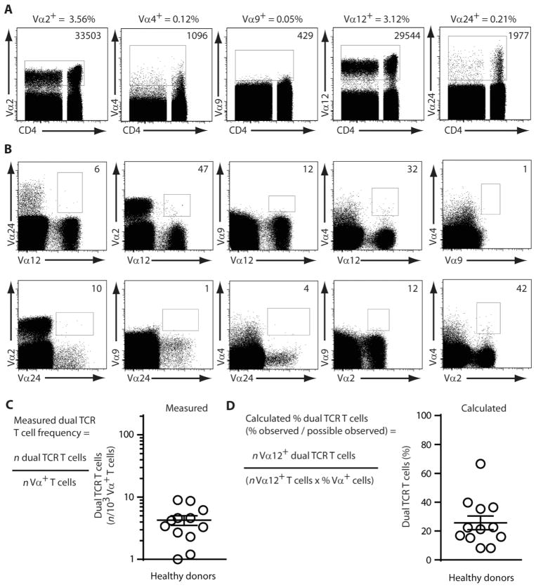
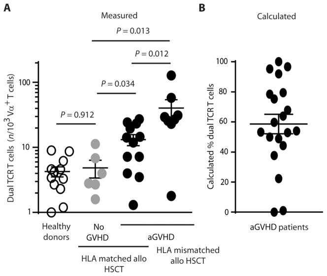
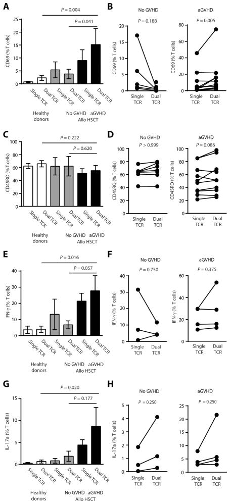
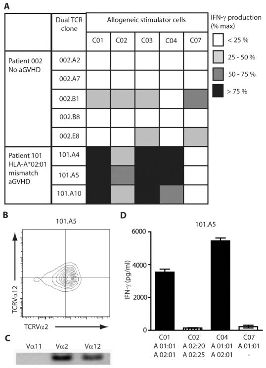
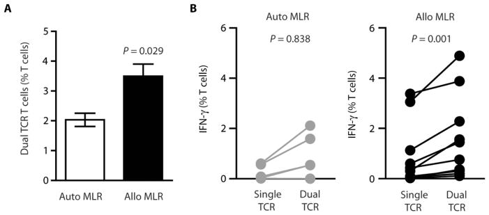
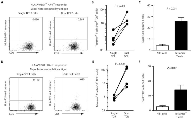
Acute graft-versus-host disease (aGVHD) results from a robust response of donor T cells transferred during hematopoietic stem cell transplantation (HSCT) to allogeneic peptide-major histocompatibility complex antigens. Previous investigations have not identified T cell subsets that selectively mediate either protective immunity or pathogenic alloreactivity. We demonstrate that the small subset of peripheral T cells that naturally express two T cell receptors (TCRs) on the cell surface contributes disproportionately to aGVHD in patients after allogeneic HSCT. Dual TCR T cells from patients with aGVHD demonstrate an activated phenotype and produce pathogenic cytokines ex vivo. Dual receptor clones from a patient with symptomatic aGVHD responded specifically to mismatched recipient human leukocyte antigens (HLAs), demonstrating pathologic alloreactivity. Human dual TCR T cells are strongly activated and expanded by allogeneic stimulation in vitro, and disproportionately contribute to the repertoire of T cells recognizing both major (HLA) and minor histocompatibility antigens, providing a mechanism for their observed activity in vivo in patients with aGVHD. These results identify dual TCR T cells as a target for focused analysis of a T cell subset mediating GVHD and as a potential prognostic indicator.


Conflict of interest statement

Competing interests: The authors declare that they have no competing interests.

Figures

Fig. 1
Fig. 1. Measurement of dual TCR T cells in peripheral blood
(A) Peripheral blood from healthy donors was analyzed for TCRVα expression by flow cytometry. PBLs were gated on live singlet CD4+ or CD8+ cells, and Vα expression was examined using simultaneous labeling with Vα2, Vα4, Vα9, Vα12, and Vα24 mAbs. Isotype controls were used to set Vα gates. Representative FACS plots from healthy donor shown, with percentages and total numbers of Vα+ T cells identified. (B) Dual TCR T cells were identified among live CD4+ and CD8+ T cells in PBLs by pairwise labeling with Vα mAbs. FACS plot of representative healthy donor PBLs is shown, with numbers of dual TCR T cells identified. (C) Dual TCR T cell numbers evaluated as a frequency of measured T cells. Total numbers of dual TCR T cells were divided by the total number of T cells labeled with Vα mAbs. Measured dual TCR T cell frequencies from healthy donors (n = 12), mean ± SEM. (D) Dual TCR T cells evaluated as a calculated percentage of the possible observable dual TCR T cells using pairwise Vα mAb analysis. Frequencies were calculated by dividing the observed number of Vα12+ dual TCR T cells by the possible number of dual TCR T cells identifiable among Vα12+ T cells (number of Vα12+ T cells multiplied by the frequency of labeling with Vα2, Vα4, Vα9, and Vα24 mAbs). Calculated dual TCR T cell frequencies from healthy donors (n = 12), mean ± SEM.
Fig. 2
Fig. 2. Dual TCR T cells are increased in patients with aGVHD
Dual TCR T cells were measured in patients after allogeneic HSCT by flow cytometry of peripheral blood as described in Fig. 1. (A) Measured frequencies of dual TCR T cells from healthy donors (n = 12, shown in Fig. 1C), allogeneic HSCT patients who did not develop aGVHD (n = 6), patients developing aGVHD after transplant with an HLA-matched graft (n = 12), and patients developing aGVHD after transplant with an HLA-mismatched graft (n = 8). Data are expressed as the number of dual TCR T cells observed per 103 T cells labeled with TCRVα mAbs. Each point represents a single patient. Bars are means ± SEM. Data were compared nonparametrically by Mann-Whitney test. (B) Dual TCR T cell frequency was calculated as described in Fig. 1 for patients with aGVHD (n = 20). Bars are means ± SEM.
Fig. 3
Fig. 3. Dual TCR T cells from patients with aGVHD are activated and produce pathogenic cytokines
(A to D) Dual TCR T cells from healthy controls (n = 9) and allogeneic HSCT patients without (n = 5) or with aGVHD (n = 11) were analyzed by flow cytometry for (A) CD69 and (C) CD45RO expression. Data are means ± SEM; groups compared nonparametrically by Mann-Whitney test. Expression of (B) CD69 and (D) CD45RO was compared on single and dual TCR T cells within samples from patients with aGVHD. Paired sample measurements indicated by connecting lines (ratio paired t test). (E to H) Production of (E) IFN-γ and (G) IL-17a by PBLs from healthy controls (n = 5) and allogeneic HSCT patients without (n = 3) or with aGVHD (n = 4) was measured by intracellular flow cytometry after 4 hours of ex vivo stimulation with PMA (0.1 μg/ml) and ionomycin (1 μM). Data are means ± SEM; Mann-Whitney test. Production of (F) IFN-γ and (H) IL-17a was compared between single and dual TCR T cells within samples. Paired sample measurements indicated by connecting lines (ratio paired t test).
Fig. 4
Fig. 4. Dual TCR T cells respond to mismatched HLA antigens in aGVHD
Dual TCR T cell clones were generated by single-cell FACS sorting of PBLs from patients after allogeneic HSCT. (A) T cell clones were tested for alloreactivity by measuring IFN-γ production by ELISA after 24 hours of culture of 5 × 10⁴ clone T cells with 1 × 10⁵ irradiated allogeneic PBLs in triplicate. Stimulation with PMA (100 ng/ml) and ionomycin (1 μM) was used as a positive control. Reactivity was assessed compared to positive controls and represented as a percentage of the maximal response. Data are averages of at least three independent experiments for each T cell clone for each condition. (B to D) Example of representative dual TCR T cell clone from patient with aGVHD. Dual TCR expression of clones was confirmed (B) by FACS, with gates determined by labeling with isotype controls, and (C) by PCR with primers specific for TCRVα gene segments. (D) Alloreactivity was assessed by ELISA measurement of culture supernatants after 24 hours of mixed lymphocyte reaction (MLR). Data are means ± SEM of representative experiment.
Fig. 5
Fig. 5. Dual TCR T cells preferentially respond to allogeneic stimulation
(A) Dual TCR T cells expand in response to allogeneic stimulation in vitro. Healthy donor PBLs were cultured for 5 days in MLR with autologous or two different allogeneic donors. Dual TCR T cell frequency was assessed by FACS analysis. Data are means ± SEM from four independent experiments (n = 4); Mann-Whitney test. (B) Comparison of IFN-γ production by single TCR or dual TCR T cells in individual cultures. IFN-γ production measured by intracellular cytokine staining after 5 days of autologous (gray symbols) or allogeneic (black symbols) MLR. Each point represents a single culture from independent experiments (n = 4), with paired sample measurements indicated by connecting lines. Data analyzed by ratio paired t test.
Fig. 6
Fig. 6. Dual TCR T cells disproportionately recognize major and minor histocompatibility antigens
T cell recognition of HLA-A*02:01–HA-1 tetramer measured by flow cytometry. (A to C) HLA-A*02:01+ HA-1 cells recognize HA-1 as a minor histocompatibility antigen. (A) Representative example of recognition of HLA-A*02:01–HA-1 tetramer by single and dual TCR T cells in a single sample. FACS plot of live singlet T cells gated on cells labeled with TCRVα mAbs. (B) Recognition of minor histocompatibility antigen tetramer by single and dual TCR T cells for individual donors (n = 4). Paired measurements indicated by connecting lines; Wilcoxon matched pairs test. (C) Comparison of average frequency of dual TCR T cells among all T cells and minor histocompatibility antigen tetramer–binding T cells. Data are means ± SEM of all donors (n = 4); Mann-Whitney test. (D to F) HLA-A*02:01 HA-1 cells recognize HLA-A*02:01 as a major histocompatibility antigen. (D) Representative example of recognition of HLA-A*02:01–HA-1 tetramer by single and dual TCR T cells in a single sample. (E) Recognition of major histocompatibility antigen tetramer by single and dual TCR T cells for individual donors (n = 4). Paired measurements indicated by connecting lines; Wilcoxon matched pairs test. (F) Comparison of average frequency of dual TCR T cells among all T cells and major histocompatibility antigen tetramer–binding T cells. Data are means ± SEM of all donors (n = 4); Mann-Whitney test.

Comment in

Similar articles

Cited by

References

    1. Shlomchik WD. Graft-versus-host disease. Nat Rev Immunol. 2007;7:340–352. - PubMed
    1. Coghill JM, Sarantopoulos S, Moran TP, Murphy WJ, Blazar BR, Serody JS. Effector CD4+ T cells, the cytokines they generate, and GVHD: Something old and something new. Blood. 2011;117:3268–3276. - PMC - PubMed
    1. Blazar BR, Murphy WJ, Abedi M. Advances in graft-versus-host disease biology and therapy. Nat Rev Immunol. 2012;12:443–458. - PMC - PubMed
    1. Bacigalupo A. Antilymphocyte/thymocyte globulin for graft versus host disease prophylaxis: Efficacy and side effects. Bone Marrow Transplant. 2005;35:225–231. - PubMed
    1. Copelan EA. Hematopoietic stem-cell transplantation. N Engl J Med. 2006;354:1813–1826. - PubMed

Publication types

MeSH terms

LinkOut - more resources