. 2013 Oct 1;22(19):3894-905.
doi: 10.1093/hmg/ddt244. Epub 2013 Jun 4.

RD3 gene delivery restores guanylate cyclase localization and rescues photoreceptors in the Rd3 mouse model of Leber congenital amaurosis 12

RD3 gene delivery restores guanylate cyclase localization and rescues photoreceptors in the Rd3 mouse model of Leber congenital amaurosis 12

Laurie L Molday et al. Hum Mol Genet. .

Abstract

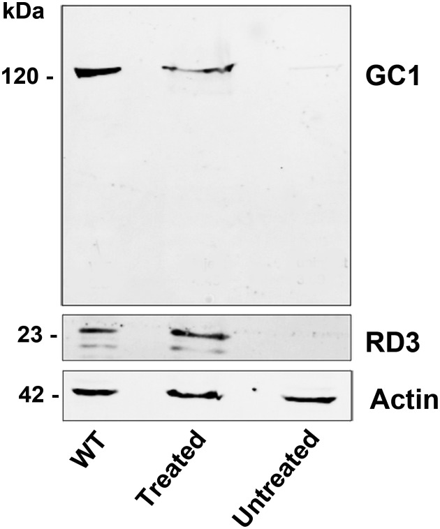
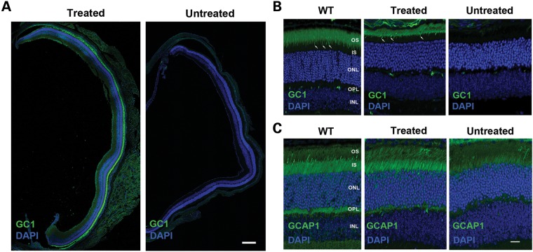
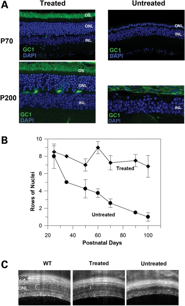
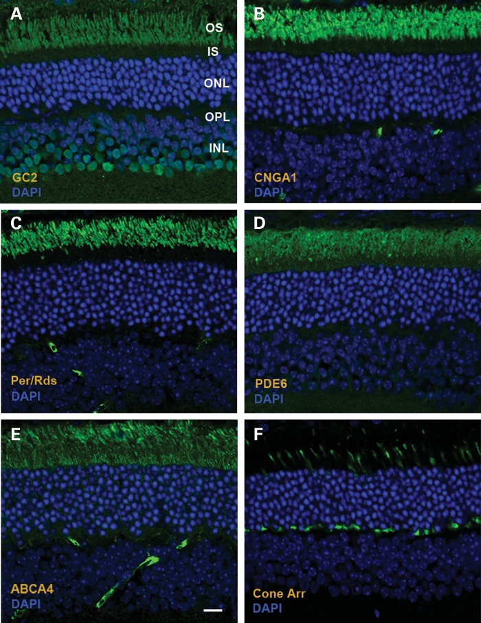
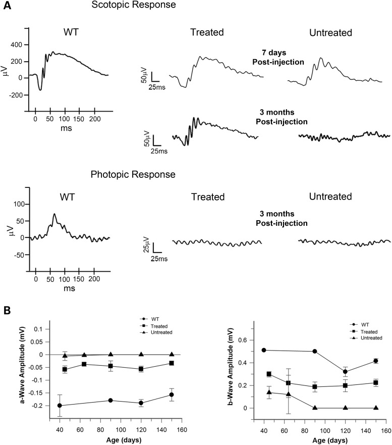
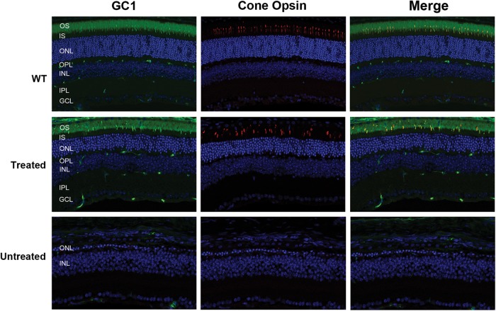
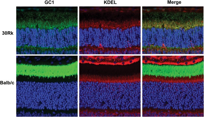
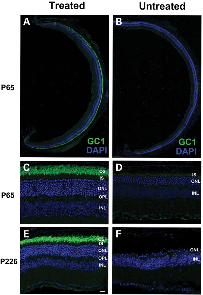
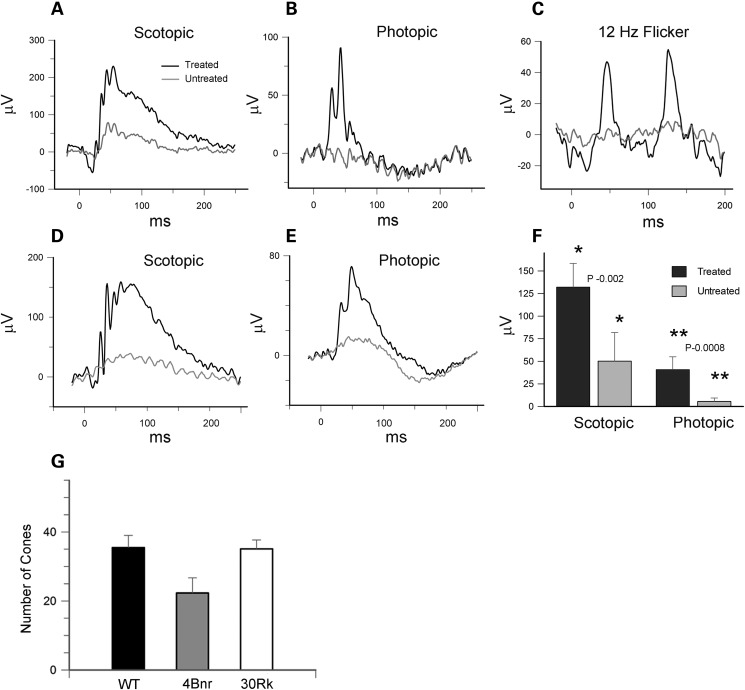
RD3 is a 23 kDa protein implicated in the stable expression of guanylate cyclase in photoreceptor cells. Truncation mutations are responsible for photoreceptor degeneration and severe early-onset vision loss in Leber congenital amaurosis 12 (LCA12) patients, the rd3 mouse and the rcd2 collie. To further investigate the role of RD3 in photoreceptors and explore gene therapy as a potential treatment for LCA12, we delivered adeno-associated viral vector (AAV8) with a Y733F capsid mutation and containing the mouse Rd3 complementary DNA (cDNA) under the control of the human rhodopsin kinase promoter to photoreceptors of 14-day-old Rb(11.13)4Bnr/J and In (5)30Rk/J strains of rd3 mice by subretinal injections. Strong RD3 transgene expression led to the translocation of guanylate cyclase from the endoplasmic reticulum (ER) to rod and cone outer segments (OSs) as visualized by immunofluorescence microscopy. Guanylate cyclase expression and localization coincided with the survival of rod and cone photoreceptors for at least 7 months. Rod and cone visual function was restored in the In (5)30Rk/J strain of rd3 mice as measured by electroretinography (ERG), but only rod function was recovered in the Rb(11.13)4Bnr/J strain, suggesting that the latter may have another defect in cone phototransduction. These studies indicate that RD3 plays an essential role in the exit of guanylate cyclase from the ER and its trafficking to photoreceptor OSs and provide a 'proof of concept' for AAV-mediated gene therapy as a potential therapeutic treatment for LCA12.


Figures

Figure 1.
Figure 1.
Expression of RD3 and GC1 by western blotting. Retinal extracts from six WT eyes, six AAV8(Y733F)-hGRK1-mRd3-treated eyes and six-untreated 4Bnr-rd3 eyes were analyzed at 35 days post-injection on SDS gels. Western blots were labeled with the Rd3–9D12 monoclonal antibody for the detection of RD3 and the GC1-8A5 monoclonal antibody for the detection of GC1. Actin labeling was used as a loading control.
Figure 2.
Figure 2.
GC1 and GCAP1 expression and localization in the photoreceptors of untreated and treated 4Bnr rd3 mice by immunofluorescence microscopy. The right eye of a 4Bnr rd3 mouse was injected (treated) with AAV8(Y733F)-hGRK1-mRd3 at P14 with the uninjected (untreated) left eye serving as a contralateral control. Retinal cryosections prepared 2 weeks post-injection (P28) were labeled for GC1 or GCAP1 (green) and counterstained with DAPI (blue) nuclear stain. (A) Retinal sections from a whole eye at low magnification. GC1 staining is present throughout the retina of the treated, but not the untreated eye. Bar 200 μm. (B) GC-labeled retinal sections at higher magnification. GC1 is correctly localized to the rod and cone (arrow) OSs of the treated eye similar to GC1 localization in WT Balb/c mouse retina. (C) GCAP1-labeled retinal sections at higher magnification. GCAP1 distribution in the treated retina is similar to that in the WT retina, whereas the untreated retina shows reduced GCAP1 expression primarily in the inner segment. Representative micrographs are shown from analysis of six mice. OS, OS layer; IS, inner segment layer; ONL, outer nuclear layer; OPL, outer plexiform layer; INL, inner nuclear layer. Bar—10 μm.
Figure 3.
Figure 3.
Prolonged GC1 expression and localization and sustained photoreceptor survival in AAV8(Y733F)-hGRK1-mRd3 treated mice. (A) Retinal sections of treated and untreated 4Bnr rd3 mice injected at P14 were labeled for GC1 at P70 and P200. Normal localization of GC1 in the OS layer of treated mice was observed with a substantial ONL indicative of photoreceptor survival. No GC1 expression and significant loss (P70) or absence (P200) of the ONL layer was observed in the untreated retina. (B) Loss in the number of rows of photoreceptor nuclei in the ONL as a function of postnatal age for treated and untreated mice. Data are the mean ± SD for n ≥ 3. (C) OCT image of WT, treated and untreated B4nr rd3 mice injected at P14 and analyzed at P105. White vertical brackets designate the ONL observed in the retinas from WT and treated mice. No ONL was observed in the untreated mice. Representative micrographs are shown from analysis of four mice.
Figure 4.
Figure 4.
Distribution of photoreceptor proteins in AAV8(Y733F)-hGRK1-mRd3-treated 4Bnr rd3 mice at 96 days post-injection (p110) as visualized by immunofluorescence microscopy. (A) GC2 labeling with an anti-GC2 polyclonal antibody; (B) Cyclic nucleotide-gated channel A1 subunit (CNGA1) labeling with the PMc 1D1 monoclonal antibody; (C) Peripherin/rds (Per/rds) labeling with the Per5H2 monoclonal antibody; (D) Rod phosphodiesterase alpha subunit (PDE6) labeling with an anti-PDE6A polyclonal antibody; (E) ABCA4 transporter labeling with the Rim 3F4 monoclonal antibody; (F) Cone arrestin labeling with a cone arrestin antibody. Representative micrographs are shown for two mice analyzed. Bar—10 μm.
Figure 5.
Figure 5.
ERGs of AAV8(Y733F)-hGRK1-mRd3-treated and -untreated 4Bnr rd3 mice. Mice were treated at P14 and their dark-adapted (scotopic) and light-adapted (photopic) ERGs were measured at various times after treatment. (A) Typical scotopic ERG ellicited at a light intensity of 2.25 cd s/m² and photopic ERG ellicited at a light intensity of 12 cd.s/m2 in the presence of background light of 30 cd/m2. Scotopic response was observed for the untreated and treated eyes at 7 days post-injection. At 3 months post-injection an ERG response was only observed for the treated eye. Photopic response was not observed at any age for the untreated or treated eyes. ERGs of WT are shown as a reference. (B) Effect of a-wave and b-wave amplitudes (mV) of the scotopic response as a function of the age of the WT, treated and untreated mice. Data are expressed as mean ± SD for n ≥ 3.
Figure 6.
Figure 6.
Presence of cone photoreceptors in the retina of AAV8(Y733F)-hGRK1-mRd3-treated and -untreated mice as visualized by immunofluorescence microscopy. Retinal cryosections from WT mice and the treated and untreated 4Bnr mice were were double labeled for GC1 and middle wavelength cone opsin. Both the WT and treated mice showed a significant number of cone photoreceptor cells with cone opsin and GC1 co-localized to the OSs (merged image). The treated mice were injected at P14  and analyzed 115 days later. No significant labeling was observed for the untreated 4Bnr mice indicative of extensive cone degeneration. Representative micrographs are shown from the analysis of six 4Bnr mice and three WT mice analyzed.
Figure 7.
Figure 7.
Localization of GC1 in the retina of untreated 21-day-old 30Rk mouse. GC1 labeling is predominantly found in the inner segment where it co-localizes with the anti-KDEL ER marker. Weak GC1 labeling is also detected in the OSs and outer plexiform layers. Labeling of a retina from WT (Balb/c) mice is shown for comparison with intense GC1 labeling detectable only in the OS and KDEL labeling in the inner segment. Representative micrographs are shown from the analysis of two WT and two 30Rk mice.
Figure 8.
Figure 8.
GC1 expression and localization in the photoreceptors of AAV8(Y733F)-hGRK1-mRd3-treated and -untreated 30Rk-rd3 mice. The treated right eye was injected at P14 with the untreated left eye serving as a contralateral control. Retinal cryosections were prepared at P65 and labeled with a polyclonal antibody to GC1 and counterstained with DAPI. (A and B) Retinal sections of a treated and untreated eye labeled for GC1. Intense endogenous GC1 immunostaining is present laterally throughout the retina of the treated eye (A) and only faint immunostaining observed in the untreated eye (B). (C and D) At higher magnification, GC1 labeling is restricted to the OS layer of the treated eye (C). Faint labeling of GC1 in the untreated eye is observed primarily in the inner segment (D). (E and F) Labeling of a treated and untreated eye at P226, ∼7 months post-injection. The retina of the treated eye showed intense GC1 labeling in the OS layer and a substantial DAPI stained ONL (E). In contrast, the untreated eye showed a single row of nuclei in the ONL (F). Representative micrographs are shown from the analysis of six mice. Bar—20 μm.
Figure 9.
Figure 9.
ERGs of 30Rk-rd3 mice treated with AAV8(Y733F)-hGRK1-mRd3 at P14 and analyzed at P65 and P150. (A) Scotopic ERG response of the AAV treated and untreated eyes ellicited at a light intensity of 2.25 cd s/m² for a 30Rk mouse at P65. (B) Photopic ERG response ellicited at a light intensity of 12 cd s/m2 in the presence of background light of 30 cd/m2 at P65. (C) The 12 Hz flicker response for the treated and untreated eyes at P65. (D and E) An example of the scotopic and photopic response for the treated and untreated eye for a 30Rk mouse at P150. (F) Differences in scotopic and photopic b-wave amplitudes for mice at 5-month post-injection. Data are expressed as mean ± SD for n = 6. Mean values within each group for the ERG response was compared using a standard paired Student's t-test. (G) The number of cones was determined for WT and AAV-treated 30Rk mice 6.5 months postinjection and 4Bnr mice 4.5 months postinjection over 300 μm length of retina. Data are expressed as mean ± SD for n = 5. A multigroup statistical test described in the Materials and Methods section was used to compare groups (P = 0.009).

References

    1. den Hollander A.I., Roepman R., Koenekoop R.K., Cremers F.P. Leber congenital amaurosis: genes, proteins and disease mechanisms. Prog. Retin. Eye Res. 2008;27:391–419. - PubMed
    1. Perrault I., Rozet J.M., Calvas P., Gerber S., Camuzat A., Dollfus H., Chatelin S., Souied E., Ghazi I., Leowski C., et al. Retinal-specific guanylate cyclase gene mutations in Leber's congenital amaurosis. Nat. Genet. 1996;14:461–464. - PubMed
    1. Yang R.B., Garbers D.L. Two eye guanylyl cyclases are expressed in the same photoreceptor cells and form homomers in preference to heteromers. J. Biol. Chem. 1997;272:13738–13742. - PubMed
    1. Baehr W., Karan S., Maeda T., Luo D.G., Li S., Bronson J.D., Watt C.B., Yau K.W., Frederick J.M., Palczewski K. The function of guanylate cyclase 1 and guanylate cyclase 2 in rod and cone photoreceptors. J. Biol. Chem. 2007;282:8837–8847. - PMC - PubMed
    1. Shyjan A.W., de Sauvage F.J., Gillett N.A., Goeddel D.V., Lowe D.G. Molecular cloning of a retina-specific membrane guanylyl cyclase. Neuron. 1992;9:727–737. - PubMed

Publication types

MeSH terms

Substances

Supplementary concepts