. 2013 Oct 15;22(20):4127-35.
doi: 10.1093/hmg/ddt261. Epub 2013 Jun 4.

Method for widespread microRNA-155 inhibition prolongs survival in ALS-model mice

Method for widespread microRNA-155 inhibition prolongs survival in ALS-model mice

Erica D Koval et al. Hum Mol Genet. .

Abstract

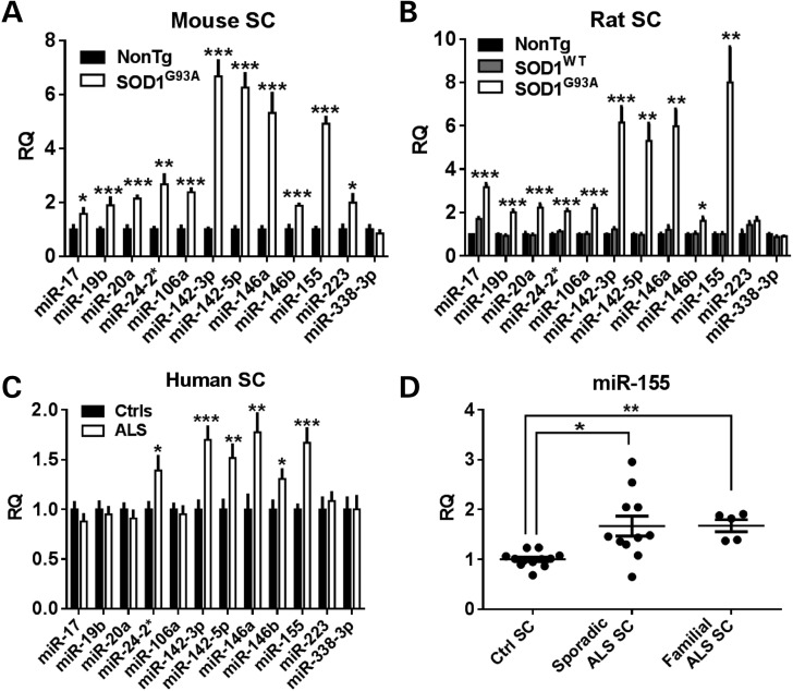
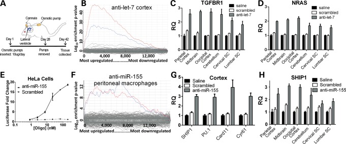
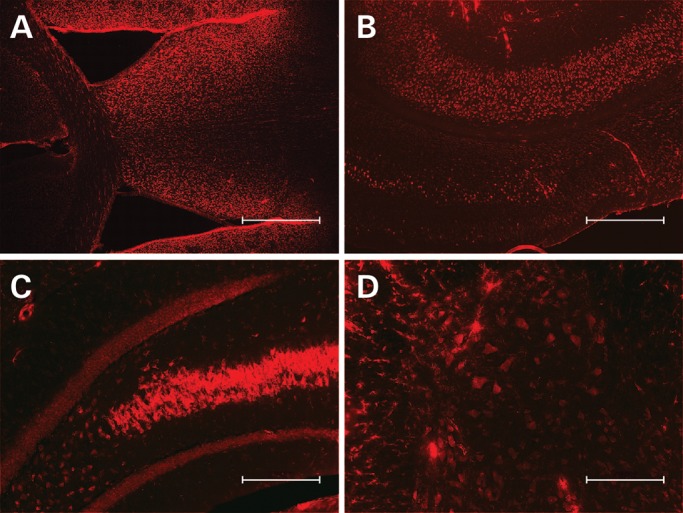
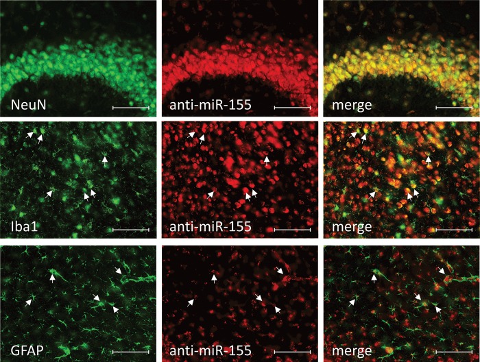
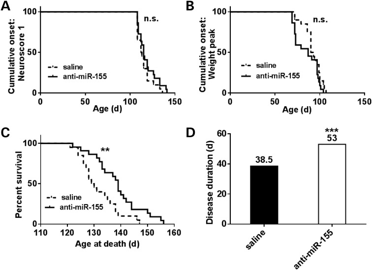
microRNAs (miRNAs) are dysregulated in a variety of disease states, suggesting that this newly discovered class of gene expression repressors may be viable therapeutic targets. A microarray of miRNA changes in ALS-model superoxide dismutase 1 (SOD1)(G93A) rodents identified 12 miRNAs as significantly changed. Six miRNAs tested in human ALS tissues were confirmed increased. Specifically, miR-155 was increased 5-fold in mice and 2-fold in human spinal cords. To test miRNA inhibition in the central nervous system (CNS) as a potential novel therapeutic, we developed oligonucleotide-based miRNA inhibitors (anti-miRs) that could inhibit miRNAs throughout the CNS and in the periphery. Anti-miR-155 caused global derepression of targets in peritoneal macrophages and, following intraventricular delivery, demonstrated widespread functional distribution in the brain and spinal cord. After treating SOD1(G93A) mice with anti-miR-155, we significantly extended survival by 10 days and disease duration by 15 days (38%) while a scrambled control anti-miR did not significantly improve survival or disease duration. Therefore, antisense oligonucleotides may be used to successfully inhibit miRNAs throughout the brain and spinal cord, and miR-155 is a promising new therapeutic target for human ALS.


Figures

Figure 1.
Figure 1.
miRNA array changes were confirmed in mouse, rat and human spinal cord samples. (A) Using individual TaqMan miRNA assays, 11 out of the 12 miRNA microarray hits were confirmed significantly increased in end-stage mouse spinal cord tissue (n = 5–6). (B) In rat lumbar spinal cord tissue, 10 out of the 12 miRNAs tested were increased in end-stage superoxide dismutase 1 (SOD1)G93A rats over non-transgenic and overexpressing SOD1WT age-matched controls (n = 4,4,4). (C) In autopsy spinal cord samples from 16 ALS patients and 12 non-ALS controls, 6 miRNAs assayed were significantly increased in the ALS tissue. (D) Specifically, both sporadic and familial ALS patients showed a significant increase in miR-155 (n = 12 controls; 1.7 × increase in n = 11 sporadic ALS; 1.7 × increase in n = 5 familial ALS; ANOVA with Tukey's post-hoc test). (SC, spinal cord. *P < 0.05, **P < 0.01, ***P < 0.001.)
Figure 2.
Figure 2.
Let-7 and miR-155 anti-miRs distribute throughout CNS and derepress target mRNAs. (A) Saline, scrambled anti-miR control or anti-let-7 was infused directly to the lateral ventricle for 28 days with an osmotic pump. At 42 days, RNA was extracted. (B) Using Affymetrix 430 2.0 mouse gene arrays and Sylamer software analysis, cortical mRNA with let-7 binding sites (blue and red lines denoting mRNAs that contain the 1–7 nt or 2–8 nt miRNA seed region compliment, respectively) was enriched amongst the upregulated (derepressed) mRNAs for anti-let-7-treated mice as normalized to scrambled-treated mice. (C and D) mRNA of two confirmed let-7 targets (TGFBR1, NRAS) were significantly increased in anti-let-7-treated mice in all regions assayed (P < 0.05 TGFBR1; P < 0.01 NRAS). (E) Hela cells were transfected with a miR-155 expression plasmid and a luciferase reporter containing two miR-155 binding sites. After 4 h, either anti-miR-155 or a scrambled control anti-miR was transfected into cells (1–200 nM). Quantified 24 h later, luciferase levels were increased in anti-miR-155-treated cells, indicating inhibition of miR-155. (F) After 3 daily IP injections of either anti-miR-155 or scrambled anti-miR control, peritoneal macrophages were isolated from adult mice, stimulated with LPS, and RNA was extracted. An Affymetrix microarray of mRNA followed by Sylamer analysis shows derepression of mRNAs that contain the heptamer seed region compliment to miR-155 (blue line denotes 1–7nt miRNA heptamer compliment, red for 2–8-nt heptamer). (G) anti-miR-155 was infused into the lateral ventricles of adult mice for 4 weeks. Two weeks later, mRNA was extracted. mRNA levels of 4 confirmed and/or predicted miR-155 targets were significantly increased in anti-miR-155-treated mice over both saline- and scrambled-treated mice [Src homology-2 domain-containing inositol 5 phosphatase 1 (SH1P1) (P < 0.001, PU.1 P < 0.001; Card11 P < 0.01, Cyr61 P < 0.001]. (H) SHIP1, a confirmed miR-155 target, was significantly increased in anti-miR-155-treated mice in all regions assayed (P < 0.05).
Figure 3.
Figure 3.
cy3-anti-miR-155 distributes from lateral cerebral ventricle throughout brain and spinal cord. Mice were treated for 2 weeks with an osmotic pump delivering 10 µg/day cy3-labeled anti-miR-155 directly into the lateral ventricle. Anti-miR-155 distributes throughout the (A) subventricular zone (4×, scale bar = 500 µm), (B) cortical layers (4×, scale bar = 500 µm), (C) hippocampus (10×, scale bar = 200 µm) and (D) spinal cord (10×, scale bar = 200 µm).
Figure 4.
Figure 4.
cy3-anti-miR-155 distributes into neurons, microglia and astrocytes. Mice were treated for 2 weeks with an osmotic pump delivering 10 µg/day cy3-labeled anti-miR-155 directly into the lateral ventricle (B, E, H). 40-µm brain slices were collected, and cell-specific antibodies were used against NeuN to mark neurons (A), Iba1 to mark microglia (D) and GFAP to mark astrocytes (G). cy3-anti-miR-155 is present in all cell types assayed (C, F, I, arrowheads mark examples of double positive cells) (20×, scale bar = 100 µm).
Figure 5.
Figure 5.
Survival, but not onset, is extended in anti-miR-155-treated ALS mice. SOD1G93A SJL mice were treated with both osmotic pumps directed to the lateral ventricles and with weekly IP injections starting at 60 days of age. Mice were weighed and neurological scores were determined biweekly. A score of 1 marked onset and was characterized by one of the following: (i) failure to fully extend legs when lifted by tail; (ii) failure to spread legs past midline when lifted by tail and (iii) marked tremors when lifted by tail. Survival was determined as when the mouse could not right itself within 30 s of being placed on either side. (A and B) Saline (n = 20) and anti-miR-155-treated mice (n = 22) had no significant difference in onset as determined by neurological score of 1 or by age at peak weight. (C and D) Anti-miR-155-treated mice had a significant extension in survival and a 38% extension in disease duration over saline (median values shown, log-rank test, *P < 0.01, **P < 0.01, ***P < 0.001).

References

    1. Lee R.C., Feinbaum R.L., Ambros V. The C. elegans heterochronic gene lin-4 encodes small RNAs with antisense complementarity to lin-14. Cell. 1993;75:843–854. - PubMed
    1. Rothstein J.D. Current hypotheses for the underlying biology of amyotrophic lateral sclerosis. Ann. Neurol. 2009;65(Suppl 1):S3–S9. - PubMed
    1. Landgraf P., Rusu M., Sheridan R., Sewer A., Iovino N., Aravin A., Pfeffer S., Rice A., Kamphorst A.O., Landthaler M., et al. A mammalian microRNA expression atlas based on small RNA library sequencing. Cell. 2007;129:1401–1414. - PMC - PubMed
    1. O'Connell R.M., Chaudhuri A.A., Rao D.S., Baltimore D. Inositol phosphatase SHIP1 is a primary target of miR-155. Proc. Natl. Acad. Sci. USA. 2009;106:7113–7118. - PMC - PubMed
    1. Wang X., Zhao Q., Matta R., Meng X., Liu X., Liu C.G., Nelin L.D., Liu Y. Inducible nitric-oxide synthase expression is regulated by mitogen-activated protein kinase phosphatase-1. J. Biol. Chem. 2009;284:27123–27134. - PMC - PubMed

Publication types

MeSH terms