. 2013 Jul 1;191(1):30-4.
doi: 10.4049/jimmunol.1300121. Epub 2013 Jun 5.

Cutting edge: CD1d restriction and Th1/Th2/Th17 cytokine secretion by human Vδ3 T cells

Affiliations

Cutting edge: CD1d restriction and Th1/Th2/Th17 cytokine secretion by human Vδ3 T cells

Bozgana A Mangan et al. J Immunol. .

Abstract

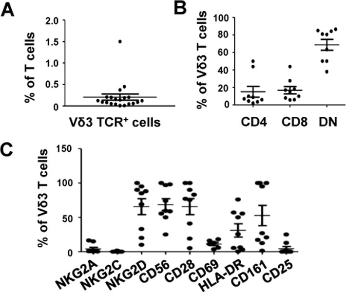
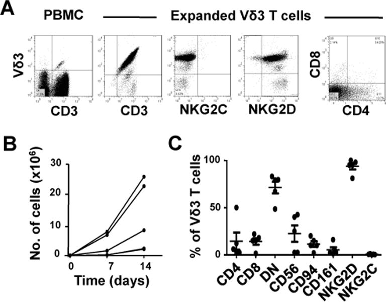
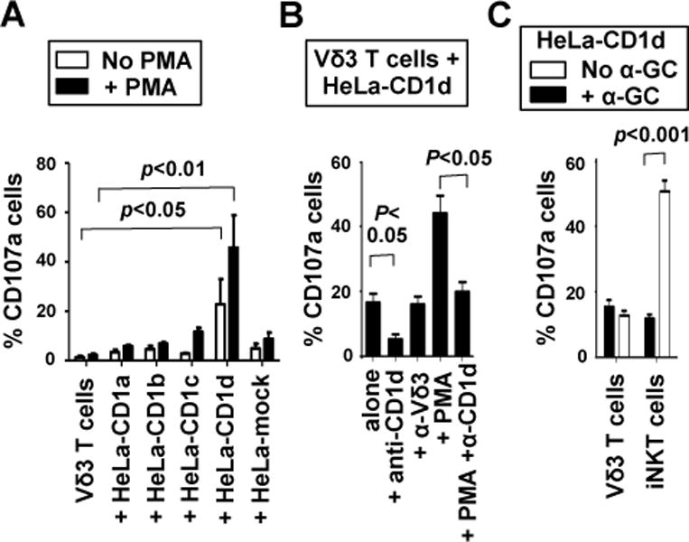
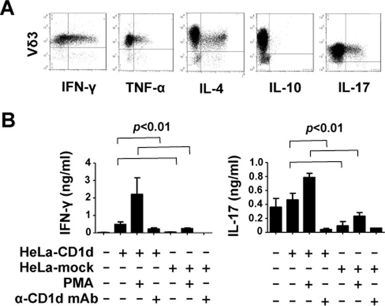
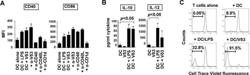
Human γδ T cells expressing the Vδ3 TCR make up a minor lymphocyte subset in blood but are enriched in liver and in patients with some chronic viral infections and leukemias. We analyzed the frequencies, phenotypes, restriction elements, and functions of fresh and expanded peripheral blood Vδ3 T cells. Vδ3 T cells accounted for ~0.2% of circulating T cells, included CD4(+), CD8(+), and CD4(-)CD8(-) subsets, and variably expressed CD56, CD161, HLA-DR, and NKG2D but neither NKG2A nor NKG2C. Vδ3 T cells were sorted and expanded by mitogen stimulation in the presence of IL-2. Expanded Vδ3 T cells recognized CD1d but not CD1a, CD1b, or CD1c. Upon activation, they killed CD1d(+) target cells, released Th1, Th2, and Th17 cytokines, and induced maturation of dendritic cells into APCs. Thus, Vδ3 T cells are glycolipid-reactive T cells with distinct Ag specificities but functional similarities to NKT cells.


Figures

Fig. 1
Fig. 1. Frequencies and phenotypes of human Vδ3 T cells
A, Scatterplot showing percentages of freshly-isolated circulating CD3+ cells from 20 healthy donors that express the Vδ3 TCR chain as detected by flow cytometry. B, Percentages of circulating Vδ3 T cells from 9 healthy donors that express CD4, CD8 and neither (DN). C, Percentages of circulating Vδ3 T cells from 9 healthy donors that express NKG2A, NKG2C, NKG2D CD56, CD28, CD69, HLA-DR, CD161 and CD25. Horizontal lines show means.
Fig. 2
Fig. 2. Ex vivo expansion of human Vδ3 T cells
A, Flow cytometric dot plots showing the expression of CD3 and the Vδ3 TCR by fresh PBMC (left panel) and by sorted and expanded Vδ3 T cells (second panel) and the expression of NKG2C (third panel), NKG2D (fourth panel) and CD4 and CD8 (right panel) by expanded Vδ3 T cells. B, Kinetics of mitogen-stimulated Vδ3 T cell expansion starting with 1,000 sorted Vδ3 T cells. C, Percentages of Vδ3 T cells expanded from 5 healthy donors that express CD4, CD8 or neither (DN), CD56, CD94, CD161, NKG2D and NKG2C. Horizontal lines show means.
Fig. 3
Fig. 3. Vδ3 T cells recognize CD1d
A, Mean percentages of Vδ3 T cells expanded from 5 donors that externalise CD107a after culturing for 4 hours in medium alone, with HeLa cells expressing CD1a, CD1b, CD1c and CD1d, and with mock-transfected HeLa cells in the absence and presence of PMA. B, mean percentages of expanded Vδ3 T cells from 3 donors that degranulate in response to HeLa-CD1d in the absence and presence of PMA and mAbs specific for CD1d (α-CD1d) or Vδ3 (α-Vδ3). C, mean percentages of Vδ3 T cells or iNKT cells from 5 donors that degranulate in response to HeLa-CD1d cells in the absence and presence of α-GC. Error bars show SEM.
Fig. 4
Fig. 4. Vδ3 T cells produce multiplecytokines upon stimulation with PMA and ionomycin or CD1d+ cells
A, Flow cytometric dot plots showing expression of IFN-γ, TNF-α, IL-4, IL-10 and IL-17 by expanded Vδ3 T cell lines after stimulation for 4 hours with PMA and ionomycin. Results are representative of data using expanded Vδ3 T cells from 6 donors. B, Mean levels of IFN-γ and IL-17 released by expanded Vδ3 T cells from 4 donors after stimulation for 24 hours with mock transfected HeLa cells or HeLa-CD1d cells in the absence or presence of PMA and blocking anti-CD1d mAb. No blocking was seen when isotype-matched control Ab was used (not shown). Error bars show SEM.
Fig. 5
Fig. 5. Vδ3 T cells induce dendritic cell (DC) maturation
A, Mean fluorescence intensities (MFI) of expression of CD40 and CD86 by monocyte-derived DC after culturing them for 3 days in medium alone, with LPS or with equal numbers of Vδ3 T cells in the absence and presence of blocking mAbs against CD1d, Vδ3, CD40 and CD40L. Results are means of 5 different DC-Vδ3 T cell combinations. Error bars show SEM. B, Levels of IL-10 and IL-12 released by DC or Vδ3 T cells cultured alone for 2 days, DC cultured with LPS and DC cultured with Vδ3 T cells. Results are means of 3 experiments using different DC and Vδ3 T cells. C, Proliferation of naïve allogeneic T cells in response to medium alone, immature DC, or DC matured for 24 hours with LPS or Vδ3 T cells. T cells were labelled with Cell Trace Violet before adding to the DC. Results show representative flow cytometry histograms (from 4 experiments) showing Cell Trace dye dilution after 6 days.

References

    1. Bendelac A, Savage PB, Teyton L. The biology of NKT cells. Annu Rev Immunol. 2007;25:297–336. - PubMed
    1. Berzins SP, Smyth MJ, Baxter AG. Presumed guilty: natural killer T cell defects and human disease. Nat Rev Immunol. 2011;11:131–142. - PubMed
    1. Le Bourhis L, Guerri L, Dusseaux M, Martin E, Soudais C, Lantz O. Mucosal-associated invariant T cells: unconventional development and function. Trends Immunol. 2011;32:212–218. - PubMed
    1. Uldrich AP, Patel O, Cameron G, Pellicci DG, Day EB, Sullivan LC, Kyparissoudis K, Kjer-Nielsen L, Vivian JP, Cao B, Brooks AG, Williams SJ, Illarionov P, Besra GS, Turner SJ, Porcelli SA, McCluskey J, Smyth MJ, Rossjohn J, Godfrey DI. A semi-invariant Vα10+ T cell antigen receptor defines a population of natural killer T cells with distinct glycolipid antigen-recognition properties. Nat Immunol. 2011;12:616–623. - PMC - PubMed
    1. Morita CT, Jin C, Sarikonda G, Wang H. Nonpeptide antigens, presentation mechanisms, and immunological memory of human Vγ2Vδ2 T cells: discriminating friend from foe through the recognition of prenyl pyrophosphate antigens. Immunol Rev. 2007;215:59–76. - PubMed

Publication types

Substances