. 2013 Aug;87(15):8687-96.
doi: 10.1128/JVI.00376-13. Epub 2013 Jun 5.

The kinase activity of ataxia-telangiectasia mutated interferes with adenovirus E4 mutant DNA replication

Affiliations

The kinase activity of ataxia-telangiectasia mutated interferes with adenovirus E4 mutant DNA replication

Dipendra Gautam et al. J Virol. 2013 Aug.

Abstract

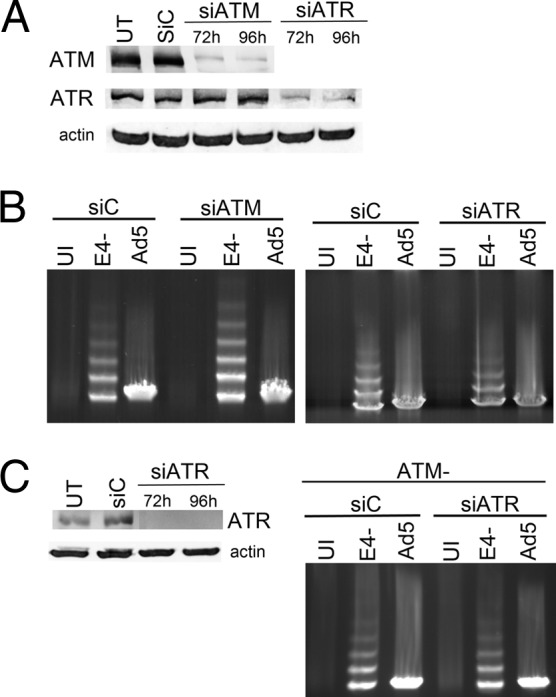
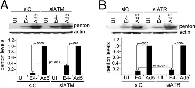
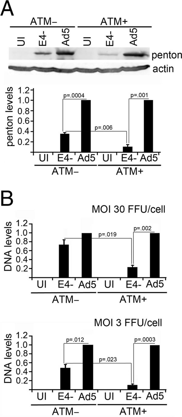
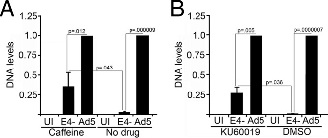
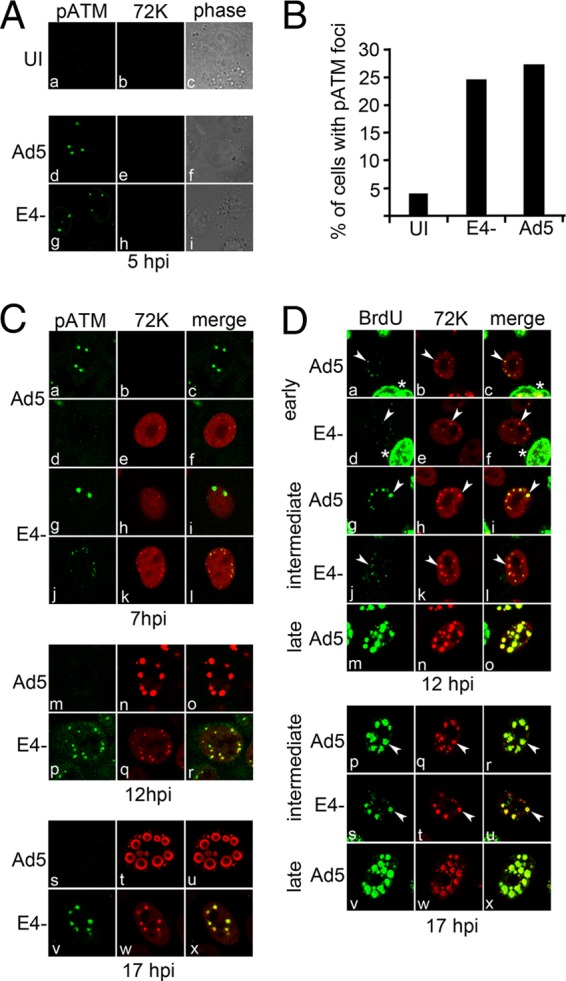
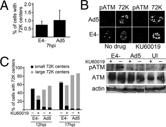
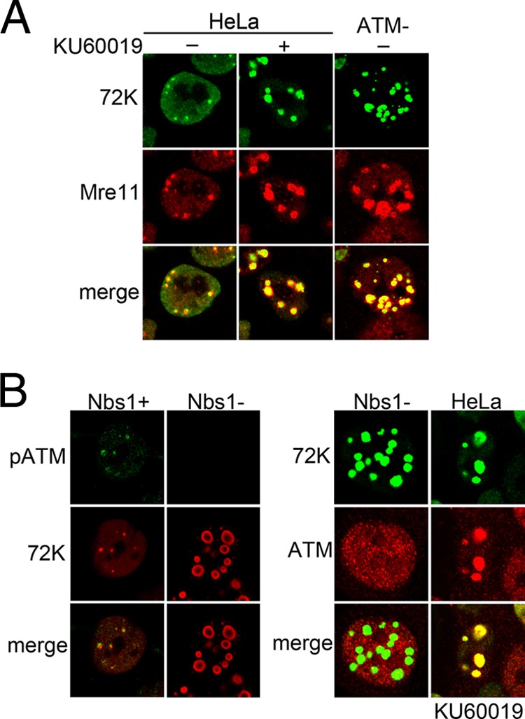
Adenovirus (Ad) mutants that lack early region 4 (E4) are unable to produce the early regulatory proteins that normally inactivate the Mre11/Rad50/Nbs1 (MRN) sensor complex, which is a critical component for the ability of cells to respond to DNA damage. E4 mutant infection therefore activates a DNA damage response, which in turn interferes with a productive viral infection. MRN complex proteins localize to viral DNA replication centers in E4 mutant-infected cells, and this complex is critical for activating the kinases ataxia-telangiectasia mutated (ATM) and ATM and Rad3-related (ATR), which phosphorylate numerous substrates important for DNA repair, cell cycle checkpoint activation, and apoptosis. E4 mutant growth defects are substantially rescued in cells lacking an intact MRN complex. We have assessed the role of the downstream ATM and ATR kinases in several MRN-dependent E4 mutant phenotypes. We did not identify a role for either ATM or ATR in "repair" of E4 mutant genomes to form concatemers. ATR was also not observed to contribute to E4 mutant defects in late protein production. In contrast, the kinase activity of ATM was important for preventing efficient E4 mutant DNA replication and late gene expression. Our results suggest that the MRN complex interferes with E4 mutant DNA replication at least in part through its ability to activate ATM.


Figures

Fig 1
Fig 1
ATM and ATR are not required for genome concatenation. HeLa cells were either untreated (UT), transfected with control siRNAs (siC), or specific siRNAs targeting either ATM (siATM) or ATR (siATR) mRNAs. (A) Transfected cells were processed for Western blotting to determine the levels of ATM or ATR at 72 and 96 hpt, or analyzed for the presence of β-actin as a control. (B) Cells transfected with the indicated siRNAs were either left uninfected (UI) or infected for 24 h with Ad5 or E4- at 30 FFU/cell from 72–96 hpt, and total DNA was analyzed by PFGE to measure the level of viral DNA concatemers. (C) siRNA knockdown of ATR was performed in ATM− cells. ATM− cells were transfected with control or ATR specific siRNA and the levels of ATR and β-actin proteins were determined by Western blotting (left panel). Total DNA from transfected cells infected with Ad5 or E4- was analyzed by PFGE to measure the level of viral DNA concatemers (right panel) as described for B.
Fig 2
Fig 2
ATM interferes with E4 mutant late protein production while ATR does not. HeLa cells were either transfected with control siRNAs (siC), or specific siRNAs targeting either ATM (siATM) or ATR (siATR) mRNAs. Cells were then either left uninfected (UI) or infected with Ad5 or E4- as described for Fig. 1, and at 24 hpi analyzed for the presence of the late protein penton by Western blotting. Representative Western blots of penton levels achieved in cells treated with siATM (A) or siATR (B) are shown in the top panels. Levels of the control protein β-actin are also shown. E4- penton protein levels were quantified by phosphorimaging analysis of Western blots from 3 independent experiments and expressed as the fraction of the level achieved by Ad5, which was set at 1 (A and B, bottom graphs). Error bars show the standard error of the mean. A one-tailed Student's t test analysis was performed on the data used to generate the graphs. Statistically significant (P < 0.05) differences between the columns are indicated. N.S. = not statistically significantly different.
Fig 3
Fig 3
ATM interferes with E4 mutant DNA replication. ATM− and isogenic ATM+ cells complemented for ATM, were infected with Ad5 or E4- at an MOI of 30 FFU/cell unless otherwise indicated. At 24 hpi cells were harvested and processed for Western blot analysis to study viral late protein levels, or total DNA was prepared and analyzed by dot blotting to quantify viral DNA levels. (A) Top panel shows a representative Western blot using an antibody that detects the viral late protein penton. Levels of the control protein β-actin are also shown. E4- penton protein levels were quantified by phosphorimaging analysis of Western blots from 3 independent experiments and expressed as the fraction of the level achieved by Ad5, which was set at 1 (bottom graph). Error bars show the standard error of the mean. Statistically significant differences between the columns are indicated, as determined by one-tailed Student's t test. (B) Graphs showing the results of dot blot quantitation of viral DNA levels in experiments performed at 30 (top graph) and 3 (bottom graph) FFU/cell. E4- DNA levels were quantified from 3 independent experiments and expressed as the fraction of the level achieved by Ad5, which was set at 1. Error bars show the standard error of the mean. Statistically significant differences between the columns are indicated, as determined by one-tailed Student's t test.
Fig 4
Fig 4
The kinase activity of ATM is important for inhibiting E4 mutant DNA replication. HeLa cells were infected with Ad5 or E4- at an MOI of 3 FFU/cell and subsequently incubated with the ATM kinase inhibitors caffeine (A) or KU60019 (B) from 2–24 hpi. Control infected cells were incubated in medium without the inhibitors but containing the same amount of DMSO solvent used for the drug treatment where indicated. Total DNA samples from 3 independent experiments were analyzed by dot blotting to measure viral DNA levels. E4- DNA levels were expressed as the fraction of the level achieved by Ad5, which was set at 1. Error bars show the standard error of the mean. Statistically significant differences between the columns are indicated, as determined by one-tailed Student's t test.
Fig 5
Fig 5
Time course showing pATM localization and BrdU incorporation relative to DNA replication centers in Ad5 and E4- infections. HeLa cells were uninfected (UI) or infected with Ad5 or E4- at 3 FFU/cell for the times indicated and then fixed and immunostained with antibodies against pATM (phosphoepitope S1981), BrdU, or the viral DNA binding protein E2-72kDa (72K) to detect viral DNA replication centers. (A) Immunofluorescence confocal micrographs showing pATM (green) and E2-72kDa (red) staining, or phase contrast images of uninfected cells or cells infected with Ad5 and E4- at 5 hpi. (B) Uninfected cells and cells infected with Ad5 or E4- were blindly scored for the presence of pATM foci at 5 hpi, and the results presented as a graph showing the percentage of cells in the culture with pATM foci. (C) Immunofluorescence confocal micrographs showing pATM (green), E2-72kDa (red), and merged staining patterns observed at 7, 12 and 17 hpi with Ad5 or E4-. (D) Ad5 or E4- infected HeLa cells were incubated with 150 mM BrdU for 1 h at either 12 or 17 hpi. Immunofluorescence confocal micrographs showing BrdU (green), E2-72kDa (red), and merged staining patterns are shown. Cells with early-, intermediate-, or late-stage replication centers were identified based on the size and shape of the E2-72kDa foci. Similarly sized replication centers in Ad5 and E4- infections are marked with white arrowheads to facilitate comparison of BrdU incorporation at these centers. Cells with intermediate- and late-stage DNA replication centers were the most prevalent phenotype in Ad5 infections while cells with early stage replication foci were the most prevalent phenotype in E4- infections at these time points. Cells marked with an asterix are examples of cells with widespread incorporation of BrdU in cellular DNA.
Fig 6
Fig 6
ATM inhibition facilitates progression of E4- DNA replication centers from small early replication foci to larger intermediate- and late-stage replication centers. HeLa cells were infected with Ad5 or E4- and then either incubated with medium containing DMSO only, or 5 μM KU60019 in DMSO to inhibit ATM kinase activity from 2 hpi until the times indicated. (A) The percentage of cells infected with Ad5 and E4- with E2-72kDa (72K) replication foci was scored at 7 hpi. (B) The top panel shows immunofluorescence micrographs of pATM and E2-72kDa staining at 17 hpi with Ad5 and E4- incubated in the presence or absence of KU60019. The bottom panel shows Western blot analysis of extracts prepared from uninfected (UI) cells or from cells infected with Ad5 or E4- at 17 hpi that were or were not treated with KU60019, and probed with antibodies against pATM, ATM, or β-actin. (C) The percentage of cells containing small (black bars) or large (gray bars) E2-72kDa containing DNA replication centers was determined in Ad5 and E4- infections that were and were not incubated with KU60019 from 2 hpi until the times indicated.
Fig 7
Fig 7
Localization of Mre11 or ATM to viral replication centers does not interfere with E4- DNA replication when the ATM kinase is inactivated. (A) HeLa or ATM− cells were infected with E4- at 3 FFU/cell and fixed for immunofluorescence at 17 hpi. The indicated infections were incubated with 5 μM KU60019 from 2–17 hpi to inhibit ATM kinase activity. Immunofluorescence micrographs showing E2-72kDa (green) and Mre11 (red) and merged staining patterns are shown. (B) Nbs1+, Nbs1−, and HeLa cells were infected with E4- at 3 FFU/cell and fixed for immunofluorescence at 17 hpi. E4- infected HeLa cells were incubated with 5 μM KU60019 from 2–17 hpi to inhibit ATM kinase activity. Immunofluorescence micrographs showing E2-72kDa (72K), pATM, ATM, and merged staining patterns are shown.

Similar articles

Cited by

References

    1. Weitzman MD, Ornelles DA. 2005. Inactivating intracellular antiviral responses during adenovirus infection. Oncogene 24:7686–7696 - PubMed
    1. Evans JD, Hearing P. 2005. Relocalization of the Mre11-Rad50-Nbs1 complex by the adenovirus E4 ORF3 protein is required for viral replication. J. Virol. 79:6207–6215 - PMC - PubMed
    1. Lakdawala SS, Schwartz RA, Ferenchak K, Carson CT, McSharry BP, Wilkinson GW, Weitzman MD. 2008. Differential requirements of the C terminus of Nbs1 in suppressing adenovirus DNA replication and promoting concatemer formation. J. Virol. 82:8362–8372 - PMC - PubMed
    1. Mathew SS, Bridge E. 2007. The cellular Mre11 protein interferes with adenovirus E4 mutant DNA replication. Virology 365:346–355 - PubMed
    1. Mathew SS, Bridge E. 2008. Nbs1-dependent binding of Mre11 to adenovirus E4 mutant viral DNA is important for inhibiting DNA replication. Virology 374:11–22 - PMC - PubMed

Publication types

LinkOut - more resources